BTemplates.com

Powered by Blogger.

Pageviews past week

Quantum mechanics

Auto News

artificial intelligence

About Me

Recommend us on Google!

Information Technology

Popular Posts

Showing posts with label Science Updates. Show all posts

Friday, June 28, 2013

Breaking habits before they start


Our daily routines can become so ingrained that we perform them automatically, such as taking the same route to work every day. Some behaviors, such as smoking or biting your fingernails, become so habitual that we can't stop even if we want to.


Although breaking habits can be hard, MIT neuroscientists have now shown that they can prevent them from taking root in the first place, in rats learning to run a maze to earn a reward. The researchers first demonstrated that activity in two distinct brain regions is necessary in order for habits to crystallize. Then, they were able to block habits from forming by interfering with activity in one of the brain regions—the infralimbic (IL) cortex, which is located in the prefrontal cortex.

The MIT researchers, led by Institute Professor Ann Graybiel, used a technique called optogenetics to block activity in the IL cortex. This allowed them to control cells of the IL cortex using light. When the cells were turned off during every maze training run, the rats still learned to run the maze correctly, but when the reward was made to taste bad, they stopped, showing that a habit had not formed. If it had, they would keep going back by habit.

"It's usually so difficult to break a habit," Graybiel says. "It's also difficult to have a habit not form when you get a reward for what you're doing. But with this manipulation, it's absolutely easy. You just turn the light on, and bingo."

Graybiel, a member of MIT's McGovern Institute for Brain Research, is the senior author of a paper describing the findings in the June 27 issue of the journal Neuron. Kyle Smith, a former MIT postdoc who is now an assistant professor at Dartmouth College, is the paper's lead author.

Patterns of habitual behavior

Previous studies of how habits are formed and controlled have implicated the IL cortex as well as the striatum, a part of the brain related to addiction and repetitive behavioral problems, as well as normal functions such as decision-making, planning and response to reward. It is believed that the motor patterns needed to execute a habitual behavior are stored in the striatum and its circuits.

Recent studies from Graybiel's lab have shown that disrupting activity in the IL cortex can block the expression of habits that have already been learned and stored in the striatum. Last year, Smith and Graybiel found that the IL cortex appears to decide which of two previously learned habits will be expressed.

"We have evidence that these two areas are important for habits, but they're not connected at all, and no one has much of an idea of what the cells are doing as a habit is formed, as the habit is lost, and as a new habit takes over," Smith says.

To investigate that, Smith recorded activity in cells of the IL cortex as rats learned to run a maze. He found activity patterns very similar to those that appear in the striatum during habit formation. Several years ago, Graybiel found that a distinctive "task-bracketing" pattern develops when habits are formed. This means that the cells are very active when the animal begins its run through the maze, are quiet during the run, and then fire up again when the task is finished.

This kind of pattern "chunks" habits into a large unit that the brain can simply turn on when the habitual behavior is triggered, without having to think about each individual action that goes into the habitual behavior.

The researchers found that this pattern took longer to appear in the IL cortex than in the striatum, and it was also less permanent. Unlike the pattern in the striatum, which remains stored even when a habit is broken, the IL cortex pattern appears and disappears as habits are formed and broken. This was the clue that the IL cortex, not the striatum, was tracking the development of the habit.

Multiple layers of control


The researchers' ability to optogenetically block the formation of new habits suggests that the IL cortex not only exerts real-time control over habits and compulsions, but is also needed for habits to form in the first place.

"The previous idea was that the habits were stored in the sensorimotor system and this cortical area was just selecting the habit to be expressed. Now we think it's a more fundamental contribution to habits, that the IL cortex is more actively making this happen," Smith says.

This arrangement offers multiple layers of control over habitual behavior, which could be advantageous in reining in automatic behavior, Graybiel says. It is also possible that the IL cortex is contributing specific pieces of the habitual behavior, in addition to exerting control over whether it occurs, according to the researchers. They are now trying to determine whether the IL cortex and the striatum are communicating with and influencing each other, or simply acting in parallel.

The study suggests a new way to look for abnormal activity that might cause disorders of repetitive behavior, Smith says. Now that the researchers have identified the neural signature of a normal habit, they can look for signs of habitual behavior that is learned too quickly or becomes too rigid. Finding such a signature could allow scientists to develop new ways to treat disorders of repetitive behavior by using deep brain stimulation, which uses electronic impulses delivered by a pacemaker to suppress abnormal brain activity.

Journal reference: Neuron

Provided by Massachusetts Institute of Technolog

Thursday, September 23, 2010

Credit Card with a Computer Inside A smarter credit card could mean new security features and other functionality.


A programmable credit card can display useful information, offer added security features, and even act as several different cards by rewriting its own magnetic strip.
Smarter card: A user has to enter a PIN to display this
card’s full number and unlock its magnetic stripe for use
either online or in-store. After a short time the display
and magnetic stripe become blank again.
Credit: Dynamics

Two types of programmable credit cards were unveiled this week at the DEMO conference in Santa Clara, California, by Dynamics, a startup based in Pittsburgh that's been developing the technology in stealth mode for three years. The company raised $5.7 million of funding last year.

The new cards are no bigger than the one in your wallet, and is actually slightly more flexible. It can display information at the press of a button, and can become several different cards by rewriting its own magnetic strip.

The "MultiAccount" card has two buttons on its face, each with an indicator light that can be pressed to record data to its magnetic strip. "One might switch the card to be your debit card, and the other your credit card," says Dynamics CEO Jeff Mullen. "These cards are exactly the same size and thickness of a conventional card, and the lithium-polymer battery inside can last four years under high usage. They're also fully waterproof, so you can put them through the washing machine."

The "Hidden" card features a keypad and black-and-white display for six of the digits in the card's unique number. Once the correct PIN is entered on the card's four buttons, the missing digits are filled in and the card's magnetic strip is populated with data. Both the digits and the strip become blank again after a short time. "If this card is lost, it's just dead plastic to anyone who finds it," says Mullen, who thinks it could help banks attract security-conscious consumers.

That may be true, says Avivah Litan, a Gartner analyst who researches security and technology in the financial sector, but "most card data is stolen electronically, in large volumes," she notes. As a result, banks "may prevent a few percentage points [of fraud], but it seems unlikely to be worth the investment for them."

The MultiAccount card may be more attractive to the financial sector than the "Hidden" card. "It could help the very large card issuers, such as Chase, that have a lot of overlap between their credit and debit accounts," says Litan. However, convincing banks to invest in an unproven technology will require the potential for a very strong effect on their bottom lines, she says.

Mullen says Dynamics's cards are significantly more expensive to produce than standard credit cards, but argues that the additional cost is offset by the benefits to a bank. "These cards are significant revenue generators for them, not cost centers," he says.

Banks already target different types of cards to particular demographics, and use reward schemes to attract new business and encourage heavy use of their products. Cards with computational smarts inside could enable more of that, Mullen argues. For example, a credit card that can suddenly act as a loyalty card might encourage customers to use a scheme that they otherwise wouldn't.

"Cards with this technology have been used in large numbers in stealth trials in the U.S. since earlier this year," says Mullen, who adds that banking partners will begin talking about their plans for the technology in coming months. A particular attraction for banks, he says, is that the cards are compatible with existing infrastructure, unlike contactless payments based on RFID chips.

"There are 16 million magnetic stripe readers in the world," he says. "It's hard to change that, but easy to upgrade your own cards without building new infrastructure."

Dynamics is also working on cards that include E Ink-style displays that remain switched on for longer periods, and the company is also investigating a card that can transfer more data. Typically only a third of the magnetic strip on a card carries the card's details, says Mullen. "You can send messages between card and reader using the rest of that area."

Monday, July 12, 2010

Surprisingly Regular Patterns in Hurricane Energy Discovered


Researchers at the Mathematics Research Centre and Universitat Autònoma de Barcelona have discovered the mathematical relation between the number of hurricanes produced in certain parts of Earth and the energy they release. The distribution is valid for all series of hurricanes under study, independent of when and where they occurred.
Image
Meteorologist puts his finger on the eye of a 
hurricane. Earth image: visibleearth.nasa.gov. 
(Credit: iStockphoto/Eric Hood)

The research, published in Nature Physics, suggests that the evolution of hurricane intensity will be very difficult to predict.

It is well known that there are fewer probabilities of a devastating hurricane developing than of a modest one. However, the exact relation between the number of hurricanes and energy released was not known until now. Researchers from the Mathematics Research Centre (CRM) and the Department of Physics of Universitat Autònoma de Barcelona have analysed data corresponding to tropical cyclones (generic name used for hurricanes) which have appeared in different parts of the planet between 1945 and 2007. Scientists have discovered that this relation corresponds to a power-law, a precise mathematical formula cyclones obey in a surprising manner, regardless of where on the planet and when they appear.

This fundamental discovery has led researchers to more general conclusions on the behaviour of hurricanes. The first conclusion states that a hurricane's dynamics can be the result of a critical process, therefore making it impossible to predict its intensity. One of the aspects traditionally studied by organisations monitoring the danger of hurricanes is the prediction of their intensity, since this determines which alert and prevention systems are to be used in populated areas. Despite the efforts of scientists and resources invested, until now results have been very poor, although predictions on hurricane trajectory have improved considerably. The fact that hurricanes follow this power-law, as do other natural phenomena where large amounts of energy are released, e.g. earthquakes, questions the ability to predict the evolution of their intensity. In these types of processes, the dynamics behind large hurricanes are the same as those producing tropical storms of less importance and range. The way in which a small storm evolves and transforms into a catastrophic hurricane depends on whether the fluctuations amplifying the storm are stronger than those which tend to dissipate it. However, there is no specific aspect pointing to which will be the dominant fluctuations, since the system at that moment is in a critical situation, i.e. on the verge of either dissipating or growing.

The second conclusion of the study is related to the effects of global warming on the behaviour of tropical cyclones: a recent increase in activities in the North Atlantic has shown to follow the same pattern as other high-activity periods in the past. Although there has been a dramatic increase in the number of hurricanes occurring in the North Atlantic since mid-1990s when compared to the period starting in the 1970s, the distribution of hurricanes in the 1950s was similar to today's activity level. Therefore, this increase cannot be explained solely on the basis of climate change. Even so, the research points to the existence of a relation between global warming and the distribution of tropical cyclones. The number of hurricanes is inversely proportional to the energy released, except for the highest values of energy, where the relation is suddenly interrupted. Researchers have observed that the cut-off point where the power-law does not represent the behaviour of hurricanes is influenced by factors such as average sea surface temperature and the El Niño phenomenon. Thus at a higher temperature, for example, the cut-off point rises to higher energy values.

The research was carried out by Álvaro Corral, researcher at Mathematics Research Centre (consortium formed by the Institute of Catalan Studies and the Catalan Government, located at the UAB Research Park; CRM is also a CERCA center); Albert Ossó, UAB student in Physics; and Dr Josep Enric Llebot, professor at the UAB Department of Physics.

Thursday, July 8, 2010

Brain's Energy Restored During Sleep


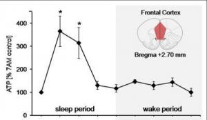
In the initial stages of sleep, energy levels increase dramatically in brain regions found to be active during waking hours, according to new research in the June 30 issue of the Journal of Neuroscience. These results suggest that a surge of cellular energy may replenish brain processes needed to function normally while awake.
Image
currency of cells, in rats increased in four key brain regions normally active during wakefulness. Shown here is the energy surge measured in the frontal cortex, a brain region associated with higher-level thinking. (Credit: Courtesy, with permission: Dworak et al. The Journal of Neuroscience 2010.)

A good night's rest has clear restorative benefits, but evidence of the actual biological processes that occur during sleep has been elusive. Radhika Basheer, PhD, and Robert McCarley, MD, of Boston V.A. Healthcare System and Harvard Medical School, proposed that brain energy levels are key to nightly restoration.

"Our finding bears on one of the perennial conundrums in biology: the function of sleep," Basheer said. "Somewhat surprisingly, there have been no modern-era studies of brain energy using the most sensitive measurements."

The authors measured levels of adenosine triphosphate (ATP), the energy currency of cells, in rats. They found that ATP levels in four key brain regions normally active during wakefulness increased when the rats were in non-REM sleep, but were accompanied by an overall decrease in brain activity. When the animals were awake, ATP levels were steady. When the rats were gently nudged to stay awake three or six hours past their normal sleep times, there was no increase in ATP.

The authors conclude that sleep is necessary for this ATP energy surge, as keeping the rats awake prevented the surge. The energy increase may then power restorative processes absent during wakefulness, because brain cells consume large amounts of energy just performing daily waking functions.

"This research provides intriguing evidence that a sleep-dependent energy surge is needed to facilitate the restorative biosynthetic processes," said Robert Greene, MD, PhD, of the University of Texas Southwestern, a sleep expert who was unaffiliated with the study. He observed that questions arise from the findings, such as the specific cause of the ATP surge. "The authors propose that the surge is related to decreases in brain cell activity during sleep, but it may be due to many other factors as well, including cellular signaling in the brain," he said.

The research was supported by the Department of Veterans Affairs, a Deutsche Forschungsgemeinschaft Fellowship, and the National Institute of Mental Health.

Wednesday, July 7, 2010

Thermal-Powered, Insect-Like Robot Crawls Into Microrobot Contenders' Ring


Robotic cars attracted attention last decade with a 100-mile driverless race across the desert competing for a $1 million prize put up by the U.S. government.
the robot
Tiny, four-sided cilia, pulsating structures that mimic 
the hairs that line the human windpipe, are arranged 
in rows along the underside of the robot. (Credit: John 
Suh, Stanford University)

The past few years have given rise to a growing number of microrobots, miniaturized mobile machines designed to perform specific tasks. And though spectators might need magnifying glasses to see the action, some think the time has come for a microrobotics challenge.

"I'd like to see a similar competition at the small scale, where we dump these microrobots from a plane and have them go off and run for days and just do what they've been told," said Karl Böhringer, a University of Washington professor of electrical engineering. "That would require quite an effort at this point, but I think it would be a great thing."

Researchers at the UW and Stanford University have developed what might one day be a pint-sized contender. Böhringer is lead author of a paper in the June issue of the Journal of Microelectromechanical Systems introducing an insectlike robot with hundreds of tiny legs.

Compared to other such robots, the UW model excels in its ability to carry heavy loads -- more than seven times its own weight -- and move in any direction.

Someday, tiny mobile devices could crawl through cracks to explore collapsed structures, collect environmental samples or do other tasks where small size is a benefit. The UW's robot weighs half a gram (roughly one-hundredth of an ounce), measures about 1 inch long by a third of an inch wide, and is about the thickness of a fingernail.

Technically it is a centipede, with 512 feet arranged in 128 sets of four. Each foot consists of an electrical wire sandwiched between two different materials, one of which expands under heat more than the other. A current traveling through the wire heats the two materials and one side expands, making the foot curl. Rows of feet shuffle along in this way at 20 to 30 times each second.

"The response time is an interesting point about these tiny devices," Böhringer said. "On your stove, it might take minutes or even tens of minutes to heat something up. But on the small scale it happens much, much faster."

The legs' surface area is so large compared to their volume that they can heat up or cool down in just 20 milliseconds.

"It's one of the strongest actuators that you can get at the small scale, and it has one of the largest ranges of motion," Böhringer said. "That's difficult to achieve at the small scale."

The microchip, the robot's body and feet, was first built in the mid 1990s at Stanford University as a prototype part for a paper-thin scanner or printer. A few years later the researchers modified it as a docking system for space satellites. Now they have flipped it over so the structures that acted like moving cilia are on the bottom, turning the chip into an insectlike robot.

"There were questions about the strength of the actuators. Will they be able to support the weight of the device?" Böhringer said. "We were surprised how strong they were. For these things that look fragile, it's quite amazing."

The tiny legs can move more than just the device. Researchers were able to pile paper clips onto the robot's back until it was carrying more than seven times its own weight. This means that the robot could carry a battery and a circuit board, which would make it fully independent. (It now attaches to nine threadlike wires that transmit power and instructions.)

Limbs pointing in four directions allow the robot flexibility of movement.

"If you drive a car and you want to be able to park it in a tight spot, you think, 'Wouldn't it be nice if I could drive in sideways,'" Böhringer said. "Our robot can do that -- there's no preferred direction."

Maneuverability is important for a robot intended to go into tight spaces.

The chip was not designed to be a microrobot, so little effort was made to minimize its weight or energy consumption. Modifications could probably take off 90 percent of the robot's weight, Böhringer said, and eliminate a significant fraction of its power needs.

As with other devices of this type, he added, a major challenge is the power supply. A battery would only let the robot run for 10 minutes, while researchers would like it to go for days.

Another is speed. Right now the UW robot moves at about 3 feet per hour -- and it's far from the slowest in the microrobot pack.

Co-authors are former UW graduate students Yegan Erdem, Yu-Ming Chen and Matthew Mohebbi; UW electrical engineering professor Robert Darling; John Suh at General Motors; and Gregory Kovacs at Stanford.

Research funding was provided by the U.S. Defense Advanced Research Projects Agency, the National Science Foundation and General Motors Co.

Sunday, June 20, 2010

Constraining the Reign of Ancient Egypt: Radiocarbon Dating Helps to Nail Down the Chronology of Kings, Researchers Say


For several thousands of years, ancient Egypt dominated the Mediterranean world -- and scholars across the globe have spent more than a century trying to document the reigns of the various rulers of Egypt's Old, Middle and New Kingdoms. Now, a detailed radiocarbon analysis of short-lived plant remains from the region is providing scientists with a long and accurate chronology of ancient Egyptian dynasties that agrees with most previous estimates but also imposes some historic revisions.
Image
Statue of the head of Ramses II at Luxor temple at night. 
(Credit: iStockphoto/Paul Vinten)

Although previous chronologies have been precise in relative ways, assigning absolute dates to specific events in ancient Egyptian history has been an extremely contentious undertaking. This new study tightly constrains those previous predictions, especially for the Old Kingdom, which was determined to be slightly older than some scholars had believed. The study will also allow for more accurate historical comparisons to surrounding areas, like Libya and Sudan, which have been subject to many radiocarbon dating techniques in the past.

Christopher Bronk Ramsey and colleagues from the Universities of Oxford and Cranfield in England, along with a team of researchers from France, Austria and Israel, collected radiocarbon measurements from 211 various plants -- obtained from museum collections in the form of seeds, baskets, textiles, plant stems and fruits -- that were directly associated with particular reigns of ancient Egyptian kings. They then combined their radiocarbon data with historical information about the order and length of each king's reign to make a complete chronology of ancient Egyptian dynasties.

Their research is published in the June 18 issue of Science, the peer-reviewed journal published by AAAS, the nonprofit science society.

"My colleague, Joanne Rowland, went to a lot of museums, explaining what we were doing and asking for their participation," Bronk Ramsey said. "The museums were all very helpful in providing material we were interested in -- especially important since export of samples from Egypt is currently prohibited. Fortunately, we only needed samples that were about the same size as a grain of wheat."

The researchers' new chronology does indicate that a few events occurred earlier than previously predicted. It suggests, for example, that the reign of Djoser in the Old Kingdom actually started between 2691 and 2625 B.C. and that the New Kingdom began between 1570 and 1544 B.C.

Bronk Ramsey and his colleagues also found some discrepancies in the radiocarbon levels of the Nile Valley, but they suggest that these are due to ancient Egypt's unusual growing season, which is concentrated in the winter months.

For the most part, the new chronology simply narrows down the various historical scenarios that researchers have been considering for ancient Egypt.

"For the first time, radiocarbon dating has become precise enough to constrain the history of ancient Egypt to very specific dates," said Bronk Ramsey. "I think scholars and scientists will be glad to hear that our small team of researchers has independently corroborated a century of scholarship in just three years."

This report by Bronk Ramsey et al. was funded by the Leverhulme Trust with additional financial support from the German-Israeli Foundation for Scientific Research and Development, NERC, CNRS, CEA, IRSN, IRD, and Ministère de La Culture.
Enhanced by Zemanta

Thursday, June 17, 2010

Inbred Sperm Fertilize Fewer Eggs, Beetle Study Finds


Inbred male sperm have been found to fertilize fewer eggs when in competition with non-inbred males, according to a new study by the University of East Anglia.
Image
Red flour beetle. (Credit: Image courtesy of 
University of East Anglia)

Research into the breeding habits of the red flour beetle, published in Proceedings of the Royal Society B, shows that the reduced fitness of inbred beetles, known as 'inbreeding depression', reveals itself in competitive scenarios.

Inbreeding is a potentially important problem in declining species across the world, and conserving genetic variation is now recognized as a priority by the International Union for Conservation of Nature. The new research is potentially vital for the successful implementation of recovery programs of inbred species.

When populations deplete or fragment, relatives can be forced into reproduction, often leading to inbreeding depression.

Led by Dr. Matt Gage, the new research into the promiscuous red flour beetle (Tribolium castaneum) measured how male reproduction responded to forced inbreeding.

After mating brothers with sisters for eight generations, the research found no changes in male fertility or mating behavior.

However, inbred male sperm fertilized fewer eggs when in competition with another non-inbred male, and sperm became more variable in size.

Dr. Gage said: "The experiment was designed to make comparisons with non-inbred control lines. Using multiple inbred lines, we measured the effects of inbreeding on pre- and post-mating success, in the absence and presence of male-male competition."

The results showed no differences between inbred and non-inbred males in terms of mating success, latency, duration, the number of mounts or persistency in a non-competitive setting.

However inbred males suffered significantly reduced sperm competitiveness, fathering an average of 15 per cent fewer offspring than non-inbred males across 330 sperm competition comparisons.

Dr. Gage said: "It seems that inbreeding depression in sperm competitiveness was caused by a decrease in either sperm quantity or quality that is critical for relative competitiveness, but still allows full male fertilization success to be achieved under benign, competition-free conditions.

"We have shown that male fertility and mating competence are not affected by inbreeding and that any decline in sperm quality under inbreeding is only detectable when sperm competition is invoked.

"One limitation to this study is that the ancestral laboratory stock we have used is likely to carry relatively reduced genetic diversity. Also insect sperm do not generally manifest cellular abnormalities akin to those commonly found in more complex mammalian sperm," he added.

The next stage of the research will explore ways that female beetles use multiple mating to generate sperm competition and thereby avoid inbreeding depression of their own fertility.

The research is part of a three-year £400,000 project funded by the Natural Environmental Research Council (NERC). The overall results will help managers of conservation and captive breeding projects recognize when inbreeding is a problem, how it progresses and how best to manage or reverse it.
Enhanced by Zemanta

Thursday, May 27, 2010

Copycat Behavior in Children Is Universal


Children learn a great deal by imitating adults. A new study of Australian preschoolers and Kalahari Bushman children finds that a particular kind of imitation -- overimitation, in which a child copies everything an adult shows them, not just the steps that lead to some outcome -- appears to be a universal human activity, rather than something the children of middle-class parents pick up. The work helps shed light on how humans develop and transmit culture.
Me
For the experiments, children were shown how to open a box -- but in a complicated way, with impractical actions thrown in. For example, the adult would drag a stick across a box, then use a stick to open the box by pulling on a knob -- which is a lot easier if you just use your fingers. Most of the children copied what the adults did, even if they'd been given the opportunity to play with the box first and figure out how it worked. This was just as true for Bushman children as for the Australian children. (Credit: iStockphoto)

Scientists "have been finding this odd effect where children will copy everything that they see an adult demonstrate to them, even if there are clear or obvious reasons why those actions would be irrelevant," says psychologist Mark Nielsen, of the University of Queensland in Australia. "It's something that we know that other primates don't do." If a chimpanzee is shown an irrelevant action, they won't copy it -- they'll skip right to the action that makes something happen.

But it's not clear that the results found in child psychology research apply to all people, Nielsen says. This research is usually done with children who live in Western cultures, whose parents are well educated and middle to upper class. And these parents are constantly teaching their children. But parents in indigenous cultures generally don't spend a lot of time teaching. "They may slow what they're doing if the child is watching, but it's not the kind of active instruction that's common in Western cultures," says Nielsen. So he teamed up with Keyan Tomaselli, an anthropologist at the University of KwaZulu-Natal in Durban, South Africa, who has worked for decades in Bushman communities in southern Africa. Their study is published in Psychological Science, a journal of the Association for Psychological Science.

For the experiments, the children were shown how to open a box -- but in a complicated way, with impractical actions thrown in. For example, the adult would drag a stick across a box, then use a stick to open the box by pulling on a knob -- which is a lot easier if you just use your fingers. Most of the children copied what the adults did, even if they'd been given the opportunity to play with the box first and figure out how it worked. This was just as true for Bushman children as for the Australian children.

But aren't the children just following the rules of what appears to be a game? "That kind of is the point," says Nielsen. "Perhaps not a game, but certainly, when I demonstrate the action, it's purposeful. So from the mind of a child, perhaps there's a reason why I'm doing this." This willingness to assume that an action has some unknown purpose, and to copy it, may be part of how humans develop and share culture, he says. "Really, we see these sorts of behaviors as being a core part of developing this human cultural mind, where we're so motivated to do things like those around us and be like those around us."

Friday, May 14, 2010

Toward Deafness Cure: Inner-Ear Cells Created


Deep inside the ear, specialized cells called hair cells detect vibrations in the air and translate them into sound. Ten years ago, Stefan Heller, PhD, professor of otolaryngology at the Stanford University School of Medicine, came up with the idea that if you could create these cells in the laboratory from stem cells, it would go a long way toward helping scientists understand the molecular basis of hearing in order to develop better treatments for deafness.

Me
Section through the organ of corti
showing inner and outer hair cells
(Credit: Courtesy of Wikimedia Commons)

After years of lab work, researchers in Heller's lab will report in the May 14 issue of Cell that they have found a way to develop mouse cells that look and act just like the animal's inner-ear hair cells -- the linchpin to our sense of hearing and balance -- in a petri dish.

If they can further perfect the recipe to generate hair cells in the millions, it could lead to significant scientific and clinical advances along the path to curing deafness in the future, they said.

"This gives us real hope that there might be some kind of therapy for regenerating hair cells," said David Corey, PhD, professor of neurobiology at Harvard University who was not involved in the study. "It could take a decade or more, but it's a possibility."

Using both embryonic stem cells from mice as well as reprogrammed mouse fibroblasts (a type of relatively undifferentiated cell found in many parts of the body), the researchers present a step-by-step guide on how to coax these cells into the sensory cells that normally reside in the inner ear.

"We knew it was really working when we saw them in the electron microscope," Heller said. "They really looked like they were more or less taken out of the ear."

Humans are born with 30,000 cochlear and vestibular hair cells per ear. (By contrast, one retina harbors about 120 million photoreceptors.) When a significant number of these cells are lost or damaged, hearing loss occurs. The major reason for hearing loss and certain balance disorders is that -- unlike other species such as birds -- humans and other mammals are unable to spontaneously regenerate these hearing cells.

As the population has aged and noise pollution has grown more severe, health experts now estimate that one in three adults over the age of 65 has developed a handicapping hearing loss due to the destruction of these limited number of hair cells.

One of the roadblocks to understanding the molecular basis of hearing is the paucity of hair cells available for study, Heller said. While researchers will ultimately need human hair cells, the mouse version is a good model for the initial phases of experimentation, he said. In addition to using mouse embryonic stem cells, the researchers used fibroblasts that had been reprogrammed to behave like stem cells: These are known as induced pluripotent stem cells, or iPS cells.

"Our study offers a protocol to generate millions of functional hair cells from a renewable source," Heller said. "We can now generate these cells and don't have to go through dozens of mice for a single experiment. This allows us to do molecular studies with much higher efficiency."

The study details how the researchers succeeded in coaxing the mouse embryonic stem cells and the iPS cells through different phases of development that occur in the womb. According to lead author Kazuo Oshima, MD, PhD, a research instructor at Stanford who works in Heller's lab, they started by turning the stem and iPS cells into the type of cells that form a young embryo's ectoderm -- the embryo's outer layer of cells that eventually differentiate into many tissues and structures, such as skin and nerve cells. Next they used specific growth factors to transform them into "otic-progenitor" cells (otic means ear). And after that, they varied the chemical soup in the dish, so that the cells clustered in a manner similar to hair cells and developed stereociliary bundles, which are also characteristic of hair cells.

"We looked at how the ear develops in an embryo, at the developmental steps, and mimicked these steps in a culture dish," Heller said.

Hair cells in the inner ear contain tiny clumps of hair-like projections, known as stereocilia. Sound vibrations cause the stereocilia to bend slightly, causing mechanical vibrations that are then converted into an electrochemical signal that the brain interprets as sound.

The cells in the petri dish, under close examination, had this same structure.

"These cells have a very intriguing structure," Heller said. "They look like they have hair tufts of stereocilia."

More importantly, further study showed that the cells also responded to mechanical stimulation by producing currents just like hair cells. Using a probe, researchers stimulated the bundles and recorded the currents that were evoked. Co-author Anthony Ricci, PhD, associate professor of otolaryngology, was responsible for this step of the work.

Heller, a leader in stem-cell based research on the inner ear, has recently been focused on two paths for possible cures for deafness: drug therapy -- which could be as simple as an application of ear drops -- and stem cell transplantation into the inner ear.

Both paths could be further advanced by the ability to develop hair-cell-like cells, he said. "We could now test thousands of drugs in a culture dish," he explained. "It is impossible to achieve such a scale in animals. Within a decade or so we could reap the benefits of this type of screening."

The lab's research into the regeneration of hair cells for transplantation into the inner ear to cure deafness will also continue.

"We made hair-cell-like cells in a petri dish," said Oshima. "This is an important step toward development of future therapies."

The study was funded by grants from the National Institute of Health, the California Institute for Regenerative Medicine and by a Neuroscience of Brain Disorders Award from the McKnight Endowment Fund for Neuroscience.

Other Stanford co-authors include postdoctoral scholars Kunyoo Shin, PhD; Mark Diensthuber, MD; and Anthony Peng, PhD.

Why Is Breast Milk Best? It's All in the Genes


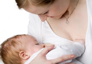
Is breast milk so different from infant formula? The ability to track which genes are operating in an infant's intestine has allowed University of Illinois scientists to compare the early development of breast-fed and formula-fed babies. They say the difference is very real.

Me
Breast milk induces genetic pathways that are quite 
different from those in formula-fed infants, new research 
has found. (Credit: iStockphoto/Oleg Kozlov)

"For the first time, we can see that breast milk induces genetic pathways that are quite different from those in formula-fed infants. Although formula makers have tried to develop a product that's as much like breast milk as possible, hundreds of genes were expressed differently in the breast-fed and formula-fed groups," said Sharon Donovan, a U of I professor of nutrition.

Although both breast-fed and formula-fed babies gain weight and seem to develop similarly, scientists have known for a long time that breast milk contains immune-protective components that make a breast-fed infant's risk lower for all kinds of illnesses, she said.

"The intestinal tract of the newborn undergoes marked changes in response to feeding. And the response to human milk exceeds that of formula, suggesting that the bioactive components in breast milk are important in this response," she noted.

"What we haven't known is how breast milk protects the infant and particularly how it regulates the development of the intestine," she said.

Understanding those differences should help formula makers develop a product that is more like the real thing, she said. The scientists hope to develop a signature gene or group of genes to use as a biomarker for breast-fed infants.

Many of the differences found by the scientists were in fundamental genes that regulate the development of the intestine and provide immune defense for the infant.

In this small proof-of-concept study, Donovan used a new technique patented by Texas A&M colleague Robert Chapkin to examine intestinal gene expression in 22 healthy infants -- 12 breast-fed, 10 formula-fed.

The technique involved isolating intestinal cells shed in the infants' stools, then comparing the expression of different genes between the two groups. Mothers in the study collected fecal samples from their babies at one, two, and three months of age. Scientists were then able to isolate high-quality genetic material, focusing on the RNA to get a gene expression or signature.

Donovan said that intestinal cells turn over completely every three days as billions of cells are made, perform their function, and are exfoliated. Examining the shed cells is a noninvasive way to examine intestinal health and see how nutrition affects intestinal development in infants.

Understanding early intestinal development is important for many reasons, she said.

"An infant's gut has to adapt very quickly. A new baby is coming out of a sterile environment, having received all its nutrients intravenously through the placenta. At that point, babies obviously must begin eating, either mother's milk or formula.

"They also start to become colonized with bacteria, so it's very important that the gut learns what's good and what's bad. The baby's body needs to be able to recognize a bad bacteria or a bad virus and fight it, but it also needs to recognize that even though a food protein is foreign, that protein is okay and the body doesn't want to develop an immune response to it," she said.

If anything goes wrong at this stage, babies can develop food allergies, inflammatory bowel disease, and even asthma. "We're very interested in frequent sampling at this early period of development," she added.

Donovan also would like to learn how bacteria in the gut differ in formula- and breast-fed babies, and this technique should make that possible. "Now we'll be able to get a complete picture of what's happening in an infant -- from the composition of the diet to the microbes in the gut and the genes that are activated along the way."

Of potential clinical importance: The gene expressed most often in breast-fed infants is involved in the cell's response to oxygen deprivation. Lack of oxygen is a factor in the development of necrotizing enterocolitis (NEC), a kind of gangrene of the intestine that can be fatal in premature babies. NEC is a leading cause of disease and death in neonatal intensive care units, with a reported 2,500 cases occurring annually in the United States and a mortality rate of 26 percent.

The study will appear in the June 2010 issue of the American Journal of Physiology, Gastrointestinal and Liver Physiology. Co-authors are Robert S. Chapkin, Chen Zhao, Ivan Ivanov, Laurie A. Davidson, Jennifer S. Goldsby, Joanne R. Lupton, and Edward R. Dougherty, all of Texas A&M University, Rose Ann Mathai and Marcia H. Monaco of the U of I, and Deshanie Rai and W. Michael Russell of Mead Johnson Nutrition. The study was funded by Mead Johnson Nutrition.
Reblog this post [with Zemanta]

Nano-Spider Molecules Behave Like Robots


A team of scientists from Columbia University, Arizona State University, the University of Michigan, and the California Institute of Technology (Caltech) have programmed an autonomous molecular "robot" made out of DNA to start, move, turn, and stop while following a DNA track.

Me
The latest installment in DNA nanotechnology has arrived: 
A molecular nanorobot dubbed a "spider" and labeled with 
green dyes traverses a substrate track built upon a DNA 
origami scaffold. It journeys towards its red-labeled goal 
by cleaving the visited substrates, thus exhibiting the 
characteristics of an autonomously moving, behavior-based 
robot at the molecular scale. (Credit: Courtesy of Paul Michelotti)

The development could ultimately lead to molecular systems that might one day be used for medical therapeutic devices and molecular-scale reconfigurable robots -- robots made of many simple units that can reposition or even rebuild themselves to accomplish different tasks.

A paper describing the work appears in the current issue of the journal Nature.

The traditional view of a robot is that it is "a machine that senses its environment, makes a decision, and then does something -- it acts," says Erik Winfree, associate professor of computer science, computation and neural systems, and bioengineering at Caltech.

Milan N. Stojanovic, a faculty member in the Division of Experimental Therapeutics at Columbia University, led the project and teamed up with Winfree and Hao Yan, professor of chemistry and biochemistry at Arizona State University and an expert in DNA nanotechnology, and with Nils G. Walter, professor of chemistry and director of the Single Molecule Analysis in Real-Time (SMART) Center at the University of Michigan in Ann Arbor, for what became a modern-day self-assembly of like-minded scientists with the complementary areas of expertise needed to tackle a tough problem.

Shrinking robots down to the molecular scale would provide, for molecular processes, the same kinds of benefits that classical robotics and automation provide at the macroscopic scale. Molecular robots, in theory, could be programmed to sense their environment (say, the presence of disease markers on a cell), make a decision (that the cell is cancerous and needs to be neutralized), and act on that decision (deliver a cargo of cancer-killing drugs).

Or, like the robots in a modern-day factory, they could be programmed to assemble complex molecular products. The power of robotics lies in the fact that once programmed, the robots can carry out their tasks autonomously, without further human intervention.

With that promise, however, comes a practical problem: how do you program a molecule to perform complex behaviors?

"In normal robotics, the robot itself contains the knowledge about the commands, but with individual molecules, you can't store that amount of information, so the idea instead is to store information on the commands on the outside," says Walter. And you do that, says Stojanovic, "by imbuing the molecule's environment with informational cues."

"We were able to create such a programmed or 'prescribed' environment using DNA origami," explains Yan. DNA origami, an invention by Caltech Senior Research Associate Paul W. K. Rothemund, is a type of self-assembled structure made from DNA that can be programmed to form nearly limitless shapes and patterns (such as smiley faces or maps of the Western Hemisphere or even electrical diagrams). Exploiting the sequence-recognition properties of DNA base pairing, DNA origami are created from a long single strand of DNA and a mixture of different short synthetic DNA strands that bind to and "staple" the long DNA into the desired shape. The origami used in the Nature study was a rectangle that was 2 nanometers (nm) thick and roughly 100 nm on each side.

The researchers constructed a trail of molecular "bread crumbs" on the DNA origami track by stringing additional single-stranded DNA molecules, or oligonucleotides, off the ends of the staples. These represent the cues that tell the molecular robots what to do -- start, walk, turn left, turn right, or stop, for example -- akin to the commands given to traditional robots.

The molecular robot the researchers chose to use -- dubbed a "spider" -- was invented by Stojanovic several years ago, at which time it was shown to be capable of extended, but undirected, random walks on two-dimensional surfaces, eating through a field of bread crumbs.

To build the 4-nm-diameter molecular robot, the researchers started with a common protein called streptavidin, which has four symmetrically placed binding pockets for a chemical moiety called biotin. Each robot leg is a short biotin-labeled strand of DNA, "so this way we can bind up to four legs to the body of our robot," Walter says. "It's a four-legged spider," quips Stojanovic. Three of the legs are made of enzymatic DNA, which is DNA that binds to and cuts a particular sequence of DNA. The spider also is outfitted with a "start strand" -- the fourth leg -- that tethers the spider to the start site (one particular oligonucleotide on the DNA origami track). "After the robot is released from its start site by a trigger strand, it follows the track by binding to and then cutting the DNA strands extending off of the staple strands on the molecular track," Stojanovic explains.

"Once it cleaves," adds Yan, "the product will dissociate, and the leg will start searching for the next substrate." In this way, the spider is guided down the path laid out by the researchers. Finally, explains Yan, "the robot stops when it encounters a patch of DNA that it can bind to but that it cannot cut," which acts as a sort of flypaper.

Although other DNA walkers have been developed before, they've never ventured farther than about three steps. "This one," says Yan, "can walk up to about 100 nanometers. That's roughly 50 steps."

"This in itself wasn't a surprise," adds Winfree, "since Milan's original work suggested that spiders can take hundreds if not thousands of processive steps. What's exciting here is that not only can we directly confirm the spiders' multistep movement, but we can direct the spiders to follow a specific path, and they do it all by themselves -- autonomously."

In fact, using atomic force microscopy and single-molecule fluorescence microscopy, the researchers were able to watch directly spiders crawling over the origami, showing that they were able to guide their molecular robots to follow four different paths.

"Monitoring this at a single molecule level is very challenging," says Walter. "This is why we have an interdisciplinary, multi-institute operation. We have people constructing the spider, characterizing the basic spider. We have the capability to assemble the track, and analyze the system with single-molecule imaging. That's the technical challenge." The scientific challenges for the future, Yan says, "are how to make the spider walk faster and how to make it more programmable, so it can follow many commands on the track and make more decisions, implementing logical behavior."

"In the current system," says Stojanovic, "interactions are restricted to the walker and the environment. Our next step is to add a second walker, so the walkers can communicate with each other directly and via the environment. The spiders will work together to accomplish a goal." Adds Winfree, "The key is how to learn to program higher-level behaviors through lower-level interactions."

Such collaboration ultimately could be the basis for developing molecular-scale reconfigurable robots -- complicated machines that are made of many simple units that can reorganize themselves into any shape -- to accomplish different tasks, or fix themselves if they break. For example, it may be possible to use the robots for medical applications. "The idea is to have molecular robots build a structure or repair damaged tissues," says Stojanovic.

"You could imagine the spider carrying a drug and bonding to a two-dimensional surface like a cell membrane, finding the receptors and, depending on the local environment," adds Yan, "triggering the activation of this drug."

Such applications, while intriguing, are decades or more away. "This may be 100 years in the future," Stojanovic says. "We're so far from that right now."

"But," Walter adds, "just as researchers self-assemble today to solve a tough problem, molecular nanorobots may do so in the future."

The other coauthors on the paper, "Molecular robots guided by prescriptive landscapes," are Kyle Lund and Jeanette Nangreave from Arizona State University; Anthony J. Manzo, Alexander Johnson-Buck, and Nicole Michelotti from the University of Michigan; Nadine Dabby from Caltech; and Steven Taylor and Renjun Pei from Columbia University. The work was supported by the National Science Foundation, the Army Research Office, the Office of Naval Research, the National Institutes of Health, the Department of Energy, the Searle Foundation, the Lymphoma and Leukemia Society, the Juvenile Diabetes Research Foundation, and a Sloan Research Fellowship.
Reblog this post [with Zemanta]

Monday, May 10, 2010

Brain's Master Switch Is Verified


The protein that has long been suspected by scientists of being the master switch allowing brains to function has now been verified by an Iowa State University researcher.
Yeon-Kyun Shin, professor of biochemistry
biophysics and molecular biology at ISU, has 
shown that the protein called synaptotagmin1 
(Syt1) is the sole trigger for the release of 
neurotransmitters in the brain using this 
instrument that allows a new technique called 
single vesicle fusion method. 
(Credit: ISU photo by Bob Elbert)

Yeon-Kyun Shin, professor of biochemistry, biophysics and molecular biology at ISU, has shown that the protein called synaptotagmin1 (Syt1) is the sole trigger for the release of neurotransmitters in the brain.

Prior to this research, Syt1 was thought to be a part of the protein structure (not the sole protein) that triggered the release of neurotransmitters at 10 parts per million of calcium.

Shin's research is published in the current issue of the journal Science.

"Syt1 was a suspect previously, but people were not able to pinpoint that it's the real one, even though there were lots and lots of different trials," said Shin.

"In this case, we are trying to show in the laboratory that it's the real one. So we excluded everything else, and included SNARE proteins -- that's the machinery of the release, and the Syt1 is a calcium-sensing timer."

Syt1 senses, at 10 ppm of calcium, and tells the SNARE complex to open the pore to allow the movement of the neurotransmitters.

Brain activity occurs when neurotransmitters move into a fusion pore.

"We are showing that this Syt1 senses the calcium at 10 ppm, and sends the signal to the SNARE complex to open the fusion pore. That is the process that we are showing right now," Shin said.

Shin and his researchers were able to pinpoint the protein using a new technique called single vesicle fusion method. Using this method, they were able to create and monitor a single fusion event.

Previous research didn't allow scientists to look at single events, and instead required detecting many events and then taking an average of those events, Shin says.

Shin, who has been looking at this brain activity for 15 years, is happy about the discovery.

"We are quite excited that for the first time we are showing that Syt1 is really what triggers the signal in the brain," he said. "This is a really important thing in terms of neurosciences. This is the heart of the molecular part of the brain function."

Shin believes his discovery may be useful in understanding brain malfunctions such as autism, epilepsy and others.

While researching brain function, Shin has previously shown that taking statin drugs to lower cholesterol may actually inhibit some brain function.