BTemplates.com

Powered by Blogger.

Pageviews past week

Quantum mechanics

Auto News

artificial intelligence

About Me

Recommend us on Google!

Information Technology

Popular Posts

Showing posts with label Conditions and Diseases. Show all posts

Tuesday, June 28, 2011

It's Not an Apple a Day After All -- It's Strawberries: Flavonoids Could Represent Two-Fisted Assault On Diabetes and Nervous System Disorders


A recent study from scientists at the Salk Institute for Biological Studies suggests that a strawberry a day (or more accurately, 37 of them) could keep not just one doctor away, but an entire fleet of them, including the neurologist, the endocrinologist, and maybe even the oncologist.
Fisetin, a naturally-occurring flavonoid found most 
abundantly in strawberries, lessens complications of diabetes
(Credit: Courtesy of the Salk Institute for Biological Studies)

Investigations conducted in the Salk Institute's Cellular Neurobiology Laboratory (CNL) will appear in the June 27, 2011, issue of PLoS ONE. The report explains that fisetin, a naturally-occurring flavonoid found most abundantly in strawberries and to a lesser extent in other fruits and vegetables, lessens complications of diabetes. Previously, the lab showed that fisetin promoted survival of neurons grown in culture and enhanced memory in healthy mice. That fisetin can target multiple organs strongly suggests that a single drug could be used to mitigate numerous medical complications.

"This manuscript describes for the first time a drug that prevents both kidney and brain complications in a type 1 diabetes mouse model," says David Schubert, Ph.D., professor and head of the Cellular Neurobiology Laboratory and one of the manuscript's co-authors. "Moreover, it demonstrates the probable molecular basis of how the therapeutic is working."

Pam Maher, Ph.D., a senior staff scientist in the CNL, is the study's corresponding author. Maher initially identified fisetin as a neuroprotective flavonoid ten years ago. "In plants, flavonoids act as sunscreens and protect leaves and fruit from insects," she explains. "As foods they are implicated in the protective effect of the 'Mediterranean Diet.'"

Other celebrity flavonoids include polyphenolic compounds in blueberries and red wine.

Although her group's focus is neurobiology, Maher and colleagues reasoned that, like other flavonoids, fisetin might ameliorate a spectrum of disorders seen in diabetic patients. To test this, they evaluated effects of fisetin supplementation in Akita mice, a very robust model of type 1 diabetes, also called childhood onset diabetes.

Akita mice exhibit increased blood sugar typical of type 1 diabetes and display pathologies seen in serious human complications of both type 1 and 2 diabetes. Those include diabetic nephropathy or kidney disease, retinopathy, and neuropathies in which patients lose touch or heat sensations.

Mice fed a fisetin-enriched diet remained diabetic, but acute kidney enlargement-or hypertrophy-seen in untreated mice was reversed, and high urine protein levels, a sure sign of kidney disease, fell. Moreover, fisetin ingestion ameliorated anxiety-related behaviors seen in diabetic mice. "Most mice put in a large area become exploratory," says Maher. "But anxious mice tend not to move around. Akita mice showed enhanced anxiety behavior, but fisetin feeding restored their locomotion to more normal levels."


The study also defines a likely molecular mechanism underlying these effects. Researchers observed that blood and brain levels of sugars affixed to proteins known as advanced glycation end-products-or AGEs-were reduced in fisetin-treated compared to untreated Akita mice. These decreases were accompanied by increased activity of the enzyme glyoxalase 1, which promotes removal of toxic AGE precursors.

The discovery of an AGE-antagonizing enzyme upregulated by fisetin is very intriguing, because substantial evidence implicates high blood AGE levels with many if not most diabetic complications. "We know that fisetin increases activity of the glyoxalase enzyme and may increase its expression," says Maher. "But what is important is that ours is the first report that any compound can enhance glyoxalase 1 activity."

Interestingly, excessively high AGE levels also correlate with inflammatory activity thought to promote some cancers. In fact, studies published by others confirm that fisetin decreases tumorigenicity of prostate cancer cells both in culture and in animal models, which if supported would represent a major added incentive to eat your strawberries.

To ingest fisetin levels equivalent to those fed Akita mice, Maher estimates that humans would have to eat 37 strawberries a day, assuming that strawberry fisetin is as readily metabolizable by humans as fisetin-spiked lab chow is by mice. Rather than through diet, Maher envisions that fisetin-like drugs could be taken as a supplement.

Schubert notes that fisetin is also effective in mouse models of Alzheimer's disease. "We and others have shown that diabetes may be a risk factor for Alzheimer's disease, making identification of a safe prophylactic like fisetin highly significant," he says.

Maher acknowledges that the public may be suffering from flavonoid-fatigue, given media coverage of the promises of these compounds. "Polyphenolics like fisetin and those in blueberry extracts are found in fruits and vegetables and are related to each other chemically," she says. "There is increasing evidence that they all work in multiple diseases. Hopefully some combination of these compounds will eventually get to the clinic."

Schubert concurs that their findings only reinforce what common sense and our mothers told us was a healthy lifestyle. "Eat a balanced diet and as much freshly prepared organic food as possible, get some exercise, keep socially and mentally active and avoid sodas with sugar and highly processed foods since they can contain high levels of AGEs," he advises.

But he also worries that hoops that must be jumped through to bring a natural product like fisetin, as opposed to a totally synthetic drug, to clinical trials are daunting because it is difficult to protect patents on natural products. "We will never know if a compound like fisetin works in humans until someone is willing to support a clinical trial."

Also contributing to this study were Richard Dargusch and Jennifer L. Ehren, Ph.D.,of the Cellular Neurobiology Laboratory, and Kumar Sharma, M.D., and Shinichi Okada, M.D., Ph.D., of the Department of Medicine at University of California, San Diego.

Funding for the study came from the Fritz B. Burns Foundation, the Juvenile Diabetes Research Foundation, the Hewitt Foundation, and the National Institutes of Health.

Saturday, March 5, 2011

Liver, Not Brain, May Be Origin of Alzheimer’s Plaques


Unexpected results from a Scripps Research Institute and ModGene, LLC study could completely alter scientists' ideas about Alzheimer's disease -- pointing to the liver instead of the brain as the source of the "amyloid" that deposits as brain plaques associated with this devastating condition. The findings could offer a relatively simple approach for Alzheimer's prevention and treatment.
New research suggests that the liver instead of the brain 
may be the source of the "amyloid" that deposits as brain 
plaques associated with Alzheimer's disease. 
(Credit: iStockphoto/David Marchal)


The study was published online March 3 in The Journal of Neuroscience Research.

In the study, the scientists used a mouse model for Alzheimer's disease to identify genes that influence the amount of amyloid that accumulates in the brain. They found three genes that protected mice from brain amyloid accumulation and deposition. For each gene, lower expression in the liver protected the mouse brain. One of the genes encodes presenilin -- a cell membrane protein believed to contribute to the development of human Alzheimer's.

"This unexpected finding holds promise for the development of new therapies to fight Alzheimer's," said Scripps Research Professor Greg Sutcliffe, who led the study. "This could greatly simplify the challenge of developing therapies and prevention."

An estimated 5.1 million Americans have Alzheimer's disease, including nearly half of people age 85 and older. By 2050, the number of people age 65 and over with this disease will range from 11 million to 16 million unless science finds a way to prevent or effectively treat it. In addition to the human misery caused by the disease, there is the unfathomable cost. A new report from the Alzheimer's Association shows that in the absence of disease-modifying treatments, the cumulative costs of care for people with Alzheimer's from 2010 to 2050 will exceed $20 trillion.

A Genetic Search-and-Find Mission

In trying to help solve the Alzheimer's puzzle, in the past few years Sutcliffe and his collaborators have focused their research on naturally occurring, inherited differences in neurological disease susceptibility among different mouse strains, creating extensive databases cataloging gene activity in different tissues, as measured by mRNA accumulation. These data offer up maps of trait expression that can be superimposed on maps of disease modifier genes.

As is the case with nearly all scientific discovery, Sutcliffe's research builds on previous findings. Several years ago, researchers at Case Western Reserve mapped three genes that modify the accumulation of pathological beta amyloid in the brains of a transgenic mouse model of Alzheimer's disease to large chromosomal regions, each containing hundreds of genes. The Case Western scientists used crosses between the B6 and D2 strains of mice, studying more than 500 progeny.

Using the results from this study, Sutcliffe turned his databases of gene expression to the mouse model of Alzheimer's, looking for differences in gene expression that correlated with differences in disease susceptibility between the B6 and D2 strains. This intensive work involved writing computer programs that identified each genetic difference that distinguished the B6 and D2 genomes, then running mathematical correlation analysis (known as regression analysis) of each difference. Correlations were made between the genotype differences (B6 or D2) and the amount of mRNA product made from each of the more than 25,000 genes in a particular tissue in the 40 recombinant inbred mouse strains. These correlations were repeated 10 times to cover 10 tissues, the liver being one of them.

"A key aspect of this work was learning how to ask questions of massive data sets to glean information about the identities of heritable modifier genes," Sutcliffe said. "This was novel and, in a sense, groundbreaking work: we were inventing a new way to identify modifier genes, putting all of these steps together and automating the process. We realized we could learn about how a transgene's pathogenic effect was being modified without studying the transgenic mice ourselves."

Looking for a Few Good Candidates

Sutcliffe's gene hunt offered up good matches, candidates, for each of the three disease modifier genes discovered by the Case Western scientists, and one of these candidates -- the mouse gene corresponding to a gene known to predispose humans carrying particular variations of it to develop early-onset Alzheimer's disease -- was of special interest to his team.

"The product of that gene, called Presenilin2, is part of an enzyme complex involved in the generation of pathogenic beta amyloid," Sutcliffe explained. "Unexpectedly, heritable expression of Presenilin2 was found in the liver but not in the brain. Higher expression of Presenilin2 in the liver correlated with greater accumulation of beta amyloid in the brain and development of Alzheimer's-like pathology."

This finding suggested that significant concentrations of beta amyloid might originate in the liver, circulate in the blood, and enter the brain. If true, blocking production of beta amyloid in the liver should protect the brain.

To test this hypothesis, Sutcliffe's team set up an in vivo experiment using wild-type mice since they would most closely replicate the natural beta amyloid-producing environment. "We reasoned that if brain amyloid was being born in the liver and transported to the brain by the blood, then that should be the case in all mice," Sutcliffe said, "and one would predict in humans, too."

The mice were administered imatinib (trade name Gleevec, an FDA-approved cancer drug), a relatively new drug currently approved for treatment of chronic myelogenous leukemia and gastrointestinal tumors. The drug potently reduces the production of beta amyloid in neuroblastoma cells transfected by amyloid precursor protein (APP) and also in cell-free extracts prepared from the transfected cells. Importantly, Gleevec has poor penetration of the blood-brain barrier in both mice and humans.

"This characteristic of the drug is precisely why we chose to use it," Sutcliffe explained. "Because it doesn't penetrate the blood-brain barrier, we were able to focus on the production of amyloid outside of the brain and how that production might contribute to amyloid that accumulates in the brain, where it is associated with disease."

The mice were injected with Gleevec twice a day for seven days; then plasma and brain tissue were collected, and the amount of beta amyloid in the blood and brain was measured. The findings: the drug dramatically reduced beta amyloid not only in the blood, but also in the brain where the drug cannot penetrate. Thus, an appreciable portion of brain amyloid must originate outside of the brain, and imatinib represents a candidate for preventing and treating Alzheimer's.

As for the future of this research, Sutcliffe says he hopes to find a partner and investors to move the work into clinical trials and new drug development.

In addition to Sutcliffe, the authors of the study, titled "Peripheral reduction of β-amyloid is sufficient to reduce brain Aβ: implications for Alzheimer's disease," include Peter Hedlund and Elizabeth Thomas of Scripps Research, and Floyd Bloom and Brian Hilbush of ModGene, LLC, which funded the project.

Thursday, February 17, 2011

Wireless Heart Implant Reduces HospitalizationsA pressure-sensing implant helps heart-failure patients stay healthy.


A wireless sensor developed by Atlanta-based CardioMEMS reduced the number of hospitalizations in patients with heart failure by 39 percent. The tiny implant monitors fluid pressure in the pulmonary artery and transmits the data wirelessly to physicians, who can adjust patients' medications accordingly.

Researchers say the sensor may significantly lower health-care costs and improve quality of life for people with congestive heart failure. The device is one of several prototypes being developed by CardioMEMS and other medical implant companies to provide continuous, personalized wireless monitors for such patients.
Pressure patrol: A new wireless sensor the size of a paper
clip measures fluid pressure in the pulmonary artery. The
metalloops on either end anchor the sensor to the artery
walls, while the self-contained transducer in the middle
takes pressure readings. The sensor is activated by radio
frequency, transmitting data wirelessly to physicians
via modem.Credit: OSU Medical Center/CardioMEMS


"I think the study shows this kind of device is incredibly useful in improving outcomes in patients and directing therapy," says Marc Jay Semigran, medical director of the Mass General Heart Failure and Cardiac Transplant Program, who was not involved in the study.

Hospitals admit 1.1 million adults each year for congestive heart failure, a condition in which pressure builds up in the circulatory system and the heart fails to pump blood adequately to the rest of the body. The American Heart Association estimates that the chronic condition costs the health-care system $29 billion per year. CardioMEMS aims to reduce that figure by providing an accurate way to continuously monitor patients after they've left the hospital.

The device is implanted in the pulmonary artery, an area that carries a low risk of clotting. It is smaller than other implants under development because it does not require a battery or a wire to take pressure readings. Two metal loops hold it to the sides of the artery, and a pressure transducer records the flow of fluids through the blood vessel. The sensor is powered externally by a receiver built into a pillow. When a patient lies on the pillow, the sensor is activated to take measurements and send them wirelessly to a computer, where physicians can review the data. In a large six-month clinical trial published this month in the Lancet, 550 patients from 64 centers across the United States were equipped with the device and instructed to take readings once a day. Patients were divided into two groups. The first took medication instructions from physicians who monitored the sensor data. The second took instructions from physicians who relied on traditional indicators like weight and blood pressure. Over the six months, patients in the first group experienced 39 percent fewer hospitalizations than those in the second.

Today, physicians often assess pulmonary pressure when initially evaluating a patient, but they do so far less frequently in follow-up evaluation. That's because the measurement requires doctors to snake a catheter into a patient's heart and inflate a balloon. However, fluid pressure changes by the day, and monitoring those fluctuations continuously is essential to treating heart failure effectively.

"Over the years, we found that pressures go up long before patients develop symptoms and call a doctor to say they're sick," says Philip Adamson, director of the Heart Failure Institute at Oklahoma Heart Hospital, the principal investigator in the CardioMEMS clinical trial. "By utilizing the pressure sensor information, we're given the ability to make changes in medications long before patients bring themselves to the doctor, and that's how we reduced hospitalizations."

Over the past few years, several companies have jockeyed to be first on the market with a continuous pressure-sensing cardiac implant. In 2007, Medtronic failed to get FDA approval for its sensor, a stopwatch-size, battery-powered implant wired to the heart. The device reduced hospitalizations by 22 percent, but FDA regulators did not consider that worth the risks associated with implanting it. Researchers also found that the wire connecting the sensor to the heart degraded over time.

CardioMEMS is currently seeking approval for its sensor from the U.S. Food and Drug Administration and has submitted results from the clinical study for FDA review. In the next two or three years, the company plans to integrate the sensor's receiver into a patient's cell phone, which will be able to instantly read pressure data and upload it for both physicians and patients to review.

Monday, September 27, 2010

Genetic "Light Switches" Control Muscle Movement The technique will improve research on neuromuscular disorders and could one day help paralyzed patients.


Using light-sensitive proteins from a single-celled alga and a tiny LED "cuff" placed on a nerve, researchers have triggered the leg muscles of mice to contract in response to millisecond pulses of light.
Light movement: This image shows a cross-section of a mouse sciatic nerve genetically engineered to produce a light-sensitive protein (shown in green). Stanford researchers used this protein to trigger muscle movements in the animal’s leg.
Credit: Nature

The study, published in the journal Nature Medicine, marks the first use of the nascent technology known as optogenetics to control muscle movements. Developed by study coauthor Karl Deisseroth, an associate professor of bioengineering and of psychiatry and behavioral science at Stanford University, optogenetics makes it possible to stimulate neurons with light by inserting the gene for a protein called channelrhodopsin-2, from a green alga. When a modified neuron is exposed to blue light, the protein initiates electrical activity inside the cell that then spreads from neuron to neuron. By controlling which neurons make the protein, as well as which cells are exposed to light, scientists can control neural activity in living animals with unprecedented precision. The paper's other senior author, Scott Delp, a professor of bioengineering, mechanical engineering, and orthopedic surgery at Stanford, says that the optical control method provides "fantastic advantages over electrical stimulation" for his study of muscles and the biomechanics of human movement.

Members of Deisseroth's lab had engineered mice to produce channelrhodopsin-2 in both the central and the peripheral nervous systems. Michael Llewellyn, a former graduate student in Delp's lab, developed a tiny, implantable LED cuff to apply light to the nerve evenly. He placed the cuff on the sciatic nerves of anesthetized mice and triggered millisecond pulses of light. This caused the leg muscles of the mice to contract. When Llewellyn compared the muscle contractions stimulated by light to those generated using a similar electrical cuff, he found that the light-triggered contractions were much more similar to normal muscle activity.

Muscles are made up of two different fibers: small, slow, fatigue-resistant fibers that are typically used for tasks that require fine motor control over longer periods, and larger, faster fibers that can produce higher forces but are more fatigue-prone. In the body, the small, slow fibers are activated first, with the large, fast fibers reserved for quick bursts of power or speed. When muscles are stimulated with electrical pulses, the fast fibers activate first. With the optogenetic switch, however, the fibers were recruited in the normal, physiological order: slow fibers first, fast fibers second. By altering the intensity of the light, Llewellyn found that he could even trigger only the slow fibers--a feat not possible with electrical stimulation.

In the near term, Delp says, the technology will improve the studies that his lab and others do on muscle activity in animal models of stroke, palsies, ALS, and other neuromuscular disorders. He also hopes that in time--a long time, he concedes--such optical switches could be used to help patients with physical disabilities caused by nerve damage such as stroke, spinal cord injury, or cerebral palsy. One possibility, he says, would be to use optical stimulation in place of functional electrical stimulation (FES), in which electrical current is applied to specific nerves or muscles to trigger muscle contractions. The U.S. Food and Drug Administration has already approved FES devices that can restore hand function and bladder control to some paralyzed people. However, FES can quickly lead to muscle fatigue. Delp hopes that, particularly with grasping functions, using optical stimulation might result in better fatigue resistance and perhaps finer muscle control.

"This is a brilliant study, really beautiful science," says Robert Kirsch, a bioengineer at Case Western Reserve University and associate director of the Cleveland Functional Electrical Stimulation Center; he was not involved in the research. "I think there are many [clinical implications]," he says, although, like Delp and Llewellyn, he notes that many high hurdles must be cleared--not least of which is developing a safe, effective way to deliver the channelrhodopsin-2 gene to nerve cells in humans. Otherwise, Kirsch says, "my one objection would be their implication that they've solved the fatigue problem with FES. I'm pretty sure that hasn't happened." Instead, Kirsch believes that most of the fatigue seen in FES patients is due to muscle atrophy and weakness that develop in the chronically paralyzed.

C.J. Heckman, a professor of physiology at Northwestern University's Feinberg School of Medicine, agrees: "It is true that a lot of the fatigue seen in FES patients is due to chronic muscle atrophy." But, he says, "if you could stimulate the muscles in the correct recruitment order repeatedly over time, you could potentially recover a lot of muscle function." This could help paralysis patients preserve their slow muscle fibers, "which would be a huge deal," Heckman says. This is because those fibers do a huge percentage of the work muscles do--everything from maintaining posture to typing on a keyboard.

Delp also thinks that stimulation-based exercise could be an important application for optical muscle control, as could helping wheelchair-bound people stand to reach for books or plates in a cabinet. "I'm not super-high on controlling locomotion"--that is, walking--"with either electrical or optical stimulation, though," Delp says. "It's an incredibly complicated command-and-control scheme that's really hard to coordinate."

In the meantime, Delp and Llewellyn have begun an effort to use a different light-sensitive protein, halorhodopsin, to inhibit motor nerves in mice, with the idea of treating or even curing muscle spasticity, often a serious side effect to brain or spinal injury. Current treatments are far from ideal; doctors may inject botulinum toxin into the affected muscles every few months to paralyze them, use oral medications such as Valium that affect the whole body instead of just the affected muscle, or, in the most severe cases, cut the nerves or tendons of the spastic muscle--a permanent treatment that leaves the patient with no control over that muscle. Delp hopes that genetically engineering the nerves with halorhodopsin might enable people to use light to reversibly relax muscles affected with spasticity.

"I think that's a great idea for treating spasticity," says Jerry Silver, a neuroscientist at Case Western. There may be some difficulties along the way, though, he says. Working with Case colleagues, Silver has started a company called LucCell to develop clinical applications of optogenetics. In one company project, scientists are trying to use halorhodopsin and other inhibitory opsins in animal models to turn off the muscle that controls the bladder sphincter; their ultimate goal is to restore bladder function to paralyzed people. Though they have seen some physiological changes in how the sphincter muscle behaves, they haven't been able to get it to relax enough. "We're learning it's easier to turn things on than turn things off," he says. Still, the team is persisting, looking for better ways to deliver the gene to nerve cells and for ways to increase production of the protein on the cell's surfaces.

"It all depends on the ability to get the transgene in the right place in the person's genome without causing problems," agrees Llewellyn. "It's the main obstacle."

Wednesday, August 18, 2010

Moderate Chocolate Consumption Linked to Lower Risks of Heart Failure, Study Finds


Middle-aged and elderly Swedish women who regularly ate a small amount of chocolate had lower risks of heart failure risks, in a study reported in Circulation: Heart Failure, a journal of the American Heart Association.
A new study has found that middle-aged and elderly Swedish women who regularly ate a small amount of chocolate had lower risks of heart failure risks. (Credit: iStockphoto)

The nine-year study, conducted among 31,823 middle-aged and elderly Swedish women, looked at the relationship of the amount of high-quality chocolate the women ate, compared to their risk for heart failure. The quality of chocolate consumed by the women had a higher density cocoa content somewhat like dark chocolate by American standards. In this study, researchers found:
 
  • Women who ate an average of one to two servings of the high-quality chocolate per week had a 32 percent lower risk of developing heart failure.

  • Those who had one to three servings per month had a 26 percent lower risk.

  • Those who consumed at least one serving daily or more didn't appear to benefit from a protective effect against heart failure.

The lack of a protective effect among women eating chocolate every day is probably due to the additional calories gained from eating chocolate instead of more nutritious foods, said Murrray Mittleman, M.D., Dr.P.H., lead researcher of the study.

"You can't ignore that chocolate is a relatively calorie-dense food and large amounts of habitual consumption is going to raise your risks for weight gain," said Mittleman, director of the Cardiovascular Epidemiology Research Unit at Harvard Medical School's Beth Israel Deaconess Medical Center in Boston. "But if you're going to have a treat, dark chocolate is probably a good choice, as long as it's in moderation."

High concentration of compounds called "flavonoids" in chocolate may lower blood pressure, among other benefits, according to mostly short-term studies. However, this is the first study to show long-term outcomes related specifically to heart failure, which can result from ongoing untreated high blood pressure.

In the observational study, researchers analyzed self-reported food-frequency questionnaire responses from participants 48-to-83-years-old in the Swedish Mammography Cohort. Combining the results with data from national Swedish hospitalization and death registries between 1998 through 2006, the researchers used multiple forms of statistical modeling to reach their conclusions on heart failure and chocolate consumption.

Mittleman said differences in chocolate quality affect the study's implications for Americans. Higher cocoa content is associated with greater heart benefits. In Sweden, even milk chocolate has a higher cocoa concentration than dark chocolate sold in the United States.

Although 90 percent of all chocolate eaten across Sweden during the study period was milk chocolate, it contained about 30 percent cocoa solids. U.S. standards only require 15 percent cocoa solids to qualify as dark chocolate. So, by comparison, American chocolate may have fewer heart benefits and more calories and fat per equivalent amounts of cocoa content compared to the chocolate eaten by the Swedish women in the study.

Also, the average serving size for Swedish women in the study ranged from 19 grams among those 62 and older, to 30 grams among those 61 and younger. In contrast, the standard American portion size is 20 grams.

"Those tempted to use these data as their rationale for eating large amounts of chocolate or engaging in more frequent chocolate consumption are not interpreting this study appropriately," said Linda Van Horn, Ph.D., R.D., immediate past chair of the American Heart Association Nutrition Committee and professor in the Department of Preventive Medicine at Northwestern University's Feinberg School of Medicine in Chicago. "This is not an 'eat all you want' take-home message, rather it's that eating a little dark chocolate can be healthful, as long as other adverse behaviors do not occur, such as weight gain or excessive intake of non-nutrient dense 'empty' calories."

Heart failure occurs among about 1 percent of Americans over age 65. A condition in which the heart can't pump enough blood to the rest of the body, heart failure rates are increasing as our aging population grows.

"Anything that helps to decrease heart failure is an important issue worth examining," Mittleman said.

Co-authors are Elizabeth Mostofsky, M.P.H.; Emily Levitan, Sc.D.; and Alicja Wolk, Dr.Med.Sci. Author disclosures and funding support are on the manuscript.

Friday, July 23, 2010

Could Diabetes Be in Your Bones?


Our bones have much greater influence on the rest of our bodies than they are often given credit for, according to two new studies in the July 23 issue of Cell, a Cell Press publication. Both studies offer new insights into the interplay between bone and blood sugar, based on signals sent via insulin and a bone-derived hormone known as osteocalcin.
Image
Insulin signaling in bone favors whole-body glucose 
homeostasis by activating osteocalcin(1) Insulin signals 
osteoblasts, bone cells responsible for bone formation, 
which (2) tell osteoclasts, bone cells responsible for 
resorption, to destroy old bone. Next (3), the acidic 
(low pH) conditions created by the osteoclasts activates 
osteocalcin inside the bone. Finally (4), the active 
osteocalcin released from bone travels to the pancreas 
and stimulates the release of more insulin. (Credit: Image 
provided by Columbia University Medical Center)

Mice whose bones can't respond to insulin develop high blood sugar and insulin resistance, both hallmarks of diabetes. Those symptoms are tied to a drop in osteocalcin. The findings suggest that osteocalcin, or perhaps a drug that targets bone, might hold promise in fighting the global epidemic of type 2 diabetes, according to the researchers.

"Our study reveals a key molecular link between bone remodeling and metabolism," said Gerard Karsenty of Columbia University.

"Bone is an organ that has to pay attention to where calories are going," added Thomas Clemens of Johns Hopkins University School of Medicine. "It talks to muscle, fat, the pancreas. It's a player in energy metabolism."

And perhaps that makes a lot of sense, Karsenty said. The remodeling of bone relies on two cell types, bone-building osteoblasts and bone-resorbing osteoclasts, making bone the only organ with a cell type that is entirely focused on destroying host tissue. "On a daily basis, the formation of bone is expensive in terms of energy," he said.

In fact, the idea that the skeleton is much more than a reservoir for calcium and phosphate isn't entirely new, the researchers said. Earlier evidence by Karsenty's group had shown links between bone and the fat hormone leptin. (Obese adults are significantly less likely to develop osteoporosis.)

Scientists also had evidence that osteoblasts might respond to insulin in important ways. Osteoblasts bear insulin receptors and when treated with insulin show signs of collagen synthesis and take up more glucose, Clemens' team notes. People with type 1 diabetes due to a lack of insulin can also develop weakened bones.

Karsenty's team describes bone as a multitasker. It has mechanical, hematopoietic (blood-producing) and metabolic functions. It also acts as an endocrine organ through the release of osteocalcin hormone, which favors glucose metabolism when in its active form.

Still, Clemens said he was surprised by what they saw after developing a mouse lacking insulin receptors only in their osteoblasts. "The mice started to get fat," he said. They showed changes in their biochemistry that were consistent with insulin resistance. They also had low osteocalcin levels and fewer osteoblasts to produce less bone.

With age, the animals became even fatter and developed more marked high blood sugar accompanied by severe glucose intolerance and insulin resistance. Those symptoms improved with osteocalcin treatment.

Karsenty's group presents independent evidence for the important role of insulin in bone for keeping glucose in check through osteocalcin, in what he refers to as a "feed-forward loop." But his group goes a step further to suggest that bone-resorbing osteoclasts (not just osteoblasts) have a place in this too.

Karsenty explains that bone-building osteoblasts actually control bone resorption by osteoclasts, a process that takes place under very acidic conditions. Those conditions would also favor the chemical modification necessary to produce active osteocalcin, which can escape bone to act as a hormone.

That could be important to those who take osteoporosis drugs designed to block bone resorption, Karsenty suggests. "It's a red flag," he said. "Osteoporotic patients treated with [bone resorption inhibitors] may be at risk of glucose intolerance."

The researchers include Mathieu Ferron, Columbia University, New York, NY; Jianwen Wei, Columbia University, New York, NY; Tatsuya Yoshizawa, Columbia University, New York, NY; Andrea Del Fattore, University of L'Aquila, L'Aquila, Italy; Ronald A. DePinho, Harvard Medical School, Boston, MA; Anna Teti, University of L'Aquila, L'Aquila, Italy; Patricia Ducy, Columbia University, New York, NY; and Gerard Karsenty, Columbia University, New York, NY.

Tuesday, July 20, 2010

Of Bugs and Brains: Gut Bacteria Affect Multiple Sclerosis


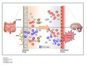
Biologists at the California Institute of Technology (Caltech) have demonstrated a connection between multiple sclerosis (MS) -- an autoimmune disorder that affects the brain and spinal cord -- and gut bacteria.
Image
In the absence of bacteria in the intestines, 
pro-inflammatory Th17 cells do not develop in 
either the gut or the central nervous system; and 
animals do not develop disease (top panel). When 
animals are colonized with symbiotic segmented 
filamentous bacteria, Th17 cell differentiation is 
induced in the gut. Th17 cells promote experimental 
autoimmune encephalomyelitis, an animal model for 
multiple sclerosis. In this way, non-pathogenic bacteria 
of the microbiota promote disease by shaping the immune 
response in both the gut and the brain (top panel). 
(Credit: Lee, Mazmanian/Caltech; modified from 
Savidge TC et al. Laboratory Investigation 2007)

The work -- led by Sarkis K. Mazmanian, an assistant professor of biology at Caltech, and postdoctoral scholar Yun Kyung Lee -- appears online the week of July 19-23 in the Proceedings of the National Academy of Sciences.

Multiple sclerosis results from the progressive deterioration of the protective fatty myelin sheath surrounding nerve cells. The loss of myelin hinders nerve cells from communicating with one another, leading to a host of neurological symptoms including loss of sensation, muscle spasms and weakness, fatigue, and pain. Multiple sclerosis is estimated to affect about half a million people in the United States alone, with rates of diagnosis rapidly increasing. There is currently no cure for MS.

Although the cause of MS is unknown, microorganisms seem to play some sort of role. "In the literature from clinical studies, there are papers showing that microbes affect MS," Mazmanian says. "For example, the disease gets worse after viral infections, and bacterial infections cause an increase in MS symptoms."

On the other hand, he concedes, "it seems counterintuitive that a microbe would be involved in a disease of the central nervous system, because these are sterile tissues."

And yet, as Mazmanian found when he began examining the multiple sclerosis literature, the suggestion of a link between bacteria and the disease is more than anecdotal. Notably, back in 1993, Caltech biochemist Leroy Hood -- who was then at the University of Washington -- published a paper describing a genetically engineered strain of mouse that developed a lab-induced form of multiple sclerosis known as experimental autoimmune encephalomyelitis, or EAE.

When Hood's animals were housed at Caltech, they developed the disease. But, oddly, when the mice were shipped to a cleaner biotech facility -- where their resident gut bacterial populations were reduced -- they didn't get sick. The question was, why? At the time, Mazmanian says, "the authors speculated that some environmental component was modulating MS in these animals." Just what that environmental component was, however, remained a mystery for almost two decades.

But Mazmanian -- whose laboratory examines the relationships between gut microbes, both harmful and helpful, and the immune systems of their mammalian hosts -- had a hunch that intestinal bacteria were the key. "As we gained an appreciation for how profoundly the gut microbiota can affect the immune system, we decided to ask if symbiotic bacteria are the missing variable in these mice with MS," he says.

To find out, Mazmanian and his colleagues tried to induce MS in animals that were completely devoid of the microbes that normally inhabit the digestive system. "Lo and behold, these sterile animals did not get sick," he says.

Then the researchers decided to see what would happen if bacteria were reintroduced to the germ-free mice. But not just any bacteria. They inoculated mice with one specific organism, an unculturable bug from a group known as segmented filamentous bacteria. In prior studies, these bacteria had been shown to lead to intestinal inflammation and, more intriguingly, to induce in the gut the appearance of a particular immune-system cell known as Th17. Th17 cells are a type of T helper cell -- cells that help activate and direct other immune system cells. Furthermore, Th17 cells induce the inflammatory cascade that leads to multiple sclerosis in animals.

"The question was, if this organism is inducing Th17 cells in the gut, will it be able to do so in the brain and central nervous system?" Mazmanian says. "Furthermore, with that one organism, can we restore to sterile animals the entire inflammatory response normally seen in animals with hundreds of species of gut bacteria?"

The answer? Yes on all counts. Giving the formerly germ-free mice a dose of one species of segmented filamentous bacteria induced Th17 not only in the gut but in the central nervous system and brain -- and caused the formerly healthy mice to become ill with MS-like symptoms.

"It definitely shows that gut microbes have a strong role in MS, because the genetics of the animals were the same. In fact, everything was the same except for the presence of those otherwise benign bacteria, which are clearly playing a role in shaping the immune system," Mazmanian says. "This study shows for the first time that specific intestinal bacteria have a significant role in affecting the nervous system during MS -- and they do so from the gut, an anatomical location very, very far from the brain."

Mazmanian and his colleagues don't, however, suggest that gut bacteria are the direct cause of multiple sclerosis, which is known to be genetically linked. Rather, the bacteria may be helping to shape the immune system's inflammatory response, thus creating conditions that could allow the disease to develop. Indeed, multiple sclerosis also has a strong environmental component; identical twins, who possess the same genome and share all of their genes, only have a 25 percent chance of sharing the disease. "We would like to suggest that gut bacteria may be the missing environmental component," he says.

For their part, Th17 cells are needed for the immune system to properly combat infection. Problems only arise when the cells are activated in the absence of infection -- just as disease can arise, Mazmanian and others suspect, when the species composition of gut bacteria become imbalanced, say, by changes in diet, because of improved hygiene (which kills off the beneficial bacteria as well as the dangerous ones), or because of stress or antibiotic use. One impact of the dysregulation of normal gut bacterial populations -- a phenomenon dubbed "dysbiosis" -- may be the rising rate of multiple sclerosis seen in recent years in more hygienic societies.

"As we live cleaner, we're not just changing our exposure to infectious agents, but we're changing our relationship with the entire microbial world, both around and inside us, and we may be altering the balance between pro- and anti-inflammatory bacteria," leading to diseases like MS, Mazmanian says. "Perhaps treatments for diseases such as multiple sclerosis may someday include probiotic bacteria that can restore normal immune function in the gut… and the brain."

The work was supported by funding from the California Institute of Technology, the Weston Havens Foundation, and the Edward Mallinckrodt, Jr. Foundation.

Thursday, July 8, 2010

Brain's Energy Restored During Sleep


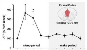
In the initial stages of sleep, energy levels increase dramatically in brain regions found to be active during waking hours, according to new research in the June 30 issue of the Journal of Neuroscience. These results suggest that a surge of cellular energy may replenish brain processes needed to function normally while awake.
Image
currency of cells, in rats increased in four key brain regions normally active during wakefulness. Shown here is the energy surge measured in the frontal cortex, a brain region associated with higher-level thinking. (Credit: Courtesy, with permission: Dworak et al. The Journal of Neuroscience 2010.)

A good night's rest has clear restorative benefits, but evidence of the actual biological processes that occur during sleep has been elusive. Radhika Basheer, PhD, and Robert McCarley, MD, of Boston V.A. Healthcare System and Harvard Medical School, proposed that brain energy levels are key to nightly restoration.

"Our finding bears on one of the perennial conundrums in biology: the function of sleep," Basheer said. "Somewhat surprisingly, there have been no modern-era studies of brain energy using the most sensitive measurements."

The authors measured levels of adenosine triphosphate (ATP), the energy currency of cells, in rats. They found that ATP levels in four key brain regions normally active during wakefulness increased when the rats were in non-REM sleep, but were accompanied by an overall decrease in brain activity. When the animals were awake, ATP levels were steady. When the rats were gently nudged to stay awake three or six hours past their normal sleep times, there was no increase in ATP.

The authors conclude that sleep is necessary for this ATP energy surge, as keeping the rats awake prevented the surge. The energy increase may then power restorative processes absent during wakefulness, because brain cells consume large amounts of energy just performing daily waking functions.

"This research provides intriguing evidence that a sleep-dependent energy surge is needed to facilitate the restorative biosynthetic processes," said Robert Greene, MD, PhD, of the University of Texas Southwestern, a sleep expert who was unaffiliated with the study. He observed that questions arise from the findings, such as the specific cause of the ATP surge. "The authors propose that the surge is related to decreases in brain cell activity during sleep, but it may be due to many other factors as well, including cellular signaling in the brain," he said.

The research was supported by the Department of Veterans Affairs, a Deutsche Forschungsgemeinschaft Fellowship, and the National Institute of Mental Health.

Thursday, July 1, 2010

Butterfly Effect Makes Brain Unreliable


Next time your brain plays tricks on you, you have an excuse: according to new research by UCL scientists published June 30 in the journal Nature, the brain is intrinsically unreliable.
Brain Intrinsically Unreliable
Researchers introduced a small perturbation into the brain, the neural equivalent of butterfly wings, and ask what would happen to the activity in the circuit. Would the perturbation grow and have a knock-on effect, thus affecting the rest of the brain, or immediately die out? (Credit: Image courtesy of University College London)

This may not seem surprising to most of us, but it has puzzled neuroscientists for decades. Given that the brain is the most powerful computing device known, how can it perform so well even though the behaviour of its circuits is variable?

A long-standing hypothesis is that the brain's circuitry actually is reliable -- and the apparently high variability is because your brain is engaged in many tasks simultaneously, which affect each other.

It is this hypothesis that the researchers at UCL tested directly. The team -- a collaboration between experimentalists at the Wolfson Institute for Biomedical Research and a theorist, Peter Latham, at the Gatsby Computational Neuroscience Unit -- took inspiration from the celebrated butterfly effect -- from the fact that the flap of a butterfly's wings in Brazil could set off a tornado in Texas. Their idea was to introduce a small perturbation into the brain, the neural equivalent of butterfly wings, and ask what would happen to the activity in the circuit. Would the perturbation grow and have a knock-on effect, thus affecting the rest of the brain, or immediately die out?

It turned out to have a huge knock-on effect. The perturbation was a single extra 'spike', or nerve impulse, introduced to a single neuron in the brain of a rat. That single extra spike caused about thirty new extra spikes in nearby neurons in the brain, most of which caused another thirty extra spikes, and so on. This may not seem like much, given that the brain produces millions of spikes every second. However, the researchers estimated that eventually, that one extra spike affected millions of neurons in the brain.

"This result indicates that the variability we see in the brain may actually be due to noise, and represents a fundamental feature of normal brain function," said lead author Dr. Mickey London, of the Wolfson Institute for Biomedical Research, UCL.

This rapid amplification of spikes means that the brain is extremely 'noisy' -- much, much noisier than computers. Nevertheless, the brain can perform very complicated tasks with enormous speed and accuracy, far faster and more accurately than the most powerful computer ever built (and likely to be built in the foreseeable future). The UCL researchers suggest that for the brain to perform so well in the face of high levels of noise, it must be using a strategy called a rate code. In a rate code, neurons consider the activity of an ensemble of many neurons, and ignore the individual variability, or noise, produced by each of them.

So now we know that the brain is truly noisy, but we still don't know why. The UCL researchers suggest that one possibility is that it's the price the brain pays for high connectivity among neurons (each neuron connects to about 10,000 others, resulting in over 8 million kilometres of wiring in the human brain). Presumably, that high connectivity is at least in part responsible for the brain's computational power. However, as the research shows, the higher the connectivity, the noisier the brain. Therefore, while noise may not be a useful feature, it is at least a by-product of a useful feature.
Enhanced by Zemanta

Wednesday, March 17, 2010

Nanoparticles: Golden Bullet for Cancer?


In a lecture he delivered in 1906, the German physician Paul Ehrlich coined the term Zuberkugel, or "magic bullet," as shorthand for a highly targeted medical treatment.
Me
Infrared images made while tumors were irradiated with a laser show that in nanocage-injected mice (left), the surface of the tumor quickly became hot enough to kill cells. In buffer-injected mice (right), the temperature barely budged. This specificity is what makes photothermal therapy so attractive as a cancer therapy. (Credit: WUSTL)

Magic bullets, also called silver bullets, because of the folkloric belief that only silver bullets can kill supernatural creatures, remain the goal of drug development efforts today.

A team of scientists at Washington University in St. Louis is currently working on a magic bullet for cancer, a disease whose treatments are notoriously indiscriminate and nonspecific. But their bullets are gold rather than silver. Literally.

The gold bullets are gold nanocages that, when injected, selectively accumulate in tumors. When the tumors are later bathed in laser light, the surrounding tissue is barely warmed, but the nanocages convert light to heat, killing the malignant cells.

In an article just published in the journal Small, the team describes the successful photothermal treatment of tumors in mice.

The team includes Younan Xia, Ph.D., the James M. McKelvey Professor of Biomedical Engineering in the School of Engineering and Applied Science, Michael J. Welch, Ph.D., professor of radiology and developmental biology in the School of Medicine, Jingyi Chen, Ph.D., research assistant professor of biomedical engineering and Charles Glaus, Ph.D., a postdoctoral research associate in the Department of Radiology.

"We saw significant changes in tumor metabolism and histology," says Welch, "which is remarkable given that the work was exploratory, the laser 'dose' had not been maximized, and the tumors were 'passively' rather than 'actively' targeted."

Why the nanocages get hot

The nanocages themselves are harmless. "Gold salts and gold colloids have been used to treat arthritis for more than 100 years," says Welch. "People know what gold does in the body and it's inert, so we hope this is going to be a nontoxic approach."

"The key to photothermal therapy," says Xia, "is the cages' ability to efficiently absorb light and convert it to heat. "

Suspensions of the gold nanocages, which are roughly the same size as a virus particle, are not always yellow, as one would expect, but instead can be any color in the rainbow.

They are colored by something called a surface plasmon resonance. Some of the electrons in the gold are not anchored to individual atoms but instead form a free-floating electron gas, Xia explains. Light falling on these electrons can drive them to oscillate as one. This collective oscillation, the surface plasmon, picks a particular wavelength, or color, out of the incident light, and this determines the color we see.

Medieval artisans made ruby-red stained glass by mixing gold chloride into molten glass, a process that left tiny gold particles suspended in the glass, says Xia.

The resonance -- and the color -- can be tuned over a wide range of wavelengths by altering the thickness of the cages' walls. For biomedical applications, Xia's lab tunes the cages to 800 nanometers, a wavelength that falls in a window of tissue transparency that lies between 750 and 900 nanometers, in the near-infrared part of the spectrum.

Light in this sweet spot can penetrate as deep as several inches in the body (either from the skin or the interior of the gastrointestinal tract or other organ systems).

The conversion of light to heat arises from the same physical effect as the color. The resonance has two parts. At the resonant frequency, light is typically both scattered off the cages and absorbed by them.

By controlling the cages' size, Xia's lab tailors them to achieve maximum absorption.

Passive targeting

"If we put bare nanoparticles into your body," says Xia, "proteins would deposit on the particles, and they would be captured by the immune system and dragged out of the bloodstream into the liver or spleen."

To prevent this, the lab coated the nanocages with a layer of PEG, a nontoxic chemical most people have encountered in the form of the laxatives GoLyTELY or MiraLAX. PEG resists the adsorption of proteins, in effect disguising the nanoparticles so that the immune system cannot recognize them.

Instead of being swept from the bloodstream, the disguised particles circulate long enough to accumulate in tumors.

A growing tumor must develop its own blood supply to prevent its core from being starved of oxygen and nutrients. But tumor vessels are as aberrant as tumor cells. They have irregular diameters and abnormal branching patterns, but most importantly, they have thin, leaky walls.

The cells that line a tumor's blood vessel, normally packed so tightly they form a waterproof barrier, are disorganized and irregularly shaped, and there are gaps between them.

The nanocages infiltrate through those gaps efficiently enough that they turn the surface of the normally pinkish tumor black.

A trial run

In Welch's lab, mice bearing tumors on both flanks were randomly divided into two groups. The mice in one group were injected with the PEG-coated nanocages and those in the other with buffer solution. Several days later the right tumor of each animal was exposed to a diode laser for 10 minutes.

The team employed several different noninvasive imaging techniques to follow the effects of the therapy. (Welch is head of the oncologic imaging research program at the Siteman Cancer Center of Washington University School of Medicine and Barnes-Jewish Hospital and has worked on imaging agents and techniques for many years.)

During irradiation, thermal images of the mice were made with an infrared camera. As is true of cells in other animals that automatically regulate their body temperature, mouse cells function optimally only if the mouse's body temperature remains between 36.5 and 37.5 degrees Celsius (98 to 101 degrees Fahrenheit).

At temperatures above 42 degrees Celsius (107 degrees Fahrenheit) the cells begin to die as the proteins whose proper functioning maintains them begin to unfold.

In the nanocage-injected mice, the skin surface temperature increased rapidly from 32 degrees Celsius to 54 degrees C (129 degrees F).

In the buffer-injected mice, however, the surface temperature remained below 37 degrees Celsius (98.6 degrees Fahrenheit).

To see what effect this heating had on the tumors, the mice were injected with a radioactive tracer incorporated in a molecule similar to glucose, the main energy source in the body. Positron emission and computerized tomography (PET and CT) scans were used to record the concentration of the glucose lookalike in body tissues; the higher the glucose uptake, the greater the metabolic activity.

The tumors of nanocage-injected mice were significantly fainter on the PET scans than those of buffer-injected mice, indicating that many tumor cells were no longer functioning.

The tumors in the nanocage-treated mice were later found to have marked histological signs of cellular damage.

Active targeting

The scientists have just received a five-year, $2,129,873 grant from the National Cancer Institute to continue their work with photothermal therapy.

Despite their results, Xia is dissatisfied with passive targeting. Although the tumors took up enough gold nanocages to give them a black cast, only 6 percent of the injected particles accumulated at the tumor site.

Xia would like that number to be closer to 40 percent so that fewer particles would have to be injected. He plans to attach tailor-made ligands to the nanocages that recognize and lock onto receptors on the surface of the tumor cells.

In addition to designing nanocages that actively target the tumor cells, the team is considering loading the hollow particles with a cancer-fighting drug, so that the tumor would be attacked on two fronts.

But the important achievement, from the point of view of cancer patients, is that any nanocage treatment would be narrowly targeted and thus avoid the side effects patients dread.

The TV and radio character the Lone Ranger used only silver bullets, allegedly to remind himself that life was precious and not to be lightly thrown away. If he still rode today, he might consider swapping silver for gold.
Reblog this post [with Zemanta]