BTemplates.com

Powered by Blogger.

Pageviews past week

Quantum mechanics

Auto News

artificial intelligence

About Me

Recommend us on Google!

Information Technology

Popular Posts

Showing posts with label Immune system. Show all posts

Tuesday, July 19, 2011

Stem Cells: Nearing Goal of Using Patient's Own Cells to Make Stem Cells to Replace Lost or Diseased Tissue


Scientists at the Salk Institute for Biological Studies have developed an improved technique for generating large numbers of blood cells from a patient's own cells. The new technique will be immediately useful in further stem cell studies, and when perfected, could be used in stem cell therapies for a wide variety of conditions including cancers and immune ailments.
Round hematopoietic (blood) cells emerge from 
differentiating human pluripotent stem cells. 
(Credit: Courtesy of Aaron Parker, Salk Institute 
for Biological Studies)

"There are further improvements that we need to make, but this takes us a significant step closer to the ultimate goal, which is to be able to take ordinary cells from a patient, induce them to become stem cells, and then use those stem cells to rebuild lost or diseased tissues, for example the patient's bone marrow," says Inder M. Verma, PhD, Irwin and Joan Jacobs Chair in Exemplary Life Science and American Cancer Society Professor of Molecular Biology at the Salk Institute Laboratory of Genetics. Verma is senior author of the report, which is published in the July edition of the journal Stem Cells.

Stem cell researchers have been racing towards this goal since 2006, when techniques for turning ordinary skin cells into induced pluripotential stem cells (iPSCs) were first reported. In principle, iPSCs mimic the embryonic stem cells (ESCs) from which organisms develop. Researchers now want to find the precise mixes and sequences of chemical compounds needed to coax iPSCs to mature into the tissue-specific stem cells of their choice. The latter are self-renewing, and can be transplanted into the body to produce the 'progenitor' cells that multiply locally and produce mature tissue cells.

However, researchers don't know yet how to induce iPSCs to become tissue-specific stem cells or mature tissue cells with high efficiency. "We've been producing these cells in quantities that are too low to enable them to be studied easily, much less used for therapies," says Aaron Parker, PhD, a former graduate student and now a postdoctoral researcher in Verma's lab. Parker is a co-lead-author of the paper, with Niels-Bjarne Woods, PhD, who was a postdoctoral researcher in the Verma lab at the outset of the project, and is now an assistant professor at Lund University in Sweden.

Like many other stem cell research laboratories, the Verma lab has been trying to find more efficient ways to turn iPSCs into blood-forming 'hematopoietic' stem cells (HSCs). These may be more valuable medically than any other tissue-specific stem cell, because they can supply not only oxygen-carrying red blood cells but also all the white blood cells of the immune system. "There would be an almost unlimited number of usages for true HSCs," says Verma.

For the present study, the research team sought to do a better job of mimicking the changing conditions that naturally direct ESCs to become HSCs in the womb. "We took seven lines of human ESCs and iPSCs, and experimented with different combinations and sequences of growth factors and other chemical compounds that are known to be present as ESCs move to the HSC state in a developing human," says Parker.


Applying cocktails of these factors, Parker and Woods and their colleagues induced the iPSCs and ESCs to form colonies of cells that bore the distinctive molecular markers of blood cells. With their best such cocktail they were able to detect blood-specific markers on 84% of their cells after three weeks. "That's a big jump in efficiency from what we saw in the field just a few years ago," says Parker.

The technique still has room for improvement. The researchers detected progenitor cells and mature cells from only one category or lineage: myeloid cells, which include red blood cells and primitive immune cells such as macrophages. "We didn't see any cells from the lymphoid lineage, meaning T-cells and B-cells," Parker says.

Another drawback was that the blood cell population they produced from ESCs and iPSCs contained short-lived progenitors and mature blood cells but no indefinitely renewing, transplantable HSCs. Their cocktail, they believed, either pushed the cells past the HSC state to the progenitor state too quickly, or made the maturing cells skip the HSC state entirely.

From this and other labs' results, the team hypothesized the existence of an intermediate, pre-hematopoietic type of stem cell, produced by ESCs and iPSCs and in turn producing HSCs. "We know that HSCs appear in a particular region of mammals during embryonic development, and our idea is that these pre-hematopoietic stem cells are there and are somehow made to mature into HSCs," says Parker. "So our lab is now going to focus on finding the precise maturation signals provided by that embryonic region to produce these true, transplantable HSCs."

Once that is done, researchers will need to make a number of further refinements to improve the safety of HSCs intended for human patients. "But we're now tantalizingly close to our ultimate goal," says Verma.

The other authors who contributed to the work were Roksana Moraghebi, of Lund University's Stem Cell Center; Margaret K. Lutz, Amy L. Firth, Kristen J. Brennand, W. Travis Berggren and Fred H. Gage of the Salk Institute Laboratory for Genetics; Juan Carlos Izpisúa Belmonte of the Salk Institute Gene Expression Laboratory; and Angel Raya of the Center of Regenerative Medicine in Barcelona, Spain.

Funding for this research was provided by the National Institutes for Health, the California Institute for Regenerative Medicine, the Leducq Foundation, the Merieux Foundation, the Ellison Medical Foundation, Ipsen/Biomeasure, Sanofi Aventis, the Prostate Cancer Foundation, the H.N. and Frances C. Berger Foundation, The Royal Physiographic Society of Sweden, the Lund University Medical Faculty, and the Lars Hierta Memorial Foundation, and the H.A. and Mary K. Chapman Charitable Trust.

Tuesday, June 28, 2011

First Patients Receive Lab-Grown Blood Vessels from Donor Cells


For the first time, blood vessels created in the lab from donor skin cells were successfully implanted in patients. Functioning blood vessels that aren't rejected by the immune system could be used to make durable shunts for kidney dialysis, and potentially to improve treatment for children with heart defects and adults needing coronary or other bypass graft surgery.
Artist's rendering of blood vessels. Researchers report 
that for the first time, blood vessels created in the lab from 
donor skin cells were successfully implanted in patients. 
(Credit: © Dario Bajurin / Fotolia)

For the first time, human blood vessels grown in a laboratory from donor skin cells have been successfully implanted into patients, according to new research presented in the American Heart Association's Emerging Science Series webinar.

While more testing is needed, such "off-the-shelf" blood vessels could soon be used to improve the process and affordability of kidney dialysis.

"Our approach could allow hundreds of thousands of patients to be treated from one master cell line," said study lead author Todd N. McAllister, Ph.D., co-founder and chief executive officer of Cytograft Tissue Engineering Inc., of Novato, Calif.

The grafts also have the potential to be used in lower limb bypass to route blood around diseased arteries, to repair congenital heart defects in pediatric patients and to fix damaged arteries in soldiers, who might otherwise lose a limb, said McAllister.


The tissue-engineered blood vessels, produced from sheets of cultured skin cells rolled around temporary support structures, were used to create access shunts between arteries and veins in the arm for kidney dialysis in three patients. These shunts, which connect an artery to a vein, provide access to the blood for dialysis. The engineered vessels were about a foot long with a diameter of 4.8 millimeters.

At follow-up exams up to eight months after implantation, none of the patients had developed an immune reaction to the implants, and the vessels withstood the high pressure and frequent needle punctures required for dialysis. Shunts created from patients' own vessels or synthetic materials are notoriously prone to failure.

Investigators previously showed that using vessels individually created from a patient's own skin cells reduced the rate of shunt complications 2.4-fold over a 3-year period. The availability of off-the-shelf vessels could avoid the expense and months-long process involved in creating custom vessels for each patient, making the technique feasible for widespread use.

Besides addressing a costly and vexing problem in kidney dialysis, off-the-shelf blood vessels might someday be used instead of harvesting patients' own vessels for bypass surgery. A larger, randomized trial of the grafts is under way for kidney dialysis, and human trials have been initiated to assess the safety and effectiveness of these grafts for lower-limb bypass.

The study will be presented in the American Heart Association's Emerging Science Series, which will be held at 1 p.m. EDT/ 12 p.m. CDT. The series is a free online webinar presentation of cutting-edge science. The Emerging Science Series provides a new venue for presenting the latest cardiovascular scientific breakthroughs several times a year, when the discoveries are ready to be presented rather than waiting for a regularly scheduled meeting. Each study is handled in a peer-reviewed process similar to late-breaking clinical trials presented at AHA's annual Scientific Sessions.

The series will include the first presentation of data from clinical trials, basic science, key updates of previously presented trials and major bench-to-bedside breakthroughs.

Co-authors are Wojciech Wystrychowski, M.D.; Lech Cierpka, M.D.; Krzysztof Zagalski, M.D.; Sergio A. Garrido, M.D.; Samuel Radochonski, B.S.; Nathalie Dusserre, Ph.D.; and Nicholas L'Heureux, Ph.D.

Wednesday, December 1, 2010

Antibacterial Soaps: Being Too Clean Can Make People Sick, Study Suggests


Young people who are overexposed to antibacterial soaps containing triclosan may suffer more allergies, and exposure to higher levels of Bisphenol A among adults may negatively influence the immune system, a new University of Michigan School of Public Health study suggests.
Young people who are overexposed to antibacterial 
soaps containing triclosan may suffer more allergies, and 
exposure to higher levels of Bisphenol A among adults 
may negatively influence the immune system, a new 
University of Michigan School of Public Health study 
suggests. (Credit: iStockphoto/Jorge Salcedo)

Triclosan is a chemical compound widely used in products such as antibacterial soaps, toothpaste, pens, diaper bags and medical devices. Bisphenol A (BPA) is found in many plastics and, for example, as a protective lining in food cans. Both of these chemicals are in a class of environmental toxicants called endocrine-disrupting compounds (EDCs), which are believed to negatively impact human health by mimicking or affecting hormones.

Using data from the 2003-2006 National Health and Nutrition Examination Survey, U-M researchers compared urinary BPA and triclosan with cytomegalovirus (CMV) antibody levels and diagnosis of allergies or hay fever in a sample of U.S. adults and children over age 6. Allergy and hay fever diagnosis and CMV antibodies were used as two separate markers of immune alterations.

"We found that people over age 18 with higher levels of BPA exposure had higher CMV antibody levels, which suggests their cell-mediated immune system may not be functioning properly," said Erin Rees Clayton, research investigator at the U-M School of Public Health and first author on the paper.

Researchers also found that people age 18 and under with higher levels of triclosan were more likely to report diagnosis of allergies and hay fever.

There is growing concern among the scientific community and consumer groups that these EDCs are dangerous to humans at lower levels than previously thought.

"The triclosan findings in the younger age groups may support the 'hygiene hypothesis,' which maintains living in very clean and hygienic environments may impact our exposure to micro-organisms that are beneficial for development of the immune system," said Allison Aiello, associate professor at the U-M School of Public Health and principal investigator on the study.

As an antimicrobial agent found in many household products, triclosan may play a role in changing the micro-organisms to which we are exposed in such a way that our immune system development in childhood is affected.

"It is possible that a person can be too clean for their own good," said Aiello, who is also a visiting associate professor of epidemiology at Harvard.

Previous animal studies indicate that BPA and triclosan may affect the immune system, but this is the first known study to look at exposure to BPA and triclosan as it relates to human immune function, Aiello said.

One surprise finding is that with BPA exposure, age seems to matter, said Rees Clayton. In people 18 or older, higher amounts of BPA were associated with higher CMV levels, but in people younger than 18 the reverse was true.

"This suggests the timing of the exposure to BPA and perhaps the quantity and length of time we are exposed to BPA may be affecting the immune system response," Rees Clayton said.

This is just the first step, she said, but a very important one. Going forward, researchers would like to study the long-term effects of BPA and triclosan in people to see if they can establish a causal relationship.

One limitation of the study is that it measured disease and exposure simultaneously and thus shows only part of the picture, Aiello said.

"It is possible, for example, that individuals who have an allergy are more hygienic because of their condition, and that the relationship we observed is, therefore, not causal or is an example of reverse causation," Aiello said.

Disclaimer: This article is not intended to provide medical advice, diagnosis or treatment.

Tuesday, July 20, 2010

Of Bugs and Brains: Gut Bacteria Affect Multiple Sclerosis


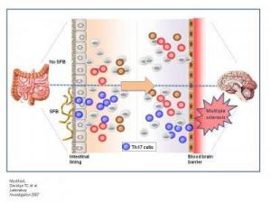
Biologists at the California Institute of Technology (Caltech) have demonstrated a connection between multiple sclerosis (MS) -- an autoimmune disorder that affects the brain and spinal cord -- and gut bacteria.
Image
In the absence of bacteria in the intestines, 
pro-inflammatory Th17 cells do not develop in 
either the gut or the central nervous system; and 
animals do not develop disease (top panel). When 
animals are colonized with symbiotic segmented 
filamentous bacteria, Th17 cell differentiation is 
induced in the gut. Th17 cells promote experimental 
autoimmune encephalomyelitis, an animal model for 
multiple sclerosis. In this way, non-pathogenic bacteria 
of the microbiota promote disease by shaping the immune 
response in both the gut and the brain (top panel). 
(Credit: Lee, Mazmanian/Caltech; modified from 
Savidge TC et al. Laboratory Investigation 2007)

The work -- led by Sarkis K. Mazmanian, an assistant professor of biology at Caltech, and postdoctoral scholar Yun Kyung Lee -- appears online the week of July 19-23 in the Proceedings of the National Academy of Sciences.

Multiple sclerosis results from the progressive deterioration of the protective fatty myelin sheath surrounding nerve cells. The loss of myelin hinders nerve cells from communicating with one another, leading to a host of neurological symptoms including loss of sensation, muscle spasms and weakness, fatigue, and pain. Multiple sclerosis is estimated to affect about half a million people in the United States alone, with rates of diagnosis rapidly increasing. There is currently no cure for MS.

Although the cause of MS is unknown, microorganisms seem to play some sort of role. "In the literature from clinical studies, there are papers showing that microbes affect MS," Mazmanian says. "For example, the disease gets worse after viral infections, and bacterial infections cause an increase in MS symptoms."

On the other hand, he concedes, "it seems counterintuitive that a microbe would be involved in a disease of the central nervous system, because these are sterile tissues."

And yet, as Mazmanian found when he began examining the multiple sclerosis literature, the suggestion of a link between bacteria and the disease is more than anecdotal. Notably, back in 1993, Caltech biochemist Leroy Hood -- who was then at the University of Washington -- published a paper describing a genetically engineered strain of mouse that developed a lab-induced form of multiple sclerosis known as experimental autoimmune encephalomyelitis, or EAE.

When Hood's animals were housed at Caltech, they developed the disease. But, oddly, when the mice were shipped to a cleaner biotech facility -- where their resident gut bacterial populations were reduced -- they didn't get sick. The question was, why? At the time, Mazmanian says, "the authors speculated that some environmental component was modulating MS in these animals." Just what that environmental component was, however, remained a mystery for almost two decades.

But Mazmanian -- whose laboratory examines the relationships between gut microbes, both harmful and helpful, and the immune systems of their mammalian hosts -- had a hunch that intestinal bacteria were the key. "As we gained an appreciation for how profoundly the gut microbiota can affect the immune system, we decided to ask if symbiotic bacteria are the missing variable in these mice with MS," he says.

To find out, Mazmanian and his colleagues tried to induce MS in animals that were completely devoid of the microbes that normally inhabit the digestive system. "Lo and behold, these sterile animals did not get sick," he says.

Then the researchers decided to see what would happen if bacteria were reintroduced to the germ-free mice. But not just any bacteria. They inoculated mice with one specific organism, an unculturable bug from a group known as segmented filamentous bacteria. In prior studies, these bacteria had been shown to lead to intestinal inflammation and, more intriguingly, to induce in the gut the appearance of a particular immune-system cell known as Th17. Th17 cells are a type of T helper cell -- cells that help activate and direct other immune system cells. Furthermore, Th17 cells induce the inflammatory cascade that leads to multiple sclerosis in animals.

"The question was, if this organism is inducing Th17 cells in the gut, will it be able to do so in the brain and central nervous system?" Mazmanian says. "Furthermore, with that one organism, can we restore to sterile animals the entire inflammatory response normally seen in animals with hundreds of species of gut bacteria?"

The answer? Yes on all counts. Giving the formerly germ-free mice a dose of one species of segmented filamentous bacteria induced Th17 not only in the gut but in the central nervous system and brain -- and caused the formerly healthy mice to become ill with MS-like symptoms.

"It definitely shows that gut microbes have a strong role in MS, because the genetics of the animals were the same. In fact, everything was the same except for the presence of those otherwise benign bacteria, which are clearly playing a role in shaping the immune system," Mazmanian says. "This study shows for the first time that specific intestinal bacteria have a significant role in affecting the nervous system during MS -- and they do so from the gut, an anatomical location very, very far from the brain."

Mazmanian and his colleagues don't, however, suggest that gut bacteria are the direct cause of multiple sclerosis, which is known to be genetically linked. Rather, the bacteria may be helping to shape the immune system's inflammatory response, thus creating conditions that could allow the disease to develop. Indeed, multiple sclerosis also has a strong environmental component; identical twins, who possess the same genome and share all of their genes, only have a 25 percent chance of sharing the disease. "We would like to suggest that gut bacteria may be the missing environmental component," he says.

For their part, Th17 cells are needed for the immune system to properly combat infection. Problems only arise when the cells are activated in the absence of infection -- just as disease can arise, Mazmanian and others suspect, when the species composition of gut bacteria become imbalanced, say, by changes in diet, because of improved hygiene (which kills off the beneficial bacteria as well as the dangerous ones), or because of stress or antibiotic use. One impact of the dysregulation of normal gut bacterial populations -- a phenomenon dubbed "dysbiosis" -- may be the rising rate of multiple sclerosis seen in recent years in more hygienic societies.

"As we live cleaner, we're not just changing our exposure to infectious agents, but we're changing our relationship with the entire microbial world, both around and inside us, and we may be altering the balance between pro- and anti-inflammatory bacteria," leading to diseases like MS, Mazmanian says. "Perhaps treatments for diseases such as multiple sclerosis may someday include probiotic bacteria that can restore normal immune function in the gut… and the brain."

The work was supported by funding from the California Institute of Technology, the Weston Havens Foundation, and the Edward Mallinckrodt, Jr. Foundation.

Monday, June 21, 2010

Gene Therapy Reverses Type 1 Diabetes in Mice


Researchers have developed an experimental cure for Type 1 diabetes, a disease that affects about one in every 400 to 600 children and adolescents.

Image
In a new study using gene therapy in mice, researchers 
say they have developed an experimental cure for 
Type 1 diabetes. (Credit: iStockphoto)

Results of the research in a mouse model of Type 1 diabetes are being presented at The Endocrine Society's 92nd Annual Meeting in San Diego.

Using gene therapy, the team from Baylor College of Medicine in Houston tried to counter the two defects that cause Type 1 diabetes: autoimmune attack and destruction of the insulin-producing beta cells. They used nonobese diabetic mice, which spontaneously develop diabetes due to autoimmunity, just as humans do with Type 1 diabetes.

"A single treatment cured about 50 percent of the diabetic mice, restoring their blood sugar to normal so that they no longer need insulin injections," said study co-author Lawrence Chan, MD, DSc, chief of Baylor's diabetes, endocrinology and metabolism division.

Type 1 diabetes occurs when the body's immune system attacks and destroys the beta cells in the pancreas, the insulin "factory" of the body. The resulting near-complete deficiency of insulin -- the hormone that controls blood sugar -- leads to a buildup of high blood sugar and thus diabetes.

In past studies of their original gene therapy, Chan's group was able to stimulate new formation of beta cells in the liver and restore insulin production and normal blood sugar levels in more than 100 mice with chemically induced diabetes. However, in nonobese diabetic mice the treatment failed to reverse Type 1 diabetes because the mouse's immune system killed the newly formed beta cells, he said.

In this research, which was funded by the National Institute of Diabetes, Kidney and Digestive Diseases, Chan said they "added to the original gene therapy approach a protective gene that shields the newly formed beta cells from autoimmune attack." The added gene was for interleukin-10, an important regulator of the immune system. Past studies showed that interleukin-10 can prevent diabetes development in mice but cannot reverse the disease once it has developed because of the lack of beta cells.

However, when the researchers combined the gene therapy with interleukin-10 into a single intravenous injection, the treatment showed a complete reversal of diabetes in half of the mice during more than 20 months' follow-up. Although the therapy did not reverse autoimmunity throughout the body, it protected the new beta cells from the local destructive effect of autoimmunity, Chan explained.

"We developed a protective 'moat' around the new beta cells," he said. "We are now developing other strategies to try to fortify the newly formed beta cells and give them better weapons in addition to the moat, in order to increase the treatment's cure rate."

Why the gene therapy did not work in all the mice is unclear. However, Chan said the treated mice that did not have improvements in their blood sugar did gain weight and lived a little longer than untreated mice.
Enhanced by Zemanta

Friday, May 14, 2010

Why Is Breast Milk Best? It's All in the Genes


Is breast milk so different from infant formula? The ability to track which genes are operating in an infant's intestine has allowed University of Illinois scientists to compare the early development of breast-fed and formula-fed babies. They say the difference is very real.

Me
Breast milk induces genetic pathways that are quite 
different from those in formula-fed infants, new research 
has found. (Credit: iStockphoto/Oleg Kozlov)

"For the first time, we can see that breast milk induces genetic pathways that are quite different from those in formula-fed infants. Although formula makers have tried to develop a product that's as much like breast milk as possible, hundreds of genes were expressed differently in the breast-fed and formula-fed groups," said Sharon Donovan, a U of I professor of nutrition.

Although both breast-fed and formula-fed babies gain weight and seem to develop similarly, scientists have known for a long time that breast milk contains immune-protective components that make a breast-fed infant's risk lower for all kinds of illnesses, she said.

"The intestinal tract of the newborn undergoes marked changes in response to feeding. And the response to human milk exceeds that of formula, suggesting that the bioactive components in breast milk are important in this response," she noted.

"What we haven't known is how breast milk protects the infant and particularly how it regulates the development of the intestine," she said.

Understanding those differences should help formula makers develop a product that is more like the real thing, she said. The scientists hope to develop a signature gene or group of genes to use as a biomarker for breast-fed infants.

Many of the differences found by the scientists were in fundamental genes that regulate the development of the intestine and provide immune defense for the infant.

In this small proof-of-concept study, Donovan used a new technique patented by Texas A&M colleague Robert Chapkin to examine intestinal gene expression in 22 healthy infants -- 12 breast-fed, 10 formula-fed.

The technique involved isolating intestinal cells shed in the infants' stools, then comparing the expression of different genes between the two groups. Mothers in the study collected fecal samples from their babies at one, two, and three months of age. Scientists were then able to isolate high-quality genetic material, focusing on the RNA to get a gene expression or signature.

Donovan said that intestinal cells turn over completely every three days as billions of cells are made, perform their function, and are exfoliated. Examining the shed cells is a noninvasive way to examine intestinal health and see how nutrition affects intestinal development in infants.

Understanding early intestinal development is important for many reasons, she said.

"An infant's gut has to adapt very quickly. A new baby is coming out of a sterile environment, having received all its nutrients intravenously through the placenta. At that point, babies obviously must begin eating, either mother's milk or formula.

"They also start to become colonized with bacteria, so it's very important that the gut learns what's good and what's bad. The baby's body needs to be able to recognize a bad bacteria or a bad virus and fight it, but it also needs to recognize that even though a food protein is foreign, that protein is okay and the body doesn't want to develop an immune response to it," she said.

If anything goes wrong at this stage, babies can develop food allergies, inflammatory bowel disease, and even asthma. "We're very interested in frequent sampling at this early period of development," she added.

Donovan also would like to learn how bacteria in the gut differ in formula- and breast-fed babies, and this technique should make that possible. "Now we'll be able to get a complete picture of what's happening in an infant -- from the composition of the diet to the microbes in the gut and the genes that are activated along the way."

Of potential clinical importance: The gene expressed most often in breast-fed infants is involved in the cell's response to oxygen deprivation. Lack of oxygen is a factor in the development of necrotizing enterocolitis (NEC), a kind of gangrene of the intestine that can be fatal in premature babies. NEC is a leading cause of disease and death in neonatal intensive care units, with a reported 2,500 cases occurring annually in the United States and a mortality rate of 26 percent.

The study will appear in the June 2010 issue of the American Journal of Physiology, Gastrointestinal and Liver Physiology. Co-authors are Robert S. Chapkin, Chen Zhao, Ivan Ivanov, Laurie A. Davidson, Jennifer S. Goldsby, Joanne R. Lupton, and Edward R. Dougherty, all of Texas A&M University, Rose Ann Mathai and Marcia H. Monaco of the U of I, and Deshanie Rai and W. Michael Russell of Mead Johnson Nutrition. The study was funded by Mead Johnson Nutrition.
Reblog this post [with Zemanta]

Friday, April 2, 2010

Carbon Nanostructures: Elixir or Poison?


A Los Alamos National Laboratory toxicologist and a multidisciplinary team of researchers have documented potential cellular damage from "fullerenes" -- soccer-ball-shaped, cage-like molecules composed of 60 carbon atoms. The team also noted that this particular type of damage might hold hope for treatment of Parkinson's disease, Alzheimer's disease, or even cancer.
Me
Los Alamos National Laboratory toxicologist Jun Gao works in his laboratory using a protective fume hood. (Credit: Photo by James R. Rickman)
The research recently appeared in Toxicology and Applied Pharmacology and represents the first-ever observation of this kind for spherical fullerenes, also known as buckyballs, which take their names from the late Buckminster Fuller because they resemble the geodesic dome concept that he popularized.

Engineered carbon nanoparticles, which include fullerenes, are increasing in use worldwide. Each buckyball is a skeletal cage of carbon about the size of a virus. They show potential for creating stronger, lighter structures or acting as tiny delivery mechanisms for designer drugs or antibiotics, among other uses. About four to five tons of carbon nanoparticles are manufactured annually.

"Nanomaterials are the 21st century revolution," said Los Alamos toxicologist Rashi Iyer, the principal research lead and coauthor of the paper. "We are going to have to live with them and deal with them, and the question becomes, 'How are we going to maximize our use of these materials and minimize their impact on us and the environment?'"

Iyer and lead author Jun Gao, also a Los Alamos toxicologist, exposed cultured human skin cells to several distinct types of buckyballs. The differences in the buckyballs lay in the spatial arrangement of short branches of molecules coming off of the main buckyball structure. One buckyball variation, called the "tris" configuration, had three molecular branches off the main structure on one hemisphere; another variation, called the "hexa" configuration, had six branches off the main structure in a roughly symmetrical arrangement; the last type was a plain buckyball.

The researchers found that cells exposed to the tris configuration underwent premature senescence -- what might be described as a state of suspended animation. In other words, the cells did not die as cells normally should, nor did they divide or grow. This arrest of the natural cellular life cycle after exposure to the tris-configured buckyballs may compromise normal organ development, leading to disease within a living organism. In short, the tris buckyballs were toxic to human skin cells.

Moreover, the cells exposed to the tris arrangement caused unique molecular level responses suggesting that tris-fullerenes may potentially interfere with normal immune responses induced by viruses. The team is now pursuing research to determine if cells exposed to this form of fullerenes may be more susceptible to viral infections.

Ironically, the discovery could also lead to a novel treatment strategy for combating several debilitating diseases. In diseases like Parkinson's or Alzheimer's, nerve cells die or degenerate to a nonfunctional state. A mechanism to induce senescence in specific nerve cells could delay or eliminate onset of the diseases. Similarly, a disease like cancer, which spreads and thrives through unregulated replication of cancer cells, might be fought through induced senescence. This strategy could stop the cells from dividing and provide doctors with more time to kill the abnormal cells.

Because of the minute size of nanomaterials, the primary hazard associated with them has been potential inhalation -- similar to the concern over asbestos exposure.

"Already, from a toxicological point of view, this research is useful because it shows that if you have the choice to use a tris- or a hexa-arrangement for an application involving buckyballs, the hexa-arrangement is probably the better choice," said Iyer. "These studies may provide guidance for new nanomaterial design and development."

These results were offshoots from a study (Shreve, Wang, and Iyer) funded to understand the interactions between buckyballs and biological membranes. Los Alamos National Laboratory has taken a proactive role by initiating a nanomaterial bioassessmnet program with the intention of keeping its nanomaterial workers safe while facilitating the discovery of high-function, low-bioimpact nanomaterials with the potential to benefit national security missions. In addition to Gao and Iyer, the LANL program includes Jennifer Hollingsworth, Yi Jiang, Jian Song, Paul Welch, Hsing Lin Wang, Srinivas Iyer, and Gabriel Montaño.

Los Alamos National Laboratory researchers will continue to attempt to understand the potential effects of exposure to nanomaterials in much the same way that Los Alamos was a worldwide leader in understanding the effects of radiation during the Lab's early history. Los Alamos workers using nanomaterials will continue to follow protocols that provide the highest degree of protection from potential exposure.

Meantime, Los Alamos research into nanomaterials provides a cautionary tale for nanomaterial use, as well as early foundations for worker protection. Right now, there are no federal regulations for the use of nanomaterials. Disclosure of use by companies or individuals is voluntary. As nanomaterial use increases, understanding of their potential hazards should also increase.
Reblog this post [with Zemanta]

Saturday, March 20, 2010

Acne Drug Prevents HIV Breakout


Johns Hopkins scientists have found that a safe and inexpensive antibiotic in use since the 1970s for treating acne effectively targets infected immune cells in which HIV, the virus that causes AIDS, lies dormant and prevents them from reactivating and replicating.
Acne Drug
Janice E. Clements, Ph.D. (Credit: Image courtesy of Johns Hopkins Medical Institutions)

The drug, minocycline, likely will improve on the current treatment regimens of HIV-infected patients if used in combination with a standard drug cocktail known as HAART (Highly Active Antiretroviral Therapy), according to research published now online and appearing in print April 15 in The Journal of Infectious Diseases. "The powerful advantage to using minocycline is that the virus appears less able to develop drug resistance because minocycline targets cellular pathways not viral proteins," says Janice Clements, Ph.D., Mary Wallace Stanton Professor of Faculty Affairs, vice dean for faculty, and professor of molecular and comparative pathobiology at the Johns Hopkins University School of Medicine.

"The big challenge clinicians deal with now in this country when treating HIV patients is keeping the virus locked in a dormant state," Clements adds. "While HAART is really effective in keeping down active replication, minocycline is another arm of defense against the virus."

Unlike the drugs used in HAART which target the virus, minocycline homes in on, and adjusts T cells, major immune system agents and targets of HIV infection. According to Clements, minocycline reduces the ability of T cells to activate and proliferate, both steps crucial to HIV production and progression toward full blown AIDS.

If taken daily for life, HAART usually can protect people from becoming ill, but it's not a cure. The HIV virus is kept at a low level but isn't ever entirely purged; it stays quietly hidden in some immune cells. If a person stops HAART or misses a dose, the virus can reactivate out of those immune cells and begin to spread.

The idea for using minocycline as an adjunct to HAART resulted when the Hopkins team learned of research by others on rheumatoid arthritis patients showing the anti-inflammatory effects of minocycline on T cells. The Hopkins group connected the dots between that study with previous research of their own showing that minocycline treatment had multiple beneficial effects in monkeys infected with SIV, the primate version of HIV. In monkeys treated with minocycline, the virus load in the cerebrospinal fluid, the viral RNA in the brain and the severity of central nervous system disease were significantly decreased. The drug was also shown to affect T cell activation and proliferation.

"Since minocycline reduced T cell activation, you might think it would have impaired the immune systems in the macaques, which are very similar to humans, but we didn't see any deleterious effect," says Gregory Szeto, a graduate student in the Department of Cellular and Molecular Medicine working in the Retrovirus Laboratory at Hopkins. "This drug strikes a good balance and is ideal for HIV because it targets very specific aspects of immune activation."

The success with the animal model prompted the team to study in test tubes whether minocycline treatment affected latency in human T cells infected with HIV. Using cells from HIV-infected humans on HAART, the team isolated the "resting" immune cells and treated half of them with minocycline. Then they counted how many virus particles were reactivated, finding completely undetectable levels in the treated cells versus detectable levels in the untreated cells.

"Minocycline reduces the capability of the virus to emerge from resting infected T cells," Szeto explains. "It prevents the virus from escaping in the one in a million cells in which it lays dormant in a person on HAART, and since it prevents virus activation it should maintain the level of viral latency or even lower it. That's the goal: Sustaining a latent non-infectious state."

The team used molecular markers to discover that minocycline very selectively interrupts certain specific signaling pathways critical for T cell activation. However, the antibiotic doesn't completely obliterate T cells or diminish their ability to respond to other infections or diseases, which is crucial for individuals with HIV.

"HIV requires T cell activation for efficient replication and reactivation of latent virus," Clement says, "so our new understanding about minocyline's effects on a T cell could help us to find even more drugs that target its signaling pathways."

The research was supported by grants from the National Institutes of Health.

Authors of the paper, in addition to Clements and Szeto, are Angela K. Brice, Sheila A. Barber and Robert F. Siliciano, all of Johns Hopkins. Also, Hung-Chih Yang of National Taiwan University Hospital.


Reblog this post [with Zemanta]

Saturday, December 19, 2009

Synthetic Red Blood Cells Developed: Red-Blood-Cell-Like Particles Carry Oxygen, Drugs, and More


Scientists at UC Santa Barbara, in collaboration with scientists at University of Michigan, have developed synthetic particles that closely mimic the characteristics and key functions of natural red blood cells, including softness, flexibility, and the ability to carry oxygen.

Biocompatible synthetic red blood cells (sRBCs) synthesized by the UCSB team, 
where the shell is composed of alternate layers of hemoglobin and BSA. 
(Scale bar, 5 microns). (Credit: Image courtesy of)


The primary function of natural red blood cells is to carry oxygen, and the synthetic red blood cells (sRBCs) do that very well, retaining 90% of their oxygen-binding capacity after a week. The sRBCs also, however, have been shown to deliver therapeutic drugs effectively and with controlled release, and to carry well-distributed contrast agents for enhanced resolution in diagnostic imaging.

Saturday, March 28, 2009

Goodbye Needle, Hello Smoothie: New Generation Oral Vaccine Uses Dairy Probiotics To Protect Against Disease


The dendritic cell (green) engulfs the lactobacilli (small blue dots),
which release the vaccine. The dendritic cells will induce the
proliferation and the activation of T and B cells which will
eliminate the infected cells. (Credit: Mansour Mohamadzadeh)

Instead of a dreaded injection with a needle, someday getting vaccinated against disease may be as pleasant as drinking a yogurt smoothie.

A researcher from the Northwestern University Feinberg School of Medicine has developed a new oral vaccine using probiotics, the healthy bacteria that are found in dairy products like yogurt and cheese. He has successfully used the approach in a preclinical study to create immunity to anthrax exposure. He also is using the method to develop a breast cancer vaccine and vaccines for various infectious diseases.

This new generation vaccine has big benefits beyond eliminating the "Ouch!" factor. Delivering the vaccine to the gut -- rather than injecting it into a muscle -- harnesses the full power of the body's primary immune force, which is located in the small intestine.

"This is potentially a great advance in the way we give vaccines to people," said Mansour Mohamadzadeh, the lead author and an associate professor of medicine in gastroenterology at the Feinberg School.

"You swallow the vaccine, and the bacteria colonize your intestine and start to produce the vaccine in your gut," Mohamadzadeh said. "Then it's quickly dispatched throughout your body. If you can activate the immune system in your gut, you get a much more powerful immune response than by injecting it. The pathogenic bacteria will be eliminated faster."

Most vaccines consist of protein and won't maintain their effectiveness after being digested by the stomach. However, the lactobacillus protects the vaccine until it is in the small intestine.

The Northwestern study was reported in a recent issue of the Proceedings of the National Academy of Science.

There are other advantages to the new oral vaccine. Probiotics, which are natural immune stimulators, eliminate the need for a chemical in traditional vaccines that inflames the immune system and triggers a local immune response. The chemical, called an adjuant, may cause side effects such as dizziness, arm swelling and vomiting. Probiotic vaccines also are inexpensive to produce.

The specially engineered vaccine gives more immune bang for the buck than an injected one because it induces a local and a systemic immune response. The vaccine targets the first line of gut immune cells called dendritic cells -- the commanders-in-chiefs of the immune system.

They engulf the vaccine then instruct the immune system's foot soldiers -- killer T-cells and B-cells -- to seek out and destroy any cells in the body infected with a particular bacterium or virus.

In the study, Mohamadzadeh fed mice the new oral anthrax vaccine, and then exposed them to anthrax bacteria. Eighty percent of the mice survived, which is comparable to the results when mice were injected with anthrax vaccine, he said.

"Their immune response was higher and more robust than with the injected vaccine," Mohamadzadeh said. The mice generated a much higher T and B immunity against the pathogenic bacteria.

Mohamadzadeh's vaccine technology can be applied to many other diseases. He is developing an oral vaccine for breast cancer using probiotics. The vaccine would use the Her2/neu breast cancer antigen, a protein highly produced by breast tumor cells, and train the immune system to destroy any cells producing Her2/neu, he said.

In addition, Mohamadzadeh has developed a "multi-tasking" cancer vaccine against breast, colon and pancreatic cancer that soon will be tested in mouse models.

The technology also can be used to develop a probiotic vaccine for HIV, hepatitis C and the flu, he said.

Terrence Barrrett, M.D., chief and professor of gastroenterology at the Feinberg School, said delivering a vaccine to the gut is the most logical route.

"Nature isn't used to seeing antigens injected into a muscle," said Barrett, who also is a physician at Northwestern Memorial Hospital. "The place where your immune system is designed to encounter and mount a defense against antigens is your gut."

======================================================

Journal reference:

  1. M. Mohamadzadeh, T. Duong, S. J. Sandwick, T. Hoover, and T. R. Klaenhammer. Dendritic cell targeting of Bacillus anthracis protective antigen expressed by Lactobacillus acidophilus protects mice from lethal challenge. Proceedings of the National Academy of Sciences, 2009; DOI: 10.1073/pnas.0900029106
Adapted from materials provided by Northwestern University.


If you like this post, buy me a beer at $3!
Reblog this post [with Zemanta]