BTemplates.com

Powered by Blogger.

Pageviews past week

Quantum mechanics

Auto News

artificial intelligence

About Me

Recommend us on Google!

Information Technology

Popular Posts

Showing posts with label Neuroscience. Show all posts

Friday, October 10, 2014

Manipulating memory with light: Scientists erase specific memories in mice


Just look into the light: not quite, but researchers at the UC Davis Center for Neuroscience and Department of Psychology have used light to erase specific memories in mice, and proved a basic theory of how different parts of the brain work together to retrieve episodic memories.
During memory retrieval, cells in the hippocampus
connect to cells in the brain cortex.
Credit: Photo illustration by Kazumasa Tanaka and 
Brian Wiltgen/UC Davis
Optogenetics, pioneered by Karl Diesseroth at Stanford University, is a new technique for manipulating and studying nerve cells using light. The techniques of optogenetics are rapidly becoming the standard method for investigating brain function.

Kazumasa Tanaka, Brian Wiltgen and colleagues at UC Davis applied the technique to test a long-standing idea about memory retrieval. For about 40 years, Wiltgen said, neuroscientists have theorized that retrieving episodic memories -- memories about specific places and events -- involves coordinated activity between the cerebral cortex and the hippocampus, a small structure deep in the brain.

"The theory is that learning involves processing in the cortex, and the hippocampus reproduces this pattern of activity during retrieval, allowing you to re-experience the event," Wiltgen said. If the hippocampus is damaged, patients can lose decades of memories.

But this model has been difficult to test directly, until the arrival of optogenetics.

Wiltgen and Tanaka used mice genetically modified so that when nerve cells are activated, they both fluoresce green and express a protein that allows the cells to be switched off by light. They were therefore able both to follow exactly which nerve cells in the cortex and hippocampus were activated in learning and memory retrieval, and switch them off with light directed through a fiber-optic cable.

They trained the mice by placing them in a cage where they got a mild electric shock. Normally, mice placed in a new environment will nose around and explore. But when placed in a cage where they have previously received a shock, they freeze in place in a "fear response."

Tanaka and Wiltgen first showed that they could label the cells involved in learning and demonstrate that they were reactivated during memory recall. Then they were able to switch off the specific nerve cells in the hippocampus, and show that the mice lost their memories of the unpleasant event. They were also able to show that turning off other cells in the hippocampus did not affect retrieval of that memory, and to follow fibers from the hippocampus to specific cells in the cortex.

"The cortex can't do it alone, it needs input from the hippocampus," Wiltgen said. "This has been a fundamental assumption in our field for a long time and Kazu’s data provides the first direct evidence that it is true."

They could also see how the specific cells in the cortex were connected to the amygdala, a structure in the brain that is involved in emotion and in generating the freezing response.

Co-authors are Aleksandr Pevzner, Anahita B. Hamidi, Yuki Nakazawa and Jalina Graham, all at the Center for Neuroscience. The work was funded by grants from the Whitehall Foundation, McKnight Foundation, Nakajima Foundation and the National Science Foundation.

Story Source:
The above story is based on materials provided by University of California - Davis. Note: Materials may be edited for content and length.

Journal Reference:
Kazumasa Z. Tanaka, Aleksandr Pevzner, Anahita B. Hamidi, Yuki Nakazawa, Jalina Graham, Brian J. Wiltgen. Cortical Representations Are Reinstated by the Hippocampus during Memory Retrieval. Neuron, 2014 DOI: 10.1016/j.neuron.2014.09.037

Friday, June 28, 2013

Breaking habits before they start


Our daily routines can become so ingrained that we perform them automatically, such as taking the same route to work every day. Some behaviors, such as smoking or biting your fingernails, become so habitual that we can't stop even if we want to.


Although breaking habits can be hard, MIT neuroscientists have now shown that they can prevent them from taking root in the first place, in rats learning to run a maze to earn a reward. The researchers first demonstrated that activity in two distinct brain regions is necessary in order for habits to crystallize. Then, they were able to block habits from forming by interfering with activity in one of the brain regions—the infralimbic (IL) cortex, which is located in the prefrontal cortex.

The MIT researchers, led by Institute Professor Ann Graybiel, used a technique called optogenetics to block activity in the IL cortex. This allowed them to control cells of the IL cortex using light. When the cells were turned off during every maze training run, the rats still learned to run the maze correctly, but when the reward was made to taste bad, they stopped, showing that a habit had not formed. If it had, they would keep going back by habit.

"It's usually so difficult to break a habit," Graybiel says. "It's also difficult to have a habit not form when you get a reward for what you're doing. But with this manipulation, it's absolutely easy. You just turn the light on, and bingo."

Graybiel, a member of MIT's McGovern Institute for Brain Research, is the senior author of a paper describing the findings in the June 27 issue of the journal Neuron. Kyle Smith, a former MIT postdoc who is now an assistant professor at Dartmouth College, is the paper's lead author.

Patterns of habitual behavior

Previous studies of how habits are formed and controlled have implicated the IL cortex as well as the striatum, a part of the brain related to addiction and repetitive behavioral problems, as well as normal functions such as decision-making, planning and response to reward. It is believed that the motor patterns needed to execute a habitual behavior are stored in the striatum and its circuits.

Recent studies from Graybiel's lab have shown that disrupting activity in the IL cortex can block the expression of habits that have already been learned and stored in the striatum. Last year, Smith and Graybiel found that the IL cortex appears to decide which of two previously learned habits will be expressed.

"We have evidence that these two areas are important for habits, but they're not connected at all, and no one has much of an idea of what the cells are doing as a habit is formed, as the habit is lost, and as a new habit takes over," Smith says.

To investigate that, Smith recorded activity in cells of the IL cortex as rats learned to run a maze. He found activity patterns very similar to those that appear in the striatum during habit formation. Several years ago, Graybiel found that a distinctive "task-bracketing" pattern develops when habits are formed. This means that the cells are very active when the animal begins its run through the maze, are quiet during the run, and then fire up again when the task is finished.

This kind of pattern "chunks" habits into a large unit that the brain can simply turn on when the habitual behavior is triggered, without having to think about each individual action that goes into the habitual behavior.

The researchers found that this pattern took longer to appear in the IL cortex than in the striatum, and it was also less permanent. Unlike the pattern in the striatum, which remains stored even when a habit is broken, the IL cortex pattern appears and disappears as habits are formed and broken. This was the clue that the IL cortex, not the striatum, was tracking the development of the habit.

Multiple layers of control


The researchers' ability to optogenetically block the formation of new habits suggests that the IL cortex not only exerts real-time control over habits and compulsions, but is also needed for habits to form in the first place.

"The previous idea was that the habits were stored in the sensorimotor system and this cortical area was just selecting the habit to be expressed. Now we think it's a more fundamental contribution to habits, that the IL cortex is more actively making this happen," Smith says.

This arrangement offers multiple layers of control over habitual behavior, which could be advantageous in reining in automatic behavior, Graybiel says. It is also possible that the IL cortex is contributing specific pieces of the habitual behavior, in addition to exerting control over whether it occurs, according to the researchers. They are now trying to determine whether the IL cortex and the striatum are communicating with and influencing each other, or simply acting in parallel.

The study suggests a new way to look for abnormal activity that might cause disorders of repetitive behavior, Smith says. Now that the researchers have identified the neural signature of a normal habit, they can look for signs of habitual behavior that is learned too quickly or becomes too rigid. Finding such a signature could allow scientists to develop new ways to treat disorders of repetitive behavior by using deep brain stimulation, which uses electronic impulses delivered by a pacemaker to suppress abnormal brain activity.

Journal reference: Neuron

Provided by Massachusetts Institute of Technolog

Wednesday, June 12, 2013

Brain-Computer Interfaces: Just Wave a Hand


Small electrodes placed on or inside the brain allow patients to interact with computers or control robotic limbs simply by thinking about how to execute those actions. This technology could improve communication and daily life for a person who is paralyzed or has lost the ability to speak from a stroke or neurodegenerative disease.

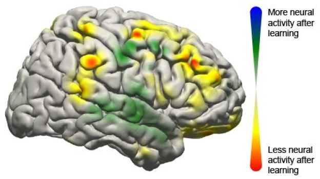
This image shows the changes that took place in the brain for all patients participating in the study using a brain-computer interface. Changes in activity were distributed widely throughout the brain.
This image shows the changes that took place in the brain for all patients participating in the study using a brain-computer interface. Changes in activity were distributed widely throughout the brain. (Credit: Jeremiah Wander, UW)
Now, University of Washington researchers have demonstrated that when humans use this technology -- called a brain-computer interface -- the brain behaves much like it does when completing simple motor skills such as kicking a ball, typing or waving a hand. Learning to control a robotic arm or a prosthetic limb could become second nature for people who are paralyzed.

"What we're seeing is that practice makes perfect with these tasks," said Rajesh Rao, a UW professor of computer science and engineering and a senior researcher involved in the study. "There's a lot of engagement of the brain's cognitive resources at the very beginning, but as you get better at the task, those resources aren't needed anymore and the brain is freed up."

Rao and UW collaborators Jeffrey Ojemann, a professor of neurological surgery, and Jeremiah Wander, a doctoral student in bioengineering, published their results online June 10 in the Proceedings of the National Academy of Sciences.

In this study, seven people with severe epilepsy were hospitalized for a monitoring procedure that tries to identify where in the brain seizures originate. Physicians cut through the scalp, drilled into the skull and placed a thin sheet of electrodes directly on top of the brain. While they were watching for seizure signals, the researchers also conducted this study.

The patients were asked to move a mouse cursor on a computer screen by using only their thoughts to control the cursor's movement. Electrodes on their brains picked up the signals directing the cursor to move, sending them to an amplifier and then a laptop to be analyzed. Within 40 milliseconds, the computer calculated the intentions transmitted through the signal and updated the movement of the cursor on the screen.

Researchers found that when patients started the task, a lot of brain activity was centered in the prefrontal cortex, an area associated with learning a new skill. But after often as little as 10 minutes, frontal brain activity lessened, and the brain signals transitioned to patterns similar to those seen during more automatic actions.

"Now we have a brain marker that shows a patient has actually learned a task," Ojemann said. "Once the signal has turned off, you can assume the person has learned it."

While researchers have demonstrated success in using brain-computer interfaces in monkeys and humans, this is the first study that clearly maps the neurological signals throughout the brain. The researchers were surprised at how many parts of the brain were involved.

"We now have a larger-scale view of what's happening in the brain of a subject as he or she is learning a task," Rao said. "The surprising result is that even though only a very localized population of cells is used in the brain-computer interface, the brain recruits many other areas that aren't directly involved to get the job done."

Several types of brain-computer interfaces are being developed and tested. The least invasive is a device placed on a person's head that can detect weak electrical signatures of brain activity. Basic commercial gaming products are on the market, but this technology isn't very reliable yet because signals from eye blinking and other muscle movements interfere too much.

A more invasive alternative is to surgically place electrodes inside the brain tissue itself to record the activity of individual neurons. Researchers at Brown University and the University of Pittsburgh have demonstrated this in humans as patients, unable to move their arms or legs, have learned to control robotic arms using the signal directly from their brain.

The UW team tested electrodes on the surface of the brain, underneath the skull. This allows researchers to record brain signals at higher frequencies and with less interference than measurements from the scalp. A future wireless device could be built to remain inside a person's head for a longer time to be able to control computer cursors or robotic limbs at home.

"This is one push as to how we can improve the devices and make them more useful to people," Wander said. "If we have an understanding of how someone learns to use these devices, we can build them to respond accordingly."

The research team, along with the National Science Foundation's Engineering Research Center for Sensorimotor Neural Engineering headquartered at the UW, will continue developing these technologies.

This research was funded by the National Institutes of Health, the NSF, the Army Research Office and the Keck Foundation. 


Share this story on Facebook, Twitter, and Google

Friday, May 24, 2013

IQ Predicted by Ability to Filter Visual Motion


A brief visual task can predict IQ, according to a new study. This surprisingly simple exercise measures the brain's unconscious ability to filter out visual movement. The study shows that individuals whose brains are better at automatically suppressing background motion perform better on standard measures of intelligence. The test is the first purely sensory assessment to be strongly correlated with IQ and may provide a non-verbal and culturally unbiased tool for scientists seeking to understand neural processes associated with general intelligence.
Intelligence is closely linked to a person's ability to filter out background movement, according to a new cognitive science study from the University of Rochester.
Intelligence is closely linked to a person's ability to filter out background movement, according to a new cognitive science study from the University of Rochester. (Credit: J. Adam Fenster, University of Rochester)

"Because intelligence is such a broad construct, you can't really track it back to one part of the brain," says Duje Tadin, a senior author on the study and an assistant professor of brain and cognitive sciences at the University of Rochester. "But since this task is so simple and so closely linked to IQ, it may give us clues about what makes a brain more efficient, and, consequently, more intelligent."

The unexpected link between IQ and motion filtering was reported online in the Cell Press journal Current Biology on May 23 by a research team lead by Tadin and Michael Melnick, a doctoral candidate in brain and cognitive sciences at the University of Rochester.

In the study, individuals watched brief video clips of black and white bars moving across a computer screen. Their sole task was to identify which direction the bars drifted: to the right or to the left. The bars were presented in three sizes, with the smallest version restricted to the central circle where human motion perception is known to be optimal, an area roughly the width of the thumb when the hand is extended. Participants also took a standardized intelligence test.

As expected, people with higher IQ scores were faster at catching the movement of the bars when observing the smallest image. The results support prior research showing that individuals with higher IQs make simple perceptual judgments swifter and have faster reflexes. "Being 'quick witted' and 'quick on the draw' generally go hand in hand," says Melnick.

But the tables turned when presented with the larger images. The higher a person's IQ, the slower they were at detecting movement. "From previous research, we expected that all participants would be worse at detecting the movement of large images, but high IQ individuals were much, much worse," says Melnick. That counter-intuitive inability to perceive large moving images is a perceptual marker for the brain's ability to suppress background motion, the authors explain. In most scenarios, background movement is less important than small moving objects in the foreground. Think about driving in a car, walking down a hall, or even just moving your eyes across the room. The background is constantly in motion.

The key discovery in this study is how closely this natural filtering ability is linked to IQ. The first experiment found a 64 percent correlation between motion suppression and IQ scores, a much stronger relationship than other sensory measures to date. For example, research on the relationship between intelligence and color discrimination, sensitivity to pitch, and reaction times have found only a 20 to 40 percent correlation. "In our first experiment, the effect for motion was so strong," recalls Tadin, "that I really thought this was a fluke."

So the group tried to disprove the findings from the initial 12-participant study conducted while Tadin was at Vanderbilt University working with co-author Sohee Park, a professor of psychology. They reran the experiment at the University of Rochester on a new cohort of 53 subjects, administering the full IQ test instead of an abbreviated version and the results were even stronger; correlation rose to 71 percent. The authors also tested for other possible explanations for their findings.

For example, did the surprising link to IQ simply reflect a person's willful decision to focus on small moving images? To rule out the effect of attention, the second round of experiments randomly ordered the different image sizes and tested other types of large images that have been shown not to elicit suppression. High IQ individuals continued to be quicker on all tasks, except the ones that isolated motion suppression. The authors concluded that high IQ is associated with automatic filtering of background motion.

"We know from prior research which parts of the brain are involved in visual suppression of background motion. This new link to intelligence provides a good target for looking at what is different about the neural processing, what's different about the neurochemistry, what's different about the neurotransmitters of people with different IQs," says Tadin.

The relationship between IQ and motion suppression points to the fundamental cognitive processes that underlie intelligence, the authors write. The brain is bombarded by an overwhelming amount of sensory information, and its efficiency is built not only on how quickly our neural networks process these signals, but also on how good they are at suppressing less meaningful information. "Rapid processing is of little utility unless it is restricted to the most relevant information," the authors conclude.

The researchers point out that this vision test could remove some of the limitations associated with standard IQ tests, which have been criticized for cultural bias. "Because the test is simple and non-verbal, it will also help researchers better understand neural processing in individuals with intellectual and developmental disabilities," says co-author Loisa Bennetto, an associate professor of psychology at the University of Rochester.

Bryan Harrison, a doctoral candidate in clinical and social psychology at the University of Rochester is also an author on the paper. The research was supported by grants from the National Institutes of Health.

Wednesday, April 17, 2013

Bad Decisions Arise from Faulty Information, Not Faulty Brain Circuits


Making decisions involves a gradual accumulation of facts that support one choice or another. A person choosing a college might weigh factors such as course selection, institutional reputation and the quality of future job prospects.
Researchers have found that it might be the information rather than the brain's decision-making process that is to blame. The researchers report that erroneous decisions tend to arise from errors, or "noise," in the information coming into the brain rather than errors in how the brain accumulates information.

But if the wrong choice is made, Princeton University researchers have found that it might be the information rather than the brain's decision-making process that is to blame. The researchers report in the journal Science that erroneous decisions tend to arise from errors, or "noise," in the information coming into the brain rather than errors in how the brain accumulates information.

These findings address a fundamental question among neuroscientists about whether bad decisions result from noise in the external information -- or sensory input -- or because the brain made mistakes when tallying that information. In the example of choosing a college, the question might be whether a person made a poor choice because of misleading or confusing course descriptions, or because the brain failed to remember which college had the best ratings.

Previous measurements of brain neurons have indicated that brain functions are inherently noisy. The Princeton research, however, separated sensory inputs from the internal mental process to show that the former can be noisy while the latter is remarkably reliable, said senior investigator Carlos Brody, a Princeton associate professor of molecular biology and the Princeton Neuroscience Institute (PNI), and a Howard Hughes Medical Institute Investigator.

"To our great surprise, the internal mental process was perfectly noiseless. All of the imperfections came from noise in the sensory processes," Brody said. Brody worked with first author Bingni Brunton, now a postdoctoral research associate in the departments of biology and applied mathematics at the University of Washington; and Matthew Botvinick, a Princeton associate professor of psychology and PNI.

The research subjects -- four college-age volunteers and 19 laboratory rats -- listened to streams of randomly timed clicks coming into both the left ear and the right ear. After listening to a stream, the subjects had to choose the side from which more clicks originated. The rats had been trained to turn their noses in the direction from which more clicks originated.

The test subjects mostly chose the correct side but occasionally made errors. By comparing various patterns of clicks with the volunteers' responses, researchers found that all of the errors arose when two clicks overlapped, and not from any observable noise in the brain system that tallied the clicks. This was true in experiment after experiment utilizing different click patterns, in humans and rats.

The researchers used the timing of the clicks and the decision-making behavior of the test subjects to create computer models that can be used to indicate what happens in the brain during decision-making. The models provide a clear window into the brain during the "mulling over" period of decision-making, the time when a person is accumulating information but has yet to choose, Brody said.

"Before we conducted this study, we did not have a way of looking at this process without inserting electrodes into the brain," Brody said. "Now thanks to our model, we have an estimation of what is going on at each moment in time during the formation of the decision."

The study suggests that information represented and processed in the brain's neurons must be robust to noise, Brody said. "In other words, the 'neural code' may have a mechanism for inherent error correction," he said.

"The new work from the Brody lab is important for a few reasons," said Anne Churchland, an assistant professor of biological sciences at Cold Spring Harbor Laboratory who studies decision-making and was not involved in the study. "First, the work was very innovative because the researchers were able to study carefully controlled decision-making behavior in rodents. This is surprising in that one might have guessed rodents were incapable of producing stable, reliable decisions that are based on complex sensory stimuli.

"This work exposed some unexpected features of why animals, including humans, sometimes make incorrect decisions," Churchland said. "Specifically, the researchers found that errors are mostly driven by the inability to accurately encode sensory information. Alternative possibilities, which the authors ruled out, included noise associated with holding the stimulus in mind, or memory noise, and noise associated with a bias toward one alternative or the other."

The work was funded by the Howard Hughes Medical Institute, Princeton University and National Institutes of Health training grants.

Monday, April 15, 2013

What Happens in the Brain to Make Music Rewarding?


A new study reveals what happens in our brain when we decide to purchase a piece of music when we hear it for the first time. The study, conducted at the Montreal Neurological Institute and Hospital -- The Neuro, McGill University and published in the journal Science on April 12, pinpoints the specific brain activity that makes new music rewarding and predicts the decision to purchase music.
A new study reveals what happens in our brain when we decide to purchase a piece of music when we hear it for the first time.
A new study reveals what happens in our brain when we decide to purchase a piece of music when we hear it for the first time. (Credit: © Warren Goldswain / Fotolia)

Participants in the study listened to 60 previously unheard music excerpts while undergoing functional resonance imaging (fMRI) scanning, providing bids of how much they were willing to spend for each item in an auction paradigm. "When people listen to a piece of music they have never heard before, activity in one brain region can reliably and consistently predict whether they will like or buy it, this is the nucleus accumbens which is involved in forming expectations that may be rewarding," says lead investigator Dr. Valorie Salimpoor, who conducted the research in Dr. Robert Zatorre's lab at The Neuro and is now at Baycrest Health Sciences' Rotman Research Institute. "What makes music so emotionally powerful is the creation of expectations. Activity in the nucleus accumbens is an indicator that expectations were met or surpassed, and in our study we found that the more activity we see in this brain area while people are listening to music, the more money they are willing to spend."

The second important finding is that the nucleus accumbens doesn't work alone, but interacts with the auditory cortex, an area of the brain that stores information about the sounds and music we have been exposed to. The more a given piece was rewarding, the greater the cross-talk between these regions. Similar interactions were also seen between the nucleus accumbens and other brain areas, involved in high-level sequencing, complex pattern recognition and areas involved in assigning emotional and reward value to stimuli.

In other words, the brain assigns value to music through the interaction of ancient dopaminergic reward circuitry, involved in reinforcing behaviours that are absolutely necessary for our survival such as eating and sex, with some of the most evolved regions of the brain, involved in advanced cognitive processes that are unique to humans.

"This is interesting because music consists of a series of sounds that when considered alone have no inherent value, but when arranged together through patterns over time can act as a reward, says Dr. Robert Zatorre, researcher at The Neuro and co-director of the International Laboratory for Brain, Music and Sound Research. "The integrated activity of brain circuits involved in pattern recognition, prediction, and emotion allow us to experience music as an aesthetic or intellectual reward."

"The brain activity in each participant was the same when they were listening to music that they ended up purchasing, although the pieces they chose to buy were all different," adds Dr. Salimpoor. "These results help us to see why people like different music -- each person has their own uniquely shaped auditory cortex, which is formed based on all the sounds and music heard throughout our lives. Also, the sound templates we store are likely to have previous emotional associations."

An innovative aspect of this study is how closely it mimics real-life music-listening experiences. Researchers used a similar interface and prices as iTunes. To replicate a real life scenario as much as possible and to assess reward value objectively, individuals could purchase music with their own money, as an indication that they wanted to hear it again. Since musical preferences are influenced by past associations, only novel music excerpts were selected (to minimize explicit predictions) using music recommendation software (such as Pandora, Last.fm) to reflect individual preferences.

The interactions between nucleus accumbens and the auditory cortex suggest that we create expectations of how musical sounds should unfold based on what is learned and stored in our auditory cortex, and our emotions result from the violation or fulfillment of these expectations. We are constantly making reward-related predictions to survive, and this study provides neurobiological evidence that we also make predictions when listening to an abstract stimulus, music, even if we have never heard the music before. Pattern recognition and prediction of an otherwise simple set of stimuli, when arranged together become so powerful as to make us happy or bring us to tears, as well as communicate and experience some of the most intense, complex emotions and thoughts.

Listen to the music excerpts used in the study: http://www.zlab.mcgill.ca/science2013/

Saturday, July 7, 2012

Diabetes Drug Makes Brain Cells Grow


The widely used diabetes drug metformin comes with a rather unexpected and alluring side effect: it encourages the growth of new neurons in the brain. The study reported in the July 6th issue of Cell Stem Cell, a Cell Press publication, also finds that those neural effects of the drug also make mice smarter.

New research finds that the widely used diabetes drug metformin comes with a rather unexpected and alluring side effect: it encourages the growth of new neurons in the brain.
New research finds that the widely used diabetes drug 
metformin comes with a rather unexpected and alluring 
side effect: it encourages the growth of new neurons in 
the brain. (Credit: iStockphoto/Guido Vrola)
The discovery is an important step toward therapies that aim to repair the brain not by introducing new stem cells but rather by spurring those that are already present into action, says the study's lead author Freda Miller of the University of Toronto-affiliated Hospital for Sick Children. The fact that it's a drug that is so widely used and so safe makes the news all that much better.

Earlier work by Miller's team highlighted a pathway known as aPKC-CBP for its essential role in telling neural stem cells where and when to differentiate into mature neurons. As it happened, others had found before them that the same pathway is important for the metabolic effects of the drug metformin, but in liver cells.

"We put two and two together," Miller says. If metformin activates the CBP pathway in the liver, they thought, maybe it could also do that in neural stem cells of the brain to encourage brain repair.

The new evidence lends support to that promising idea in both mouse brains and human cells. Mice taking metformin not only showed an increase in the birth of new neurons, but they were also better able to learn the location of a hidden platform in a standard maze test of spatial learning.

While it remains to be seen whether the very popular diabetes drug might already be serving as a brain booster for those who are now taking it, there are already some early hints that it may have cognitive benefits for people with Alzheimer's disease. It had been thought those improvements were the result of better diabetes control, Miller says, but it now appears that metformin may improve Alzheimer's symptoms by enhancing brain repair.

Miller says they now hope to test whether metformin might help repair the brains of those who have suffered brain injury due to trauma or radiation therapies for cancer.

Sunday, March 18, 2012

Brain Imaging Study Finds Evidence of Basis for Caregiving Impulse


Distinct patterns of activity -- which may indicate a predisposition to care for infants-- appear in the brains of adults who view an image of an infant face -- even when the child is not theirs, according to a study by researchers at the National Institutes of Health and in Germany, Italy, and Japan.
Researchers have found that distinct patterns of activity --
which may indicate a predisposition to care for infants --
appear in the brains of adults who view an image of an
infant face -- even when the child is not theirs.
(Credit: © Jamey Ekins / Fotolia)

Seeing images of infant faces appeared to activate in the adult's brains circuits that reflect preparation for movement and speech as well as feelings of reward.

The findings raise the possibility that studying this activity will yield insights into care giving behavior, but also in cases of child neglect or abuse.

"These adults have no children of their own. Yet images of a baby's face triggered what we think might be a deeply embedded response to reach out and care for that child," said senior author Marc H. Bornstein, Ph.D., head of the Child and Family Research Section of the Eunice Kennedy Shriver National Institute of Child Health and Human Development, the NIH institute that collaborated on the study.

While the researchers recorded participants' brain activity, the participants did not speak or move. Yet their brain activity was typical of patterns preceding such actions as picking up or talking to an infant, the researchers explained. The activity pattern could represent a biological impulse that governs adults' interactions with small children.

From their study results, the researchers concluded that this pattern is specific to seeing human infants. The pattern did not appear when the participants looked at photos of adults or of animals -- even baby animals.

Along with Dr. Bornstein, the research was carried out by first author Andrea Caria, Ph.D., of the University of Tuebingen, in Germany; Paola Venuti of the Department of Cognitive Science of University of Trento in Italy; Gianluca Esposito of the RIKEN Brain Science Institute in Saitama, Japan; researchers from the Max Planck Institute for Biological Cybernetics and Eberhard Karls University, in Tuebingen, Germany.

Their findings appear in the journal NeuroImage.

To collect the data, the researchers showed seven men and nine women a series of images while recording their brain activity with a functional magnetic resonance imaging scanner. In the scanner, participants viewed images of puppy and kitten faces, full-grown dogs and cats, human infants and adults.

When the researchers compared the areas and strength of brain activity in response to each kind of image, they found that infant images evoked more activity than any of the other images in brain areas associated with three main functions:
  • Premotor and preverbal activity -- The researchers documented increased activity in the premotor cortex and the supplemental motor area, which are regions of the brain directly under the crown of the head. These regions orchestrate brain impulses preceding speech and movement but before movement takes place.
  • Facial recognition -- Activity in the fusiform gyrus -- on each side of the brain, about where the ears are -- is associated with processing of information about faces. Activity the researchers detected in the fusiform gyrus may indicate heightened attention to the movement and expressions on an infant's face, the researchers said.
  • Emotion and reward -- Activity deep in the brain areas known as the insula and the cingulate cortex indicated emotional arousal, empathy, attachment and feelings linked to motivation and reward, the researchers said. Other studies have documented a similar pattern of activity in the brains of parents responding to their own infants.
Participants also rated how they felt when viewing adult and infant faces. They reported feeling more willing to approach, smile at, and communicate with an infant than an adult. They also recorded feeling happier when viewing images of infants.

Taken together, the researchers contend, the findings suggest a readiness to interact with infants that previously has been only inferred, and only from parents. Such brain activity in nonparents could indicate that the biological makeup of humans includes a mechanism to ensure that infants survive and receive the care they need to grow and develop.

However, signs of readiness to care for a child that appear in the brains of some or even most adults do not necessarily mean the same patterns will appear in the brains of all adults, Dr. Bornstein said. "It's equally important to investigate what's happening in the brains of those who have neglected or abused children," he said. "Additional studies could help us confirm and understand what appears to be a parenting instinct in adults, both when the instinct functions and when it fails to function."

Wednesday, August 17, 2011

Seeing eye to eye is key to copying, say scientists


Imitation may be the sincerest form of flattery but how do our brains decide when and who we should copy? Researchers from The University of Nottingham have found that the key may lie in an unspoken invitation communicated through eye contact.


In a study published this week in the Journal of Neuroscience, a team of scientists from the University's School of Psychology show that eye contact seems to act as an invitation for mimicry, triggering mechanisms in the frontal region of the brain that control imitation.

The results could be the first clues to understanding why some people, such as children with autism, struggle to grasp when they are expected to copy the actions of others in social situations.

Dr Antonia Hamilton, who led the research, said: "Many studies have looked at copying and imitation in terms of 'mirror neurons', which are believed to be specialised parts of the human brain that implement imitation. However, we also know that imitation is carefully controlled — people don't imitate everything they see, and only copy what's important.

"Our previous research has shown that when somebody makes eye contact with you, you are more likely to copy them. So eye contact seems to act as a message that says "Copy me now". This recent study aimed to see what happens to that signal in the brain."


The team of psychologists, which also included doctoral student Yin Wang and Dr Richard Ramsey, used functional magnetic resonance imaging (fMRI) to scan the brains of volunteers while they watched videos of an actress who sometimes would make eye contact with them while opening or closing her hand. The participant was told they should open their own hand whenever they saw the actress move her hand so in some trials the participant was copying the actress and in other trials they were not.

Because previous behavioural measurement such as response time revealed that the participant unconsciously copied the actress faster when the actress provided eye contact, the scientists analysed the brain imaging data to find which brain areas controlled the decision to copy. The analysis used a new mathematical method called dynamic causal modelling to compute the information processing in the brain, which has never been applied to imitation before.

The data showed that mirror neuron brain regions do play a role in the copying task. More importantly though, it revealed that these regions are controlled by the medial prefrontal cortex, an area of the brain associated with planning complex cognitive behaviours, expressing personality, decision-making and responding to social situations.

Dr Hamilton added: "Previous studies have shown that this medial prefrontal brain region is active in many social situations but responds less in people with autism, which explains why children on the autistic spectrum might not copy at the right time.

"Understanding the control of imitation has implications for many other areas of psychology too. For example, are teenagers whose prefrontal cortex is less developed more easily led to copy risky, dangerous or illegal behaviour such as imitating rioters? Could increasing the amount of eye contact between children and teachers lead to better learning by imitation? Would better control of imitation help children with autism to more effectively learn and interact? We plan further research to address these questions."

Provided by University of Nottingham


Friday, August 5, 2011

How Vampire Bats Find Veins


Heat-sensing protein channels in vampire bats allow the flying mammals to find the best place to sink their teeth into their prey.
The common vampire bat (Desmodus rotundus)

Researchers have discovered an infrared-sensing protein channel that allows vampire bats to identify the hottest part of the animal—veins close to the skin’s surface that carry 38 degree-Celsius (100° F) blood, and presumably the best spot for feeding.

The channel is a variant of TRPV1, a heat-sensing protein channel that is triggered by high temperatures that could potentially cause injury, according to the study published today (August 3) in Nature, and is distinct from the heat sensor used by snakes—the only other non-insect animals that are known to detect heat by sensing infrared radiation.

“Infrared [detection] allows these guys, in pitch black, to hunt down warm-blooded prey,” said zoologist Bill Schutt, assistant professor at Long Island University, who was not involved in the research. Here, the researchers identified a modification in a common heat-sensing protein channel that lowered its temperature threshold so that it is more attuned to an animal’s body heat, he added.

The common vampire bat was appropriately named after the myth of Dracula—it feeds at night and lives solely on a diet of blood, every day or two consuming up to half its weight in the vital substance from large mammals, especially sleeping livestock. The bats first use echolocation to detect their prey, but once they are within 20 centimeters of their target, they use infrared sensors in specialized pits around their noses to zero in on the best place to feed.

In a previous study, physiologist David Julius at the University of California, San Francisco, and colleagues found that infrared detection by snakes—which, like bats, use nerves located in facial pits to detect their prey—is mediated by a cell-surface protein channel called transient receptor potential cation channel A1 (TRPA1). The channel is actually insensitive to heat in most organisms, but had evolved the capability in snakes, leading the group to suspect that a similar transformation may have given vampire bats their ability to sense infrared.


To see if this was the case, Julius and his collaborators at the University of California, San Francisco, the Venezuelan Institute of Scientific Investigation (IVIC), and the Carnegie Institution in Baltimore, Maryland, compared gene expression in vampire bats’ heat-sensing nerves, called trigeminal ganglia, with expression in a nerve cluster near the spine, called dorsal root ganglia (DRG). They also compared these expression patterns to those of the ganglia in four bat species that do not have infrared sensory abilities.

To their surprise, they did not observe any differences in transcription of the TRPA1-coding gene, nor of any other genes. Instead, they discovered that the protein TRPV1—a heat-sensing protein channel normally triggered by temperatures over 43° C (110° F)—existed in two different isoforms—an approximately 850-amino-acid version and one that was 62 amino acids shorter. The short form, which resulted from alternative splicing of the transcribed mRNA, made up as much as half of the TRPV1 found in the trigeminal ganglia of vampire bats, whereas it comprised only a small percentage of the TRPV1 in the DRG. It was similarly low in both types of nerve clusters in the other bat species, suggesting that the short form may play a role in infrared detection.

To test this hypothesis, the researchers expressed one of the two TRPV1 isoforms in human kidney cells and in frog oocytes grown in vitro, and measured their temperature sensitivity using calcium imaging and electrophysiological assays, respectively. As expected, cells producing the long isoform were activated at 40 degrees Celsius (104° F). Cells producing the short isoform, on the other hand, were activated at just 30 degrees (86° F)—a drop that allows the protein to respond to the warmth of the vampire bats’ prey.

“This is a big jump in understanding how these animals locate their prey,” said Brock Fenton, a biology professor at the University of Western Ontario and author of an accompanying Nature News and Views article. While the longer isoform maintains its normal function of detecting potentially harmful high temperatures, the shorter isoform in the trigeminal nerves of the common vampire bat allows the animals to detect lower temperatures, such as the body heat of their mammalian prey.

“Basically, evolution tweaked a system in vampires bats that was already being used to sense temperatures,” said Schutt, author of the 2008 book Dark Banquet: Blood and the Curious Lives of Blood-Feeding Creatures, turning it into a useful hunting tool.

This is in contrast to the pit viper, whose infrared-sensing ability evolved from a different type of channel not involved in heat detection, but in the detection of noxious smells, added Fenton. The different evolutionary strategies employed by these two lineages “is an example of how plastic our sensory systems can be,” he said.


Tuesday, July 12, 2011

Researchers connect neurons to computers to decipher the enigmatic code of neuronal circuits


Machine logic is based on human logic. But although a computer processor can be dissembled and dissected in logical steps, the same is not true for the way our brains process information, says Mark Shein of Tel Aviv University's School of Electrical Engineering.

Doctoral student Shein and his supervisors, Prof. Yael Hanein of the School of Electrical Engineering and Prof. Eshel Ben-Jacob of the School of Physics and Astronomy, want to understand the brain's logic. They have developed a new kind of a lab-on-a-chip platform that may help neuroscientists understand one of the deepest mysteries of our brain –– how neuronal networks communicate and work together. The chip was recently described in an issue of the journal PLoS ONE.

Within it, Shein has applied advanced mathematical and engineering techniques to connect neurons with electronics and understand how neuronal networks communicate. Hoping to answer ultimate questions about how our neuronal circuits work, the researchers believe their tool can be also used to test new drugs. It might also advance artificial intelligence and aid scientists in rewiring artificial limbs to our brain.

Shedding light on a black box

There are relatively simple neural "firing" patterns that can be measured with sensory organs like the ears or eyes, but researchers know little about deep thought processes. Could the brain's electrical signals reveal the basis of thought itself?

"When we look at the neuronal networks operating in the ears or eyes, we have some idea about the coding schemes they utilize," explains Shein. A researcher can apply a stimulus such as a bright light, for example, and then monitor responses in the eye's neurons. But for more complex processes, like "thinking" or operating different sensory inputs and outputs together, "we are basically looking into a black box," he says.

The brain is composed of a daunting number of circuits interconnected with other countless circuits, so understanding of how they function has been close to impossible. But using engineered brain tissue in a Petri dish, Shein's device allows researchers to see what's happening to well-defined neural circuits under different conditions. The result is an active circuitry of neurons on a man-made chip. With it they can look for patterns in bigger networks of neurons, to see if there are any basic elements for information coding.


Investigating the activity of single neurons is not enough to understand how a network functions. With nanotechnological systems and tools, now researchers can explore activity patterns of many neurons simultaneously. In particular, they can investigate how several groups of neurons communicate with each other, says Shein.

The hierarchy of the brain

With these network engineering techniques, the scientists cultured different sized networks of neuronal clusters. Once they looked at these groups, they found rich and surprising behaviors which could not be predicted from what scientists know about single neurons.

The researchers were also able to measure patterns from nerve activity, at nodes where a number of nerves converged into networks. What they detected appears to show that neural networks have a hierarchical structure — large networks are composed of smaller sub-networks. This observation, and a unique setup using electrodes and living nerves, allowed them to create hierarchical networks in a dish.

The brain's circuits work like codes. They can see the patterns in the networks and simplify them, or control connectivity between cells to see how the neuronal network responds to various chemicals and conditions, the scientists report. One theory, proposed by Prof. Ben-Jacob, is that the human brain stores memories like a holograph of an image: small neural networks contain information about the whole brain, but only at a very low resolution.

So far the researchers are able to reveal that clusters of as few as 40 cells can serve as a minimal but sufficient functional network. This cluster is capable of sustaining neural network activity and communicating with other clusters. What this means exactly will be the next question.

Provided by Tel Aviv University


Related articles

Friday, December 10, 2010

Brains Wired So We Can Better Hear Ourselves


Like the mute button on the TV remote control, our brains filter out unwanted noise so we can focus on what we're listening to. But when it comes to following our own speech, a new brain study from the University of California, Berkeley, shows that instead of one homogenous mute button, we have a network of volume settings that can selectively silence and amplify the sounds we make and hear.
Activity in the auditory cortex when we speak and listen 
is amplified in some regions of the brain and muted in 
others. In this image, the black line represents muting 
activity when we speak. (Credit: Courtesy 
of Adeen Flinker)

Neuroscientists from UC Berkeley, UCSF and Johns Hopkins University tracked the electrical signals emitted from the brains of hospitalized epilepsy patients. They discovered that neurons in one part of the patients' hearing mechanism were dimmed when they talked, while neurons in other parts lit up.

Their findings, published Dec. 8, 2010 in the Journal of Neuroscience, offer new clues about how we hear ourselves above the noise of our surroundings and monitor what we say. Previous studies have shown a selective auditory system in monkeys that can amplify their self-produced mating, food and danger alert calls, but until this latest study, it was not clear how the human auditory system is wired.

"We used to think that the human auditory system is mostly suppressed during speech, but we found closely knit patches of cortex with very different sensitivities to our own speech that paint a more complicated picture," said Adeen Flinker, a doctoral student in neuroscience at UC Berkeley and lead author of the study.

"We found evidence of millions of neurons firing together every time you hear a sound right next to millions of neurons ignoring external sounds but firing together every time you speak," Flinker added. "Such a mosaic of responses could play an important role in how we are able to distinguish our own speech from that of others."

While the study doesn't specifically address why humans need to track their own speech so closely, Flinker theorizes that, among other things, tracking our own speech is important for language development, monitoring what we say and adjusting to various noise environments.

"Whether it's learning a new language or talking to friends in a noisy bar, we need to hear what we say and change our speech dynamically according to our needs and environment," Flinker said.

He noted that people with schizophrenia have trouble distinguishing their own internal voices from the voices of others, suggesting that they may lack this selective auditory mechanism. The findings may be helpful in better understanding some aspects of auditory hallucinations, he said.

Moreover, with the finding of sub-regions of brain cells each tasked with a different volume control job -- and located just a few millimeters apart -- the results pave the way for a more detailed mapping of the auditory cortex to guide brain surgery.

In addition to Flinker, the study's authors are Robert Knight, director of the Helen Wills Neuroscience Institute at UC Berkeley; neurosurgeons Edward Chang, Nicholas Barbaro and neurologist Heidi Kirsch of the University of California, San Francisco; and Nathan Crone, a neurologist at Johns Hopkins University in Maryland.

The auditory cortex is a region of the brain's temporal lobe that deals with sound. In hearing, the human ear converts vibrations into electrical signals that are sent to relay stations in the brain's auditory cortex where they are refined and processed. Language is mostly processed in the left hemisphere of the brain.

In the study, researchers examined the electrical activity in the healthy brain tissue of patients who were being treated for seizures. The patients had volunteered to help out in the experiment during lulls in their treatment, as electrodes had already been implanted over their auditory cortices to track the focal points of their seizures.

Researchers instructed the patients to perform such tasks as repeating words and vowels they heard, and recorded the activity. In comparing the activity of electrical signals discharged during speaking and hearing, they found that some regions of the auditory cortex showed less activity during speech, while others showed the same or higher levels.

"This shows that our brain has a complex sensitivity to our own speech that helps us distinguish between our vocalizations and those of others, and makes sure that what we say is actually what we meant to say," Flinker said.

Disclaimer: This article is not intended to provide medical advice, diagnosis or treatment. Views expressed here do not necessarily reflect those of Science Updates or its staff.

Saturday, November 27, 2010

Do Brain's 'Traffic Lights' Direct Our Actions?


In every waking minute, we have to make decisions -- sometimes within a split second. Neuroscientists at the Bernstein Center Freiburg have now discovered a possible explanation how the brain chooses between alternative options. The key lies in extremely fast changes in the communication between single nerve cells.
The timing of exciting (red curve) and inhibiting 
(blue curve) signals could be a way to control the 
"traffic flow" of activity in the brain. (Illustration: 
Bernstein Center Freiburg) (Credit: Illustration 
courtesy of Bernstein Center Freiburg)

The traffic light changes from green to orange -- should I push down the accelerator a little bit further or rather hit the brakes? Our daily lives present a long series of decisions we have to make, and sometimes we only have a split second at our disposal. Often the problem of decision-making entails the selection of one set of brain processes over multiple others seeking access to same resources. Several mechanisms have been suggested how the brain might solve this problem. However, up to now, it is a mystery what exactly happens when during a rapid choice between two options.

In the current issue of the Journal of Neuroscience, Jens Kremkow, Arvind Kumar, and Ad Aertsen from the Bernstein Center Freiburg propose a mechanism how the brain can choose between possible actions -- already at the level of single nerve cells.

As the structure and activity of the brain are just too complex to answer this question through a simple biological experiment, the scientists constructed a network of neurons in the computer. An important aspect of the model in this context is the property of nerve cells to influence the activity of other nerve cells, either in an excitatory or inhibitory manner. In the constructed network, two groups of neurons acted as the senders of two different signals. Further downstream in the network, another group of neurons, the "gate" neurons, were to control which of the signals would be transmitted onward.

As the cells within the network were connected both with exciting and inhibiting neurons, the signals reached the gate as excitatory and, after a short delay, inhibitory activity. In their simulations, the scientists found that the key for the gate neurons' "decision" in favour of one signal over the other was the time delay of the inhibitory signal relative to the excitatory signal. If the delay was set to be very small, the activity of the cells in the gate was quenched too quickly for the signal to be propagated.

Conversely, a larger delay caused the gate to open for the signal. Results from neurophysiological experiments have already shown that a change in delay properties is possible in real neurons. These findings therefore support the hypothesis of Kremkow and colleagues that such temporal gating can form the basis for selecting one of several alternative options in our brain.

Admin's Note: This article is not intended to provide medical advice, diagnosis or treatment.

Thursday, October 21, 2010

Human Brain Can 'See' Shapes With Sound : See No Shape, Touch No Shape, Hear a Shape? New Way of 'Seeing' the World


Scientists at The Montreal Neurological Institute and Hospital -- The Neuro, McGill University have discovered that our brains have the ability to determine the shape of an object simply by processing specially-coded sounds, without any visual or tactile input. Not only does this new research tell us about the plasticity of the brain and how it perceives the world around us, it also provides important new possibilities for aiding those who are blind or with impaired vision.
New research shows that the human brain is able to 
determine the shape of an object simply by processing 
specially-coded sounds, without any visual or tactile input. 
(Credit: iStockphoto/Sergey Chushkin)

Shape is an inherent property of objects existing in both vision and touch but not sound. Researchers at The Neuro posed the question 'can shape be represented by sound artificially?' "The fact that a property of sound such as frequency can be used to convey shape information suggests that as long as the spatial relation is coded in a systematic way, shape can be preserved and made accessible -- even if the medium via which space is coded is not spatial in its physical nature," says Jung-Kyong Kim, PhD student in Dr. Robert Zatorre's lab at The Neuro and lead investigator in the study.

In other words, similar to our ocean-dwelling dolphin cousins who use echolocation to explore their surroundings, our brains can be trained to recognize shapes represented by sound and the hope is that those with impaired vision could be trained to use this as a tool. In the study, blindfolded sighted participants were trained to recognize tactile spatial information using sounds mapped from abstract shapes. Following training, the individuals were able to match auditory input to tactually discerned shapes and showed generalization to new auditory-tactile or sound-touch pairings.

"We live in a world where we perceive objects using information available from multiple sensory inputs," says Dr. Zatorre, neuroscientist at The Neuro and co-director of the International Laboratory for Brain Music and Sound Research. "On one hand, this organization leads to unique sense-specific percepts, such as colour in vision or pitch in hearing. On the other hand our perceptual system can integrate information present across different senses and generate a unified representation of an object. We can perceive a multisensory object as a single entity because we can detect equivalent attributes or patterns across different senses." Neuroimaging studies have identified brain areas that integrate information coming from different senses -- combining input from across the senses to create a complete and comprehensive picture.

The results from The Neuro study strengthen the hypothesis that our perception of a coherent object or event ultimately occurs at an abstract level beyond the sensory input modes in which it is presented. This research provides important new insight into how our brains process the world as well as new possibilities for those with impaired senses.

The study was published in the journal Experimental Brain Research. The research was supported by grants from the Canadian Institutes of Health Research and the Natural Sciences and Engineering Research Council of Canada.

Editor's Note: This article is not intended to provide medical advice, diagnosis or treatment.

Wednesday, August 11, 2010

Brains Wiring: More Like the Internet Than a Pyramid?


The brain has been mapped to the smallest fold for at least a century, but still no one knows how all the parts talk to each other.
Image
New research suggests that the distributed network 
of the Internet may be a better model for the human 
brain than a top-down hierarchy. 
(Credit: iStockphoto/Henrik Jonsson)

A study in Proceedings of the National Academy of Sciences answers that question for a small area of the rat brain and in so doing takes a big step toward revealing the brain's wiring.

The network of brain connections was thought too complex to describe, but molecular biology and computing methods have improved to the point that the National Institutes of Health have announced a $30 million plan to map the human "connectome."

The study shows the power of a new method for tracing brain circuits.

USC College neuroscientists Richard H. Thompson and Larry W. Swanson used the method to trace circuits running through a "hedonic hot spot" related to food enjoyment.

The circuits showed up as patterns of circular loops, suggesting that at least in this part of the rat brain, the wiring diagram looks like a distributed network.

Neuroscientists are split between a traditional view that the brain is organized as a hierarchy, with most regions feeding into the "higher" centers of conscious thought, and a more recent model of the brain as a flat network similar to the Internet.

"We started in one place and looked at the connections. It led into a very complicated series of loops and circuits. It's not an organizational chart. There's no top and bottom to it," said Swanson, a member of the National Academy of Sciences and the Milo Don and Lucille Appleman Professor of Biological Sciences at USC College.

The circuit tracing method allows the study of incoming and outgoing signals from any two brain centers. It was invented and refined by Thompson over eight years. Thompson is a research assistant professor of biological sciences at the College.

Most other tracing studies at present focus only on one signal, in one direction, at one location.

"[We] can look at up to four links in a circuit, in the same animal at the same time. That was our technical innovation," Swanson said.

The Internet model would explain the brain's ability to overcome much local damage, Swanson said.

"You can knock out almost any single part of the Internet and the rest of it works."

Likewise, Swanson said, "There are usually alternate pathways through the nervous system. It's very hard to say that any one part is absolutely essential."

Swanson first argued for the distributed model of the brain in his acclaimed book Brain Architecture: Understanding the Basic Plan (Oxford University Press, 2003).

The PNAS study appears to support his view.

"There is an alternate model. It's not proven, but let's rethink the traditional way of regarding how the brain works," he said.

"The part of the brain you think with, the cortex, is very important, but it's certainly not the only part of the nervous system that determines our behavior."

The research described in the PNAS study was supported by the National Institute of Neurological Disorders and Stroke in the National Institutes of Health.