BTemplates.com

Powered by Blogger.

Pageviews past week

Quantum mechanics

Auto News

artificial intelligence

About Me

Recommend us on Google!

Information Technology

Popular Posts

Showing posts with label Nanotechnology. Show all posts

Saturday, September 13, 2014

Incredibly light, strong materials recover original shape after being smashed


Materials scientists have developed a method for creating new structural materials by taking advantage of the unusual properties that solids can have at the nanometer scale. They have used the method to produce a ceramic (e.g., a piece of chalk or a brick) that contains about 99.9 percent air yet is incredibly strong and can recover its original shape after being smashed by more than 50 percent.

This sequence shows how the Greer Lab's three-dimensional,
ceramic nanolattices can recover after being compressed by
more than 50 percent. Clockwise, from left to right, an alumina
nanolattice before compression, during compression, fully
compressed, and recovered following compression.
Credit: Lucas Meza/Caltech

Imagine a balloon that could float without using any lighter-than-air gas. Instead, it could simply have all of its air sucked out while maintaining its filled shape. Such a vacuum balloon, which could help ease the world's current shortage of helium, can only be made if a new material existed that was strong enough to sustain the pressure generated by forcing out all that air while still being lightweight and flexible. 

Caltech materials scientist Julia Greer and her colleagues are on the path to developing such a material and many others that possess unheard-of combinations of properties. For example, they might create a material that is thermally insulating but also extremely lightweight, or one that is simultaneously strong, lightweight, and nonbreakable -- properties that are generally thought to be mutually exclusive.

Greer's team has developed a method for constructing new structural materials by taking advantage of the unusual properties that solids can have at the nanometer scale, where features are measured in billionths of meters. In a paper published in the September 12 issue of the journal Science, the Caltech researchers explain how they used the method to produce a ceramic (e.g., a piece of chalk or a brick) that contains about 99.9 percent air yet is incredibly strong, and that can recover its original shape after being smashed by more than 50 percent.

"Ceramics have always been thought to be heavy and brittle," says Greer, a professor of materials science and mechanics in the Division of Engineering and Applied Science at Caltech. "We're showing that in fact, they don't have to be either. This very clearly demonstrates that if you use the concept of the nanoscale to create structures and then use those nanostructures like LEGO to construct larger materials, you can obtain nearly any set of properties you want. You can create materials by design."

The researchers use a direct laser writing method called two-photon lithography to "write" a three-dimensional pattern in a polymer by allowing a laser beam to crosslink and harden the polymer wherever it is focused. The parts of the polymer that were exposed to the laser remain intact while the rest is dissolved away, revealing a three-dimensional scaffold. That structure can then be coated with a thin layer of just about any kind of material -- a metal, an alloy, a glass, a semiconductor, etc. Then the researchers use another method to etch out the polymer from within the structure, leaving a hollow architecture.

The applications of this technique are practically limitless, Greer says. Since pretty much any material can be deposited on the scaffolds, the method could be particularly useful for applications in optics, energy efficiency, and biomedicine. For example, it could be used to reproduce complex structures such as bone, producing a scaffold out of biocompatible materials on which cells could proliferate.

In the latest work, Greer and her students used the technique to produce what they call three-dimensional nanolattices that are formed by a repeating nanoscale pattern. After the patterning step, they coated the polymer scaffold with a ceramic called alumina (i.e., aluminum oxide), producing hollow-tube alumina structures with walls ranging in thickness from 5 to 60 nanometers and tubes from 450 to 1,380 nanometers in diameter.

Greer's team next wanted to test the mechanical properties of the various nanolattices they created. Using two different devices for poking and prodding materials on the nanoscale, they squished, stretched, and otherwise tried to deform the samples to see how they held up.

They found that the alumina structures with a wall thickness of 50 nanometers and a tube diameter of about 1 micron shattered when compressed. That was not surprising given that ceramics, especially those that are porous, are brittle. However, compressing lattices with a lower ratio of wall thickness to tube diameter -- where the wall thickness was only 10 nanometers -- produced a very different result.

"You deform it, and all of a sudden, it springs back," Greer says. "In some cases, we were able to deform these samples by as much as 85 percent, and they could still recover."

To understand why, consider that most brittle materials such as ceramics, silicon, and glass shatter because they are filled with flaws -- imperfections such as small voids and inclusions. The more perfect the material, the less likely you are to find a weak spot where it will fail. Therefore, the researchers hypothesize, when you reduce these structures down to the point where individual walls are only 10 nanometers thick, both the number of flaws and the size of any flaws are kept to a minimum, making the whole structure much less likely to fail.

"One of the benefits of using nanolattices is that you significantly improve the quality of the material because you're using such small dimensions," Greer says. "It's basically as close to an ideal material as you can get, and you get the added benefit of needing only a very small amount of material in making them."

The Greer lab is now aggressively pursuing various ways of scaling up the production of these so-called meta-materials.

Story Source: http://www.sciencedaily.com/releases/2014/09/140911135450.htm

Wednesday, April 16, 2014

Lithium-sulfur batteries last longer with nanomaterial-packed cathode


Electric vehicles could travel farther and more renewable energy could be stored with lithium-sulfur batteries that use a unique powdery nanomaterial.
Pacific Northwest National Laboratory developed
a nickel-based metal organic framework, shown here
in an illustration, to hold onto polysulfide molecules
in the cathodes of lithium-sulfur batteries and extend
the batteries' lifespans. The colored spheres in this i
mage represent the 3D material's tiny pores into with
the polysulfides become trapped.
Credit: Pacific Northwest National Laboratory
Researchers added the powder, a kind of nanomaterial called a metal organic framework, to the battery's cathode to capture problematic polysulfides that usually cause lithium-sulfur batteries to fail after a few charges. A paper describing the material and its performance was published online April 4 in the American Chemical Society journal Nano Letters.

"Lithium-sulfur batteries have the potential to power tomorrow's electric vehicles, but they need to last longer after each charge and be able to be repeatedly recharged," said materials chemist Jie Xiao of the Department of Energy's Pacific Northwest National Laboratory. "Our metal organic framework may offer a new way to make that happen."

Today's electric vehicles are typically powered by lithium-ion batteries. But the chemistry of lithium-ion batteries limits how much energy they can store. As a result, electric vehicle drivers are often anxious about how far they can go before needing to charge. One promising solution is the lithium-sulfur battery, which can hold as much as four times more energy per mass than lithium-ion batteries. This would enable electric vehicles to drive farther on a single charge, as well as help store more renewable energy. The down side of lithium-sulfur batteries, however, is they have a much shorter lifespan because they can't currently be charged as many times as lithium-ion batteries.

Energy Storage 101

The reason can be found in how batteries work. Most batteries have two electrodes: one is positively charged and called a cathode, while the second is negative and called an anode. Electricity is generated when electrons flow through a wire that connects the two. To control the electrons, positively charged atoms shuffle from one electrode to the other through another path: the electrolyte solution in which the electrodes sit.

The lithium-sulfur battery's main obstacles are unwanted side reactions that cut the battery's life short. The undesirable action starts on the battery's sulfur-containing cathode, which slowly disintegrates and forms molecules called polysulfides that dissolve into the liquid electrolyte. Some of the sulfur—an essential part of the battery's chemical reactions—never returns to the cathode. As a result, the cathode has less material to keep the reactions going and the battery quickly dies.

New materials for better batteries

Researchers worldwide are trying to improve materials for each battery component to increase the lifespan and mainstream use of lithium-sulfur batteries. For this research, Xiao and her colleagues honed in on the cathode to stop polysulfides from moving through the electrolyte.

Many materials with tiny holes have been examined to physically trap polysulfides inside the cathode. Metal organic frameworks are porous, but the added strength of PNNL's material is its ability to strongly attract the polysulfide molecules.

The framework's positively charged nickel center tightly binds the polysulfide molecules to the cathodes. The result is a coordinate covalent bond that, when combined with the framework's porous structure, causes the polysulfides to stay put.

"The MOF's highly porous structure is a plus that further holds the polysulfide tight and makes it stay within the cathode," said PNNL electrochemist Jianming Zheng.

Nanomaterial is key

Metal organic frameworks—also called MOFs—are crystal-like compounds made of metal clusters connected to organic molecules, or linkers. Together, the clusters and linkers assemble into porous 3-D structures. MOFs can contain a number of different elements. PNNL researchers chose the transition metal nickel as the central element for this particular MOF because of its strong ability to interact with sulfur.

During lab tests, a lithium-sulfur battery with PNNL's MOF cathode maintained 89 percent of its initial power capacity after 100 charge-and discharge cycles. Having shown the effectiveness of their MOF cathode, PNNL researchers now plan to further improve the cathode's mixture of materials so it can hold more energy. The team also needs to develop a larger prototype and test it for longer periods of time to evaluate the cathode's performance for real-world, large-scale applications.

PNNL is also using MOFs in energy-efficient adsorption chillers and to develop new catalysts to speed up chemical reactions.

"MOFs are probably best known for capturing gases such as carbon dioxide," Xiao said. "This study opens up lithium-sulfur batteries as a new and promising field for the nanomaterial."

This research was funded by the Department of Energy's Office of Energy Efficiency and Renewable Energy. Researchers analyzed chemical interactions on the MOF cathode with instruments at EMSL, DOE's Environmental Molecular Sciences Laboratory at PNNL.

In January, a Nature Communications paper by Xiao and some of her PNNL colleagues described another possible solution for lithium-sulfur batteries: developing a hybrid anode that uses a graphite shield to block polysulfides.

Source: http://phys.org/news/2014-04-lithium-sulfur-batteries-longer-nanomaterial-packed-cathode.html

Sunday, June 23, 2013

Beyond Silicon: Transistors, No Semiconductors


For decades, electronic devices have been getting smaller, and smaller, and smaller. It's now possible -- even routine -- to place millions of transistors on a single silicon chip.

Electrons flash across a series of gold quantum dots on boron nitride nanotubes. Michigan Tech scientists made the quantum-tunneling device, which acts like a transistor at room temperature, without using semiconducting materials.
Electrons flash across a series of gold quantum dots on boron 
nitride nanotubes. Michigan Tech scientists made the quantum
-tunneling device, which acts like a transistor at room 
temperature, without using semiconducting materials. 
(Credit: Yoke Khin Yap graphic)
But transistors based on semiconductors can only get so small. "At the rate the current technology is progressing, in 10 or 20 years, they won't be able to get any smaller," said physicist Yoke Khin Yap of Michigan Technological University. "Also, semiconductors have another disadvantage: they waste a lot of energy in the form of heat."

Scientists have experimented with different materials and designs for transistors to address these issues, always using semiconductors like silicon. Back in 2007, Yap wanted to try something different that might open the door to a new age of electronics.

"The idea was to make a transistor using a nanoscale insulator with nanoscale metals on top," he said. "In principle, you could get a piece of plastic and spread a handful of metal powders on top to make the devices, if you do it right. But we were trying to create it in nanoscale, so we chose a nanoscale insulator, boron nitride nanotubes, or BNNTs for the substrate."

Yap's team had figured out how to make virtual carpets of BNNTs,which happen to be insulators and thus highly resistant to electrical charge. Using lasers, the team then placed quantum dots (QDs) of gold as small as three nanometers across on the tops of the BNNTs, forming QDs-BNNTs. BNNTs are the perfect substrates for these quantum dots due to their small, controllable, and uniform diameters, as well as their insulating nature. BNNTs confine the size of the dots that can be deposited.

In collaboration with scientists at Oak Ridge National Laboratory (ORNL), they fired up electrodes on both ends of the QDs-BNNTs at room temperature, and something interesting happened. Electrons jumped very precisely from gold dot to gold dot, a phenomenon known as quantum tunneling.

"Imagine that the nanotubes are a river, with an electrode on each bank. Now imagine some very tiny stepping stones across the river," said Yap. "The electrons hopped between the gold stepping stones. The stones are so small, you can only get one electron on the stone at a time. Every electron is passing the same way, so the device is always stable."

Yap's team had made a transistor without a semiconductor. When sufficient voltage was applied, it switched to a conducting state. When the voltage was low or turned off, it reverted to its natural state as an insulator.

Furthermore, there was no "leakage": no electrons from the gold dots escaped into the insulating BNNTs, thus keeping the tunneling channel cool. In contrast, silicon is subject to leakage, which wastes energy in electronic devices and generates a lot of heat.

Other people have made transistors that exploit quantum tunneling, says Michigan Tech physicist John Jaszczak, who has developed the theoretical framework for Yap's experimental research. However, those tunneling devices have only worked in conditions that would discourage the typical cellphone user.

"They only operate at liquid-helium temperatures," said Jaszczak.

The secret to Yap's gold-and-nanotube device is its submicroscopic size: one micron long and about 20 nanometers wide. "The gold islands have to be on the order of nanometers across to control the electrons at room temperature," Jaszczak said. "If they are too big, too many electrons can flow." In this case, smaller is truly better: "Working with nanotubes and quantum dots gets you to the scale you want for electronic devices."

"Theoretically, these tunneling channels can be miniaturized into virtually zero dimension when the distance between electrodes is reduced to a small fraction of a micron," said Yap.

Yap has filed for a full international patent on the technology.

Friday, June 1, 2012

Building Molecular 'Cages' to Fight Disease


UCLA biochemists have designed specialized proteins that assemble themselves to form tiny molecular cages hundreds of times smaller than a single cell. The creation of these miniature structures may be the first step toward developing new methods of drug delivery or even designing artificial vaccines.

This is a molecular cage created by designing specialized protein puzzle pieces. Every color represents a separate protein, where cylindrical segments indicate rigid parts and ribbon-like segments indicate flexible parts of each protein chain. The grey sphere in the protein cage was placed there to indicate the empty space in the middle of the container and is not part of the molecular structure. (Credit: Todd Yeates, Yen-Ting Lai/UCLA Chemistry and Biochemistry)
This is a molecular cage created by designing 
specialized protein puzzle pieces. Every color 
represents a separate protein, where cylindrical 
segments indicate rigid parts and ribbon-like 
segments indicate flexible parts of each protein 
chain. The grey sphere in the protein cage was 
placed there to indicate the empty space in the 
middle of the container and is not part of the 
molecular structure. (Credit: Todd Yeates, Yen-
Ting Lai/UCLA Chemistry and Biochemistry)
"This is the first decisive demonstration of an approach that can be used to combine protein molecules together to create a whole array of nanoscale materials," said Todd Yeates, a UCLA professor of chemistry and biochemistry and a member of the UCLA-DOE Institute of Genomics and Proteomics and the California NanoSystems Institute at UCLA.

Published June 1 in the journal Science, the research could be utilized to create cages from any number of different proteins, with potential applications across the fields of medicine and molecular biology.

UCLA graduate student Yen-Ting Lai, lead author of the study, used computer models to identify two proteins that could be combined to form perfectly shaped three-dimensional puzzle pieces. Twelve of these specialized pieces fit together to create a molecular cage a mere fraction of the size of a virus.

"If you just connect two random proteins together, you expect to get an irregular network," said Yeates, senior author of the study. "In order to control the geometry, the idea was to make a rigid link holding the two proteins in place as if they were parts of a toy puzzle."

The specifically designed proteins intermesh to form a hollow lattice that could act as a vessel for drug delivery, he said.

"In principle, it would be possible to attach a recognition sequence for cancer cells on the outside of the cage, with a toxin or some other 'magic bullet' contained inside," said Yeates. "That way, the drug could be delivered directly to certain targets like tumor cells."

At this stage, the assembled protein cages are porous enough that a drug placed inside would likely leak out during the delivery process, Lai said. His next project will involve constructing a new molecular cage with an interior that will be better sealed.

Another use for the versatile protein structures might be as artificial vaccines. Some traditional vaccines use an inactive surface protein from a virus to trick the body's immune system into thinking it is under attack. This method isn't always effective, because sometimes the protein in question doesn't look enough like the virus to trigger a strong response from the body's defenders.

However, by decorating the surface of a molecular cage with segments of virus-derived proteins, the tiny structures might better mimic a virus, stimulating an immune response even stronger than a traditional vaccine and better protecting the human recipient from illness.

Before these protein structures can be used in medical applications, the molecular containers themselves must be constructed from human-like proteins, rather than the currently employed bacterial proteins that the human body might immediately clear from circulation, Yeates said.

"Our first challenge will be repeating these kinds of designs with molecules that are less likely to generate a host immune response," he said. "Generally, we want to use proteins that look like human proteins so the body does not recognize them as foreign."

The idea of building complex, self-assembled protein structures has been Yeates' ambition since he published a paper outlining preliminary work on this method in 2001. Yet the concept remained on the back burner for 10 years until Yen-Ting Lai joined Yeates' research group. With three master's degrees -- in structural biology, bioinformatics and biomedical engineering -- Lai had the right combination of skills to bring the research to fruition, Yeates said.

This project is federally funded by the National Science Foundation. Other co-authors include UCLA senior staff scientist Duilio Cascio.

A second breakthrough


A second paper co-authored by Yeates creates similarly designed molecular cages using multiple copies of the same protein as building blocks. The scientists control the shape of the cage by computing the sequence of amino acids necessary to link the proteins together at the correct angles. The research, also published May 31 in Science, resulted from a collaboration between the UCLA team and professor David Baker at the University of Washington.

This alternative method represents a more versatile approach because it requires only one type of protein to form a structure, Yeates said. However, devising different kinds of links between the identical proteins remains a major challenge. Lead author Neil King, a postdoctoral scholar at the University of Washington and a former student of Yeates, took the numerous computer-generated possibilities and tested each version experimentally until he found one which produced the right behavior.

This research is federally funded by the National Institutes of Health and the U.S. Department of Energy. Other UCLA co-authors include senior staff researcher Michael Sawaya.

Sunday, July 17, 2011

Future of energy storage with Graphite + Water


A combination of two ordinary materials – graphite and water – could produce energy storage systems that perform on par with lithium ion batteries, but recharge in a matter of seconds and have an almost indefinite lifespan.
Graphene sheets. Credit: Gengping Jiang

Dr. Dan Li, of the Monash University Department of Materials Engineering, and his research team have been working with a material called graphene, which could form the basis of the next generation of ultrafast energy storage systems.

“Once we can properly manipulate this material, your iPhone, for example, could charge in a few seconds, or possibly faster.” said Dr. Li.

Graphene is the result of breaking down graphite, a cheap, readily available material commonly used in pencils, into layers one atom thick. In this form, it has remarkable properties.

Graphene is strong, chemically stable, an excellent conductor of electricity and, importantly, has an extremely high surface area.

Dr. Li said these qualities make graphene highly suitable for energy storage applications.


“The reason graphene isn’t being used everywhere is that these very thin sheets, when stacked into a usable macrostructure, immediately bond together, reforming graphite. When graphene restacks, most of the surface area is lost and it doesn’t behave like graphene anymore.”

Now, Dr. Li and his team have discovered the key to maintaining the remarkable properties of separate graphene sheets: water. Keeping graphene moist – in gel form – provides repulsive forces between the sheets and prevents re-stacking, making it ready for real-world application.

“The technique is very simple and can easily be scaled up. When we discovered it, we thought it was unbelievable. We’re taking two basic, inexpensive materials – water and graphite – and making this new nanomaterial with amazing properties,” said Dr. Li.

When used in energy devices, graphene gel significantly outperforms current carbon-based technology, both in terms of the amount of charge stored and how fast the charges can be delivered.

Dr. Li said the benefits of developing this new nanotechnology extend beyond consumer electronics.

“High-speed, reliable and cost-effective energy storage systems are critical for the future viability of electricity from renewable resources. These systems are also the key to large-scale adoption of electrical vehicles.

“Graphene gel is also showing promise for use in water purification membranes, biomedical devices and sensors.”

Provided by Monash University

Monday, July 11, 2011

U of T researchers build an antenna for light


University of Toronto researchers have derived inspiration from the photosynthetic apparatus in plants to engineer a new generation of nanomaterials that control and direct the energy absorbed from light.

Their findings are reported in a forthcoming issue of Nature Nanotechnology, which will be released on July 10, 2011.

The U of T researchers, led by Professors Shana Kelley and Ted Sargent, report the construction of what they term "artificial molecules."

"Nanotechnologists have for many years been captivated by quantum dots – particles of semiconductor that can absorb and emit light efficiently, and at custom-chosen wavelengths," explained co-author Kelley, a Professor at the Leslie Dan Faculty of Pharmacy, the Department of Biochemistry in the Faculty of Medicine, and the Department of Chemistry in the Faculty of Arts & Science. "What the community has lacked – until now – is a strategy to build higher-order structures, or complexes, out of multiple different types of quantum dots. This discovery fills that gap."

The team combined its expertise in DNA and in semiconductors to invent a generalized strategy to bind certain classes of nanoparticles to one another.

"The credit for this remarkable result actually goes to DNA: its high degree of specificity – its willingness to bind only to a complementary sequence – enabled us to build rationally-engineered, designer structures out of nanomaterials," said Sargent, a Professor in The Edward S. Rogers Sr. Department of Electrical & Computer Engineering at the University of Toronto, who is also the Canada Research Chair in Nanotechnology. "The amazing thing is that our antennas built themselves – we coated different classes of nanoparticles with selected sequences of DNA, combined the different families in one beaker, and nature took its course. The result is a beautiful new set of self-assembled materials with exciting properties."


Traditional antennas increase the amount of an electromagnetic wave – such as a radio frequency – that is absorbed, and then funnel that energy to a circuit. The U of T nanoantennas instead increased the amount of light that is absorbed and funneled it to a single site within their molecule-like complexes. This concept is already used in nature in light harvesting antennas, constituents of leaves that make photosynthesis efficient. "Like the antennas in radios and mobile phones, our complexes captured dispersed energy and concentrated it to a desired location. Like the light harvesting antennas in the leaves of a tree, our complexes do so using wavelengths found in sunlight," explained Sargent.

"Professors Kelley and Sargent have invented a novel class of materials with entirely new properties. Their insight and innovative research demonstrates why the University of Toronto leads in the field of nanotechnology," said Professor Henry Mann, Dean of the Leslie Dan Faculty of Pharmacy.

"This is a terrific piece of work that demonstrates our growing ability to assemble precise structures, to tailor their properties, and to build in the capability to control these properties using external stimuli," noted Paul S. Weiss, Fred Kavli Chair in NanoSystems Sciences at UCLA and Director of the California NanoSystems Institute.

Kelley explained that the concept published in today's Nature Nanotechnology paper is a broad one that goes beyond light antennas alone.

"What this work shows is that our capacity to manipulate materials at the nanoscale is limited only by human imagination. If semiconductor quantum dots are artificial atoms, then we have rationally synthesized artificial molecules from these versatile building blocks."

Thursday, June 23, 2011

Researchers clarify properties of 'confined' water within single-walled carbon nanotube pores


Water and ice may not be among the first things that come to mind when you think about single-walled carbon nanotubes (SWCNTs), but a Japan-based research team hoping to get a clearer understanding of the phase behavior of confined water in the cylindrical pores of carbon nanotubes zeroed in on confined water's properties and made some surprising discoveries.
This global temperature-diameter (T-D) phase diagram
of water inside SWCNTs shows that, depending on
the water content, hollow or filled ice will form. On the
right, hollow- and filled-ice nanotubes can be calculated
at low temperature for SWCNTs with diameters indicated
with (a) and (b) in the lower portion of the phase diagram.
Credit: Yutaka Maniwa

The team, from Tokyo Metropolitan University, Nagoya University, Japan Science and Technology Agency, and National Institute of Advanced Industrial Science and Technology, describes their findings in the American Institute of Physics' Journal of Chemical Physics.

Although carbon nanotubes consist of hydrophobic (water repelling) graphene sheets, experimental studies on SWCNTs show that water can indeed be confined in open-ended carbon nanotubes.

This discovery gives us a deeper understanding of the properties of nanoconfined water within the pores of SWCNTs, which is a key to the future of nanoscience. It's anticipated that nanoconfined water within carbon nanotubes can open the door to the development of a variety of nifty new nanothings—nanofiltration systems, molecular nanovalves, molecular water pumps, nanoscale power cells, and even nanoscale ferroelectric devices.

"When materials are confined at the atomic scale they exhibit unusual properties not otherwise observed, due to the so-called 'nanoconfinement effect.' In geology, for example, nanoconfined water provides the driving force for frost heaves in soil, and also for the swelling of clay minerals," explains Yutaka Maniwa, a professor in the Department of Physics at Tokyo Metropolitan University. "We experimentally studied this type of effect for water using SWCNTs."


Water within SWCNTs in the range of 1.68 to 2.40 nanometers undergoes a wet-dry type of transition when temperature is decreased. And the team discovered that when SWCNTs are extremely narrow, the water inside forms tubule ices that are quite different from any bulk ices known so far. Strikingly, their melting point rises as the SWCNT diameter decreases—contrary to that of bulk water inside a large-diameter capillary. In fact, tubule ice occurred even at room temperature inside SWCNTs.

"We extended our studies to the larger diameter SWCNTs up to 2.40 nanometers and successfully proposed a global phase behavior of water," says Maniwa. "This phase diagram (See Figure) covers a crossover from microscopic to macroscopic regions. In the macroscopic region, a novel wet-dry transition was newly explored at low temperature."

Results such as these contribute to a greater understanding of fundamental science because nanoconfined water exists and plays a vital role everywhere on Earth—including our bodies. "Understanding the nanoconfined effect on the properties of materials is also crucial to develop new devices, such as proton-conducting membranes and nanofiltration," Maniwa notes.

Next up, the team plans to investigate the physical properties of confined water discovered so far inside SWCNTs (such as dielectricity and proton conduction). They will pursue this to obtain a better understanding of the molecular structure and transport properties in biological systems.

Provided by American Institute of Physics

Sunday, April 24, 2011

Functioning Synapse Created Using Carbon Nanotubes: Devices Might Be Used in Brain Prostheses or Synthetic Brains


Engineering researchers the University of Southern California have made a significant breakthrough in the use of nanotechnologies for the construction of a synthetic brain. They have built a carbon nanotube synapse circuit whose behavior in tests reproduces the function of a neuron, the building block of the brain.
This image shows nanotubes used in synthetic 
synapse and apparatus used to create them. 
(Credit: USC Viterbi School of Engineering)

The team, which was led by Professor Alice Parker and Professor Chongwu Zhou in the USC Viterbi School of Engineering Ming Hsieh Department of Electrical Engineering, used an interdisciplinary approach combining circuit design with nanotechnology to address the complex problem of capturing brain function.

In a paper published in the proceedings of the IEEE/NIH 2011 Life Science Systems and Applications Workshop in April 2011, the Viterbi team detailed how they were able to use carbon nanotubes to create a synapse.

Carbon nanotubes are molecular carbon structures that are extremely small, with a diameter a million times smaller than a pencil point. These nanotubes can be used in electronic circuits, acting as metallic conductors or semiconductors.

"This is a necessary first step in the process," said Parker, who began the looking at the possibility of developing a synthetic brain in 2006. "We wanted to answer the question: Can you build a circuit that would act like a neuron? The next step is even more complex. How can we build structures out of these circuits that mimic the function of the brain, which has 100 billion neurons and 10,000 synapses per neuron?"

Parker emphasized that the actual development of a synthetic brain, or even a functional brain area is decades away, and she said the next hurdle for the research centers on reproducing brain plasticity in the circuits.

The human brain continually produces new neurons, makes new connections and adapts throughout life, and creating this process through analog circuits will be a monumental task, according to Parker.

She believes the ongoing research of understanding the process of human intelligence could have long-term implications for everything from developing prosthetic nanotechnology that would heal traumatic brain injuries to developing intelligent, safe cars that would protect drivers in bold new ways.

For Jonathan Joshi, a USC Viterbi Ph.D. student who is a co-author of the paper, the interdisciplinary approach to the problem was key to the initial progress. Joshi said that working with Zhou and his group of nanotechnology researchers provided the ideal dynamic of circuit technology and nanotechnology.

"The interdisciplinary approach is the only approach that will lead to a solution. We need more than one type of engineer working on this solution," said Joshi. "We should constantly be in search of new technologies to solve this problem."

Thursday, September 30, 2010

Making Music on a Microscopic Scale


Strings a fraction of the thickness of a human hair, with microscopic weights to pluck them: Researchers and students from the MESA+ Institute for Nanotechnology of the University of Twente in The Netherlands have succeeded in constructing the first musical instrument with dimensions measured in mere micrometres -- a 'micronium' -- that produces audible tones. A composition has been specially written for the instrument.
Image of the chip containing six mass-spring systems (i.e. six tones). (Credit: Image courtesy of University of Twente)

Earlier musical instruments with these minimal dimensions only produced tones that are inaudible to humans. But thanks to ingenious construction techniques, students from the University of Twente have succeeded in producing scales that are audible when amplified. To do so, they made use of the possibilities offered by micromechanics: the construction of moving structures with dimensions measured in micrometres (a micrometre is a thousandth of a millimetre). These miniscule devices can be built thanks to the ultra-clean conditions in a 'clean room', and the advanced etching techniques that are possible there.

"You can see comparable technology used in the Wii games computer for detecting movement, or in sensors for airbags," says PhD student Johan Engelen, who devised and led the student project.

Tuning

The tiny musical instrument is made up of springs that are only a tenth of the thickness of a human hair, and vary in length from a half to a whole millimetre. A mass of a few dozen micrograms is hung from these springs. The mass is set in motion by so-called 'comb drives': miniature combs that fit together precisely and shift in relation to each other, so 'plucking' the springs and creating sounds. The mass vibrates with a maximum deflection of just a few micrometres. This minimal movement can be accurately measured, and produces a tone. Each tone has its own mass spring system, and six tones fit on a microchip. By combining a number of chips, a wider range of tones can be achieved. "The tuning process turned out to be the greatest challenge," says Engelen. "We can learn a lot from this project for the construction of other moving structures. Above all, this is a great project for introducing students to micromechanics and clean room techniques."

The micronium played a leading role at the opening of a two-day scientific conference on micromechanics in the Atak music venue in Enschede on September 27 and 28. A composition has been specially written for the instrument: 'Impromptu No. 1 for Micronium' by Arvid Jense, who is studying MediaMusic at the conservatorium in Enschede.

A scientific paper -- 'A musical instrument in MEMS' -- has also been devoted to the instrument, and this will be presented to the conference by Johan Engelen. The project was carried out by the Transducers Science and Technology group led by Professor Miko Elwenspoek. The group forms a part of the MESA+ Institute for Nanotechnology of the University of Twente.

Wednesday, July 7, 2010

Magnetic Nanoparticles Control Animal Behavior


Clusters of heated, magnetic nanoparticles targeted to cell membranes can remotely control ion channels, neurons and even animal behavior, according to a paper published by University at Buffalo physicists in Nature Nanotechnology.
Image
Research on magnetic nanoparticles by UB doctoral student Heng Huang (right) and UB physics professor Arnd Pralle could lead to disease treatments that remotely manipulate proteins or cells. (Credit: Image courtesy of University at Buffalo)

The research could have broad application, potentially resulting in innovative cancer treatments that remotely manipulate selected proteins or cells in specific tissues, or improved diabetes therapies that remotely stimulate pancreatic cells to release insulin.

The work also could be applied to the development of new therapies for some neurological disorders, which result from insufficient neuro-stimulation.

"By developing a method that allows us to use magnetic fields to stimulate cells both in vitro and in vivo, this research will help us unravel the signaling networks that control animal behavior," says Arnd Pralle, PhD, assistant professor of physics in the UB College of Arts and Sciences and senior/corresponding author on the paper.

The UB researchers demonstrated that their method could open calcium ion channels, activate neurons in cell culture and even manipulate the movements of the tiny nematode, C. elegans.

"We targeted the nanoparticles near what is the 'mouth' of the worms, called the amphid," explains Pralle. "You can see in the video that the worms are crawling around; once we turn on the magnetic field, which heats up the nanoparticles to 34 degrees Celsius, most of the worms reverse course. We could use this method to make them go back and forth. Now we need to find out which other behaviors can be controlled this way." The video is available on YouTube at: http://www.youtube.com/watch?v=u9MqrLcLaCk

The worms reversed course once their temperature reached 34 degrees Celsius, Pralle says, the same threshold that in nature provokes an avoidance response. That's evidence, he says, that the approach could be adapted to whole-animal studies on innovative new pharmaceuticals.

The method the UB team developed involves heating nanoparticles in a cell membrane by exposing them to a radiofrequency magnetic field; the heat then results in stimulating the cell.

"We have developed a tool to heat nanoparticles and then measure their temperature," says Pralle, noting that not much is known about heat conduction in tissue at the nanoscale.

"Our method is important because it allows us to only heat up the cell membrane. We didn't want to kill the cell," he said. "While the membrane outside the cell heats up, there is no temperature change in the cell."

Measuring just six nanometers, the particles can easily diffuse between cells. The magnetic field is comparable to what is employed in magnetic resonance imaging. And the method's ability to activate cells uniformly across a large area indicates that it also will be feasible to use it in in vivo whole body applications, the scientists report.

In the same paper, the UB scientists also report their development of a fluorescent probe to measure that the nanoparticles were heated to 34 degrees Celsius.

"The fluorescence intensity indicates the change in temperature," says Pralle, "it's kind of a nanoscale thermometer and could allow scientists to more easily measure temperature changes at the nanoscale."

Pralle and his co-authors are active in the Molecular Recognition in Biological Systems and Bioinformatics and the Integrated Nanostructure Systems strategic strengths, identified by the UB 2020 strategic planning process.

In addition to Pralle, who has an adjunct position in the Department of Physiology and Biophysics in UB's School of Medicine and Biomedical Sciences, co-authors are Heng Huang and Savas Delikanli, both doctoral students in the UB Department of Physics, Hao Zeng, PhD, associate professor in the physics department, and Denise M. Ferkey, PhD, assistant professor in the UB Department of Biological Sciences.

The research was supported by the National Science Foundation and the UB 2020 Interdisciplinary Research Development Fund.

Tuesday, June 22, 2010

Battery Power Capacity Boost from Nanotubes?


Batteries might gain a boost in power capacity as a result of a new finding from researchers at MIT. They found that using carbon nanotubes for one of the battery's electrodes produced a significant increase -- up to tenfold -- in the amount of power it could deliver from a given weight of material, compared to a conventional lithium-ion battery. Such electrodes might find applications in small portable devices, and with further research might also lead to improved batteries for larger, more power-hungry applications.

Image
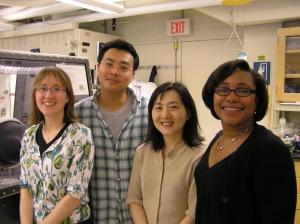
From left, students Betar Gallant and Seung Woo 
Lee and professors Yang Shao-Horn and Paula 
Hammond, in one of the labs where they did 
research on the use of carbon nanotubes 
in lithium batteries. (Credit: Image courtesy of MIT)

To produce the powerful new electrode material, the team used a layer-by-layer fabrication method, in which a base material is alternately dipped in solutions containing carbon nanotubes that have been treated with simple organic compounds that give them either a positive or negative net charge. When these layers are alternated on a surface, they bond tightly together because of the complementary charges, making a stable and durable film.

The findings, by a team led by Associate Professor of Mechanical Engineering and Materials Science and Engineering Yang Shao-Horn, in collaboration with Bayer Chair Professor of Chemical Engineering Paula Hammond, are reported in a paper published June 20 in the journal Nature Nanotechnology. The lead authors are chemical engineering student Seung Woo Lee PhD '10 and postdoctoral researcher Naoaki Yabuuchi.

Batteries, such as the lithium-ion batteries widely used in portable electronics, are made up of three basic components: two electrodes (called the anode, or negative electrode, and the cathode, or positive electrode) separated by an electrolyte, an electrically conductive material through which charged particles, or ions, can move easily. When these batteries are in use, positively charged lithium ions travel across the electrolyte to the cathode, producing an electric current; when they are recharged, an external current causes these ions to move the opposite way, so they become embedded in the spaces in the porous material of the anode.

In the new battery electrode, carbon nanotubes -- a form of pure carbon in which sheets of carbon atoms are rolled up into tiny tubes -- "self-assemble" into a tightly bound structure that is porous at the nanometer scale (billionths of a meter). In addition, the carbon nanotubes have many oxygen groups on their surfaces, which can store a large number of lithium ions; this enables carbon nanotubes for the first time to serve as the positive electrode in lithium batteries, instead of just the negative electrode.

This "electrostatic self-assembly" process is important, Hammond explains, because ordinarily carbon nanotubes on a surface tend to clump together in bundles, leaving fewer exposed surfaces to undergo reactions. By incorporating organic molecules on the nanotubes, they assemble in a way that "has a high degree of porosity while having a great number of nanotubes present," she says.

Lithium batteries with the new material demonstrate some of the advantages of both capacitors, which can produce very high power outputs in short bursts, and lithium batteries, which can provide lower power steadily for long periods, Lee says. The energy output for a given weight of this new electrode material was shown to be five times greater than for conventional capacitors, and the total power delivery rate was 10 times that of lithium-ion batteries, the team says. This performance can be attributed to good conduction of ions and electrons in the electrode, and efficient lithium storage on the surface of the nanotubes.

In addition to their high power output, the carbon nanotube electrodes showed very good stability over time. After 1,000 cycles of charging and discharging a test battery, there was no detectable change in the material's performance.

The electrodes the team produced had thicknesses up to a few microns, and the improvements in energy delivery only were seen at high-power output levels. In future work, the team aims to produce thicker electrodes and extend the improved performance to low-power outputs as well, they say. In its present form, the material might have applications for small, portable electronic devices, says Shao-Horn, but if the reported high power capability were demonstrated in a much thicker form -- with thicknesses of hundreds of microns rather than just a few -- it might eventually be suitable for other applications such as hybrid cars.

While the electrode material was produced by alternately dipping a substrate into two different solutions -- a relatively time-consuming process -- Hammond suggests that the process could be modified by instead spraying the alternate layers onto a moving ribbon of material, a technique now being developed in her lab. This could eventually open the possibility of a continuous manufacturing process that could be scaled up to high volumes for commercial production, and could also be used to produce thicker electrodes with a greater power capacity. "There isn't a real limit" on the potential thickness, Hammond says. "The only limit is the time it takes to make the layers," and the spraying technique can be up to 100 times faster than dipping, she says.

Lee says that while carbon nanotubes have been produced in limited quantities so far, a number of companies are currently gearing up for mass production of the material, which could help to make it a viable material for large-scale battery manufacturing.

Friday, May 14, 2010

Nano-Spider Molecules Behave Like Robots


A team of scientists from Columbia University, Arizona State University, the University of Michigan, and the California Institute of Technology (Caltech) have programmed an autonomous molecular "robot" made out of DNA to start, move, turn, and stop while following a DNA track.

Me
The latest installment in DNA nanotechnology has arrived: 
A molecular nanorobot dubbed a "spider" and labeled with 
green dyes traverses a substrate track built upon a DNA 
origami scaffold. It journeys towards its red-labeled goal 
by cleaving the visited substrates, thus exhibiting the 
characteristics of an autonomously moving, behavior-based 
robot at the molecular scale. (Credit: Courtesy of Paul Michelotti)

The development could ultimately lead to molecular systems that might one day be used for medical therapeutic devices and molecular-scale reconfigurable robots -- robots made of many simple units that can reposition or even rebuild themselves to accomplish different tasks.

A paper describing the work appears in the current issue of the journal Nature.

The traditional view of a robot is that it is "a machine that senses its environment, makes a decision, and then does something -- it acts," says Erik Winfree, associate professor of computer science, computation and neural systems, and bioengineering at Caltech.

Milan N. Stojanovic, a faculty member in the Division of Experimental Therapeutics at Columbia University, led the project and teamed up with Winfree and Hao Yan, professor of chemistry and biochemistry at Arizona State University and an expert in DNA nanotechnology, and with Nils G. Walter, professor of chemistry and director of the Single Molecule Analysis in Real-Time (SMART) Center at the University of Michigan in Ann Arbor, for what became a modern-day self-assembly of like-minded scientists with the complementary areas of expertise needed to tackle a tough problem.

Shrinking robots down to the molecular scale would provide, for molecular processes, the same kinds of benefits that classical robotics and automation provide at the macroscopic scale. Molecular robots, in theory, could be programmed to sense their environment (say, the presence of disease markers on a cell), make a decision (that the cell is cancerous and needs to be neutralized), and act on that decision (deliver a cargo of cancer-killing drugs).

Or, like the robots in a modern-day factory, they could be programmed to assemble complex molecular products. The power of robotics lies in the fact that once programmed, the robots can carry out their tasks autonomously, without further human intervention.

With that promise, however, comes a practical problem: how do you program a molecule to perform complex behaviors?

"In normal robotics, the robot itself contains the knowledge about the commands, but with individual molecules, you can't store that amount of information, so the idea instead is to store information on the commands on the outside," says Walter. And you do that, says Stojanovic, "by imbuing the molecule's environment with informational cues."

"We were able to create such a programmed or 'prescribed' environment using DNA origami," explains Yan. DNA origami, an invention by Caltech Senior Research Associate Paul W. K. Rothemund, is a type of self-assembled structure made from DNA that can be programmed to form nearly limitless shapes and patterns (such as smiley faces or maps of the Western Hemisphere or even electrical diagrams). Exploiting the sequence-recognition properties of DNA base pairing, DNA origami are created from a long single strand of DNA and a mixture of different short synthetic DNA strands that bind to and "staple" the long DNA into the desired shape. The origami used in the Nature study was a rectangle that was 2 nanometers (nm) thick and roughly 100 nm on each side.

The researchers constructed a trail of molecular "bread crumbs" on the DNA origami track by stringing additional single-stranded DNA molecules, or oligonucleotides, off the ends of the staples. These represent the cues that tell the molecular robots what to do -- start, walk, turn left, turn right, or stop, for example -- akin to the commands given to traditional robots.

The molecular robot the researchers chose to use -- dubbed a "spider" -- was invented by Stojanovic several years ago, at which time it was shown to be capable of extended, but undirected, random walks on two-dimensional surfaces, eating through a field of bread crumbs.

To build the 4-nm-diameter molecular robot, the researchers started with a common protein called streptavidin, which has four symmetrically placed binding pockets for a chemical moiety called biotin. Each robot leg is a short biotin-labeled strand of DNA, "so this way we can bind up to four legs to the body of our robot," Walter says. "It's a four-legged spider," quips Stojanovic. Three of the legs are made of enzymatic DNA, which is DNA that binds to and cuts a particular sequence of DNA. The spider also is outfitted with a "start strand" -- the fourth leg -- that tethers the spider to the start site (one particular oligonucleotide on the DNA origami track). "After the robot is released from its start site by a trigger strand, it follows the track by binding to and then cutting the DNA strands extending off of the staple strands on the molecular track," Stojanovic explains.

"Once it cleaves," adds Yan, "the product will dissociate, and the leg will start searching for the next substrate." In this way, the spider is guided down the path laid out by the researchers. Finally, explains Yan, "the robot stops when it encounters a patch of DNA that it can bind to but that it cannot cut," which acts as a sort of flypaper.

Although other DNA walkers have been developed before, they've never ventured farther than about three steps. "This one," says Yan, "can walk up to about 100 nanometers. That's roughly 50 steps."

"This in itself wasn't a surprise," adds Winfree, "since Milan's original work suggested that spiders can take hundreds if not thousands of processive steps. What's exciting here is that not only can we directly confirm the spiders' multistep movement, but we can direct the spiders to follow a specific path, and they do it all by themselves -- autonomously."

In fact, using atomic force microscopy and single-molecule fluorescence microscopy, the researchers were able to watch directly spiders crawling over the origami, showing that they were able to guide their molecular robots to follow four different paths.

"Monitoring this at a single molecule level is very challenging," says Walter. "This is why we have an interdisciplinary, multi-institute operation. We have people constructing the spider, characterizing the basic spider. We have the capability to assemble the track, and analyze the system with single-molecule imaging. That's the technical challenge." The scientific challenges for the future, Yan says, "are how to make the spider walk faster and how to make it more programmable, so it can follow many commands on the track and make more decisions, implementing logical behavior."

"In the current system," says Stojanovic, "interactions are restricted to the walker and the environment. Our next step is to add a second walker, so the walkers can communicate with each other directly and via the environment. The spiders will work together to accomplish a goal." Adds Winfree, "The key is how to learn to program higher-level behaviors through lower-level interactions."

Such collaboration ultimately could be the basis for developing molecular-scale reconfigurable robots -- complicated machines that are made of many simple units that can reorganize themselves into any shape -- to accomplish different tasks, or fix themselves if they break. For example, it may be possible to use the robots for medical applications. "The idea is to have molecular robots build a structure or repair damaged tissues," says Stojanovic.

"You could imagine the spider carrying a drug and bonding to a two-dimensional surface like a cell membrane, finding the receptors and, depending on the local environment," adds Yan, "triggering the activation of this drug."

Such applications, while intriguing, are decades or more away. "This may be 100 years in the future," Stojanovic says. "We're so far from that right now."

"But," Walter adds, "just as researchers self-assemble today to solve a tough problem, molecular nanorobots may do so in the future."

The other coauthors on the paper, "Molecular robots guided by prescriptive landscapes," are Kyle Lund and Jeanette Nangreave from Arizona State University; Anthony J. Manzo, Alexander Johnson-Buck, and Nicole Michelotti from the University of Michigan; Nadine Dabby from Caltech; and Steven Taylor and Renjun Pei from Columbia University. The work was supported by the National Science Foundation, the Army Research Office, the Office of Naval Research, the National Institutes of Health, the Department of Energy, the Searle Foundation, the Lymphoma and Leukemia Society, the Juvenile Diabetes Research Foundation, and a Sloan Research Fellowship.
Reblog this post [with Zemanta]