BTemplates.com

Powered by Blogger.

Pageviews past week

Quantum mechanics

Auto News

artificial intelligence

About Me

Recommend us on Google!

Information Technology

Popular Posts

Showing posts with label Health. Show all posts

Tuesday, September 27, 2011

From Your Heart to Your iPhone


A new app gets data from an implanted device and can share it with the patient, doctors, and family.

A smart-phone app under development for heart-failure patients allows them to keep track of the pressure inside their heart as measured by an implanted sensor. That data could help patients adjust their medication to maintain a healthy pressure, much as diabetics do with insulin and blood sugar readings.

Called Pam+ (for "patient advisory module"), the app is being developed by researchers at the University of Southern California in collaboration with medical device maker St. Jude Medical. The researchers hope it will help patients better manage their health and reduce hospitalizations, which are responsible for much of the $40 million in health-care costs linked to heart failure.

In congestive heart failure, pressure builds up in the circulatory system and the heart fails to pump blood adequately to the rest of the body. Fluid pressure changes by the day, and monitoring those fluctuations continuously is essential to treating heart failure effectively. A number of implanted devices are now under development to monitor this pressure, giving patients and doctors real time data.

The PAM+ app works in conjunction with an external device—developed by St. Jude and currently in clinical tests—that is placed over the heart, where it charges the implanted sensor and downloads data from it.

The data is forwarded to a server at St. Jude that analyzes it and returns, via the app, the latest readings and information about ongoing trends. A patient who has regularly monitored his or her heart pressure over a week will see a graph of pressure readings along with the message "Your heart thanks you." Users can easily share their data with their health-care team and family.


"We want patients to be able to access data but also to be rewarded and encouraged on a daily basis, so they don't feel like their whole life is a diet," says Leslie Saxon, a cardiologist and director of the Center for Body Computing at USC, who helped develop the device.

Previous research conducted by Saxon showed that remote monitoring can improve the health of heart-failure patients and lower health-care costs. She unveiled a prototype of the app at the Body Computing conference in Los Angeles today.

Users get points for monitoring their pressure—points that might eventually be tied to iTunes or Amazon credit. "Even a traditional payer would love to reward this type of behavior," says Saxon.

She believes an app like this can also change the nature of doctors' visits. Rather than a physician giving a patient the latest test results, taken at a few points in time, the patient can show the doctor measurements of heart pressure over weeks and months, and together they can discuss the trends these reveal.

Scientists discover an organizing principle for our sense of smell


The fact that certain smells cause us pleasure or disgust would seem to be a matter of personal taste. But new research at the Weizmann Institute shows that odors can be rated on a scale of pleasantness, and this turns out to be an organizing principle for the way we experience smell. The findings, which appeared today in Nature Neuroscience, reveal a correlation between the response of certain nerves to particular scents and the pleasantness of those scents. Based on this correlation, the researchers could tell by measuring the nerve responses whether a subject found a smell pleasant or unpleasant.

Our various sensory organs are have evolved patterns of organization that reflect the type of input they receive. Thus the receptors in the retina, in the back of the eye, are arranged spatially for efficiently mapping out visual coordinates. The structure of the inner ear, on the other hand, is set up according to a tonal scale. But the organizational principle for our sense of smell has remained a mystery: Scientists have not even been sure if there is a scale that determines the organization of our smell organ, much less how the arrangement of smell receptors on the membranes in our nasal passages might reflect such a scale.

A team headed by Prof. Noam Sobel of the Weizmann Institute's Neurobiology Department set out to search for the principle of organization for smell. Hints that the answer could be tied to pleasantness had been seen in research labs around the world, including that of Sobel, who had previously found a connection between the chemical structure of an odor molecule and its place on a pleasantness scale. Sobel and his team thought that smell receptors in the nose – of which there are some 400 subtypes – could be arranged on the nasal membrane according to this scale. This hypothesis goes against the conventional view, which claims that the various smell receptors are mixed -- distributed evenly, but randomly, around the membrane.


In the experiment, the researchers inserted electrodes into the nasal passages of volunteers and measured the nerves' responses to different smells in various sites. Each measurement actually captured the response of thousands of smell receptors, as these are densely packed on the membrane. The scientists found that the strength of the nerve signal varies from place to place on the membrane. It appeared that the receptors are not evenly distributed, but rather, that they are grouped into distinct sites, each engaging most strongly with a particular type of scent. Further investigation showed that the intensity of a reaction was linked to the odor's place on the pleasantness scale. A site where the nerves reacted strongly to a certain agreeable scent also showed strong reactions to other pleasing smells and vice versa: The nerves in an area with a high response to an unpleasant odor reacted similarly to other disagreeable smells. The implication is that a pleasantness scale is, indeed, an organizing principle for our smell organ.

But does our sense of smell really work according to this simple principle? Natural odors are composed of a large number of molecules – roses, for instance, release 172 different odor molecules. Nonetheless, says Sobel, the most dominant of those determine which sites on the membrane will react the most strongly, while the other substances make secondary contributions to the scent.

'We uncovered a clear correlation between the pattern of nerve reaction to various smells and the pleasantness of those smells. As in sight and hearing, the receptors for our sense of smell are spatially organized in a way that reflects the nature of the sensory experience,' says Sobel. In addition, the findings confirm the idea that our experience of smells as nice or nasty is hardwired into our physiology, and not purely the result of individual preference. Sobel doesn't discount the idea that individuals may experience smells differently. He theorizes that cultural context and personal experience may cause a certain amount of reorganization in smell perception over a person's lifetime.

More information: DOI: 10.1038/nn.2926

Sunday, July 24, 2011

Engineering a new face after injury


Today, surgeons face many limitations when it comes to helping a patient who suffers from a severe craniofacial injury, or an injury pertaining to the skull and the face. Most often a result of cancer or war-related circumstances, the injury is both psychologically and physically damaging.

Evolution of a patient's recovery from facial injury through the use of topological optimization. Credit: Hanlon, Beckman ITG, University of Illinois

Will the patients ever recover their appearance? Or more importantly, recover their ability to speak, breathe or eat correctly again?

Rebuilding the delicate facial bone structure of an individual is a complicated procedure. The surgeon constructs a facial frame with bone from other parts of the body (called autologous tissue), in order to guarantee the functionality of the specialized organs responsible for vital roles such as breathing, seeing, communicating and eating. Since there are no analogous bone structures to a person's face, the procedure depends on experience and skill. As Glaucio Paulino, program director of the mechanics of materials program at the National Science Foundation (NSF), noted, this procedure does not always generate the desired outcome.

"The middle of the face is the most complicated part of the human skeleton," said Paulino. "What makes the reconstruction more complicated is the fact that the bones are small, delicate, highly specialized and located in a region highly susceptible to contamination by bacteria."

Facial bones are unique and using bone tissue extracted from different parts of the body, such as the bones of the forearm, isn't the most effective form of recovery.

"The patient may be improved, but still suffer from significant deformity," said Paulino.

Implementation of loads, boundary conditions and different cavity constraints to a design domain and the consequent optimized results. Credit: Glaucio H. Paulino

In contrast, topological optimization is a feasible alternative to make such a recovery possible.

Topological optimization isn't native to the surgery room--it's a mathematical method that uses given loads, the applied force on an area, and boundary conditions or spatial limits, to optimize a specific structure's layout. Imagine a building grid in which you can determine where there should be material and where there shouldn't. Moreover, you can express loads and supports that would affect certain parts of this block of material. Your final result is an optimized structure that fits your established constraints.

This mathematical method is successfully used to engineer spaceships and airplanes. The Airbus 380 wing, for example, was designed with topological optimization. Today, extensive research is underway to apply topological optimization to the engineering of future high-rise buildings. Paulino is responsible for some of the recent advances in this field.


Together with Alok Sutradhar and Michael Miller, from the Ohio State University Medical Center, and Tam Nguyen, from the department of civil and environmental engineering at the University of Illinois, Paulino is studying how to bring topological optimization to the surgery room. With the recent advances in tissue engineering, Paulino believes that the method can be used to construct patient-specific bone frames.

"The key idea is to have a technique that is tailored for the specific patient. It's not one formula that fits all. People are different, therefore, you cannot have one solution for all patients," said Paulino.

Final optimized result with denture inserted into the craniofacial skeleton. Credit: Glaucio H. Paulino

Engineering a face

In an experiment, researchers explored the creation of a three-dimensional structure for a patient with severe gunshot injury. After selecting a design domain from the craniofacial skeleton, supports, loads and cavity constraints (areas with no bone, such as eye cavities) were applied. Topological optimization generated many possible structures to fit the patient-specific requirements.


Watch this video to see the process of creating a structure for a patient with severe gunshot injury using topological optimization. Although the results did not necessarily resemble the natural bone structure, they would preserve the vital functions of facial organs while providing a safe platform for prosthetics and plastic surgery.

The process will "show surgeons their alternatives before going into the operating room," said Paulino.

At the moment, such structures would be built using titanium, which is light and strong. Unfortunately, titanium may cause infections because it's foreign to the body. With future advances in tissue engineering, however, molding human bone tissue into a structure is a possibility. Researchers are still investigating how to ensure that the bone structure created through this process, a living tissue, will maintain the desired shape after implanted in the patient.

Paulino and his team of researchers hope to continue translating applicable concepts between different fields, such as engineering and medicine, to make innovative discoveries. With the development of tissue engineering and topological optimization, in the future, complete recovery from craniofacial injuries will hopefully be enabled by a routine procedure in the surgery room.

Provided by National Science Foundation

Tuesday, June 28, 2011

It's Not an Apple a Day After All -- It's Strawberries: Flavonoids Could Represent Two-Fisted Assault On Diabetes and Nervous System Disorders


A recent study from scientists at the Salk Institute for Biological Studies suggests that a strawberry a day (or more accurately, 37 of them) could keep not just one doctor away, but an entire fleet of them, including the neurologist, the endocrinologist, and maybe even the oncologist.
Fisetin, a naturally-occurring flavonoid found most 
abundantly in strawberries, lessens complications of diabetes
(Credit: Courtesy of the Salk Institute for Biological Studies)

Investigations conducted in the Salk Institute's Cellular Neurobiology Laboratory (CNL) will appear in the June 27, 2011, issue of PLoS ONE. The report explains that fisetin, a naturally-occurring flavonoid found most abundantly in strawberries and to a lesser extent in other fruits and vegetables, lessens complications of diabetes. Previously, the lab showed that fisetin promoted survival of neurons grown in culture and enhanced memory in healthy mice. That fisetin can target multiple organs strongly suggests that a single drug could be used to mitigate numerous medical complications.

"This manuscript describes for the first time a drug that prevents both kidney and brain complications in a type 1 diabetes mouse model," says David Schubert, Ph.D., professor and head of the Cellular Neurobiology Laboratory and one of the manuscript's co-authors. "Moreover, it demonstrates the probable molecular basis of how the therapeutic is working."

Pam Maher, Ph.D., a senior staff scientist in the CNL, is the study's corresponding author. Maher initially identified fisetin as a neuroprotective flavonoid ten years ago. "In plants, flavonoids act as sunscreens and protect leaves and fruit from insects," she explains. "As foods they are implicated in the protective effect of the 'Mediterranean Diet.'"

Other celebrity flavonoids include polyphenolic compounds in blueberries and red wine.

Although her group's focus is neurobiology, Maher and colleagues reasoned that, like other flavonoids, fisetin might ameliorate a spectrum of disorders seen in diabetic patients. To test this, they evaluated effects of fisetin supplementation in Akita mice, a very robust model of type 1 diabetes, also called childhood onset diabetes.

Akita mice exhibit increased blood sugar typical of type 1 diabetes and display pathologies seen in serious human complications of both type 1 and 2 diabetes. Those include diabetic nephropathy or kidney disease, retinopathy, and neuropathies in which patients lose touch or heat sensations.

Mice fed a fisetin-enriched diet remained diabetic, but acute kidney enlargement-or hypertrophy-seen in untreated mice was reversed, and high urine protein levels, a sure sign of kidney disease, fell. Moreover, fisetin ingestion ameliorated anxiety-related behaviors seen in diabetic mice. "Most mice put in a large area become exploratory," says Maher. "But anxious mice tend not to move around. Akita mice showed enhanced anxiety behavior, but fisetin feeding restored their locomotion to more normal levels."


The study also defines a likely molecular mechanism underlying these effects. Researchers observed that blood and brain levels of sugars affixed to proteins known as advanced glycation end-products-or AGEs-were reduced in fisetin-treated compared to untreated Akita mice. These decreases were accompanied by increased activity of the enzyme glyoxalase 1, which promotes removal of toxic AGE precursors.

The discovery of an AGE-antagonizing enzyme upregulated by fisetin is very intriguing, because substantial evidence implicates high blood AGE levels with many if not most diabetic complications. "We know that fisetin increases activity of the glyoxalase enzyme and may increase its expression," says Maher. "But what is important is that ours is the first report that any compound can enhance glyoxalase 1 activity."

Interestingly, excessively high AGE levels also correlate with inflammatory activity thought to promote some cancers. In fact, studies published by others confirm that fisetin decreases tumorigenicity of prostate cancer cells both in culture and in animal models, which if supported would represent a major added incentive to eat your strawberries.

To ingest fisetin levels equivalent to those fed Akita mice, Maher estimates that humans would have to eat 37 strawberries a day, assuming that strawberry fisetin is as readily metabolizable by humans as fisetin-spiked lab chow is by mice. Rather than through diet, Maher envisions that fisetin-like drugs could be taken as a supplement.

Schubert notes that fisetin is also effective in mouse models of Alzheimer's disease. "We and others have shown that diabetes may be a risk factor for Alzheimer's disease, making identification of a safe prophylactic like fisetin highly significant," he says.

Maher acknowledges that the public may be suffering from flavonoid-fatigue, given media coverage of the promises of these compounds. "Polyphenolics like fisetin and those in blueberry extracts are found in fruits and vegetables and are related to each other chemically," she says. "There is increasing evidence that they all work in multiple diseases. Hopefully some combination of these compounds will eventually get to the clinic."

Schubert concurs that their findings only reinforce what common sense and our mothers told us was a healthy lifestyle. "Eat a balanced diet and as much freshly prepared organic food as possible, get some exercise, keep socially and mentally active and avoid sodas with sugar and highly processed foods since they can contain high levels of AGEs," he advises.

But he also worries that hoops that must be jumped through to bring a natural product like fisetin, as opposed to a totally synthetic drug, to clinical trials are daunting because it is difficult to protect patents on natural products. "We will never know if a compound like fisetin works in humans until someone is willing to support a clinical trial."

Also contributing to this study were Richard Dargusch and Jennifer L. Ehren, Ph.D.,of the Cellular Neurobiology Laboratory, and Kumar Sharma, M.D., and Shinichi Okada, M.D., Ph.D., of the Department of Medicine at University of California, San Diego.

Funding for the study came from the Fritz B. Burns Foundation, the Juvenile Diabetes Research Foundation, the Hewitt Foundation, and the National Institutes of Health.

Wednesday, June 15, 2011

A pulse no longer necessary for life


While most people connect a pulse and a heartbeat to life, Dr. Billy Cohn and Dr. Bud Frazier from the Texas Heart Institute have found a way to keep the blood circulating and extend the life of patients while taking away their pulse.
This X-ray image shows the dual turbinelike 
blood pumps that replaced patient's heart. 
Image: Texas Heart Institute

Researchers have spent years trying to perfect an artificial heart that does not break down, wear out, or cause blood clots and infections. However, Cohn and Frazier have developed an artificial heart, of sorts, that seems to do the trick. The only catch is it isn’t a heart. There is no heartbeat. There is no pulse. If a patient had one of their new hearts, the patient would appear dead. Attaching an EKG would return a flat-line.

The new device uses technology that has been used to aid failing hearts since the 1980s. A ventricular assist device, or VAD, is a circulatory device designed to assist either the right or left ventricle of the heart. The VADs have a rotor of blades that circulate and push the blood forward in a continuous flow.

While VADs are typically used to help one section of the heart, Cohn and Frazier hooked two of these VADs together so they would essentially work as both sides of the heart. They began working on calves and currently have an 8-month-old calf named Abigail who has no heart. Her heart was removed and in its place the doctors inserted their new pump device. Abigail is a healthy and active young calf, however, according to any medical cardiac tests, she would appear dead.

Cohn and Frazier, after testing on 38 calves, wanted to take this new pump one step further and test it on a human patient. This is where Craig Lewis, a 55-year-old man who was dying from amyloidosis comes in. His heart had become so damaged from the disease that doctors had only given him about 12 hours to live. Lewis and his wife agreed to let the doctors try the new artificial heart pump to try and extend his life, if even for a short time. The doctors inserted the new pumps and Lewis did recover and had another month of life before the disease took other organs. His new heart however worked flawlessly.

Cohn and Frazier still have much work to do before the new heart will be available. A final design must be determined, a manufacturer must be found and they must apply for FDA approval. Results show amazing promise and may be the new future in artificial hearts.


Saturday, April 9, 2011

Is Beauty Found in the Whites of the Eyes? 'Red Eyes' Associated With the Sad and Unattractive


Beauty is said to be in the eye of the beholder, but a new study reveals that the reverse is also true; unattractiveness is in the eye of the beheld. Research published in Ethology finds that people with bloodshot eyes are considered sadder, unhealthier and less attractive than people whose eye whites are untinted, a cue which is uniquely human.
Eye images of a young adult female (left) and male (right). In the top images the sclera are white and in the bottom images the eyes have been digitally altered. In the study subjects rated 200 such images. (Credit: R.Provine)

"Red, 'bloodshot' eyes are prominent in medical diagnoses and in folk culture," said lead author Dr. Robert R. Provine from the University of Maryland, Baltimore County. "We wanted to know if they influence the everyday behaviour and attitudes of those who view them, and if they trigger perceptions of attractiveness."

Bloodshot eyes occur when the small blood vessels of the usually transparent conjunctiva membrane on the surface of the eye become enlarged and congested with blood, giving a red tint to the underlying sclera, the "white" of the eyes. Redness of the sclera is believed to be a general but important sign of a person's emotional and biological state.

"If you met a friend with bloodshot eyes it may be unclear whether you should offer sympathy or medical assistance because red eyes may be a result of weeping, allergies or infectious diseases," said Provine. "Comments from our colleagues also suggest that red eyes prompt feelings of discomfort, ranging from increased monitoring of their own eyes to a hint of sympathetic tearing."

In the first empirical test to discover the perceptions and behavioural implications of red eyes Dr. Provine's team tested 208 volunteer students from the University of Maryland, Baltimore County. The volunteers composed of 93 males and 115 females, with an average age of 20.6 years.

The volunteers were shown 200 images of eyes, half with clear white sclera and half with sclera tinted red by digital image processing. The volunteers were asked how sad, healthy or attractive the owners of the eyes were. The results revealed that people with reddened eyes appear sadder, less healthy, and less attractive compared to those with whiter, untinted eyes.

This is the first study to demonstrate that eye redness is perceived as a cue of emotion. Humans appear to be the only species which uses eye colouration as an indicator of either health or emotion. This is because other primates lack the background of white sclera necessary to make the reddened conjunctiva visible.

Sclera colour provides even casual, untrained observers with a quick estimate of the emotional and health status of an individual and the study's ratings of attractiveness suggest that this information does influence our behaviour.

"Standards of beauty vary across cultures, however, youth and healthiness are always in fashion because they are associated with reproductive fitness," said Provine. "Traits such as long, lustrous hair and smooth or scar-free skin are cues of youth and offer the beholder a partial record of health.

Now clear eye whites join these traits as a universal standard for the perception of beauty and a cue of health and reproductive fitness. Given this discovery, eye drops that 'get the red out' can be considered beauty aids."

Saturday, March 5, 2011

Liver, Not Brain, May Be Origin of Alzheimer’s Plaques


Unexpected results from a Scripps Research Institute and ModGene, LLC study could completely alter scientists' ideas about Alzheimer's disease -- pointing to the liver instead of the brain as the source of the "amyloid" that deposits as brain plaques associated with this devastating condition. The findings could offer a relatively simple approach for Alzheimer's prevention and treatment.
New research suggests that the liver instead of the brain 
may be the source of the "amyloid" that deposits as brain 
plaques associated with Alzheimer's disease. 
(Credit: iStockphoto/David Marchal)


The study was published online March 3 in The Journal of Neuroscience Research.

In the study, the scientists used a mouse model for Alzheimer's disease to identify genes that influence the amount of amyloid that accumulates in the brain. They found three genes that protected mice from brain amyloid accumulation and deposition. For each gene, lower expression in the liver protected the mouse brain. One of the genes encodes presenilin -- a cell membrane protein believed to contribute to the development of human Alzheimer's.

"This unexpected finding holds promise for the development of new therapies to fight Alzheimer's," said Scripps Research Professor Greg Sutcliffe, who led the study. "This could greatly simplify the challenge of developing therapies and prevention."

An estimated 5.1 million Americans have Alzheimer's disease, including nearly half of people age 85 and older. By 2050, the number of people age 65 and over with this disease will range from 11 million to 16 million unless science finds a way to prevent or effectively treat it. In addition to the human misery caused by the disease, there is the unfathomable cost. A new report from the Alzheimer's Association shows that in the absence of disease-modifying treatments, the cumulative costs of care for people with Alzheimer's from 2010 to 2050 will exceed $20 trillion.

A Genetic Search-and-Find Mission

In trying to help solve the Alzheimer's puzzle, in the past few years Sutcliffe and his collaborators have focused their research on naturally occurring, inherited differences in neurological disease susceptibility among different mouse strains, creating extensive databases cataloging gene activity in different tissues, as measured by mRNA accumulation. These data offer up maps of trait expression that can be superimposed on maps of disease modifier genes.

As is the case with nearly all scientific discovery, Sutcliffe's research builds on previous findings. Several years ago, researchers at Case Western Reserve mapped three genes that modify the accumulation of pathological beta amyloid in the brains of a transgenic mouse model of Alzheimer's disease to large chromosomal regions, each containing hundreds of genes. The Case Western scientists used crosses between the B6 and D2 strains of mice, studying more than 500 progeny.

Using the results from this study, Sutcliffe turned his databases of gene expression to the mouse model of Alzheimer's, looking for differences in gene expression that correlated with differences in disease susceptibility between the B6 and D2 strains. This intensive work involved writing computer programs that identified each genetic difference that distinguished the B6 and D2 genomes, then running mathematical correlation analysis (known as regression analysis) of each difference. Correlations were made between the genotype differences (B6 or D2) and the amount of mRNA product made from each of the more than 25,000 genes in a particular tissue in the 40 recombinant inbred mouse strains. These correlations were repeated 10 times to cover 10 tissues, the liver being one of them.

"A key aspect of this work was learning how to ask questions of massive data sets to glean information about the identities of heritable modifier genes," Sutcliffe said. "This was novel and, in a sense, groundbreaking work: we were inventing a new way to identify modifier genes, putting all of these steps together and automating the process. We realized we could learn about how a transgene's pathogenic effect was being modified without studying the transgenic mice ourselves."

Looking for a Few Good Candidates

Sutcliffe's gene hunt offered up good matches, candidates, for each of the three disease modifier genes discovered by the Case Western scientists, and one of these candidates -- the mouse gene corresponding to a gene known to predispose humans carrying particular variations of it to develop early-onset Alzheimer's disease -- was of special interest to his team.

"The product of that gene, called Presenilin2, is part of an enzyme complex involved in the generation of pathogenic beta amyloid," Sutcliffe explained. "Unexpectedly, heritable expression of Presenilin2 was found in the liver but not in the brain. Higher expression of Presenilin2 in the liver correlated with greater accumulation of beta amyloid in the brain and development of Alzheimer's-like pathology."

This finding suggested that significant concentrations of beta amyloid might originate in the liver, circulate in the blood, and enter the brain. If true, blocking production of beta amyloid in the liver should protect the brain.

To test this hypothesis, Sutcliffe's team set up an in vivo experiment using wild-type mice since they would most closely replicate the natural beta amyloid-producing environment. "We reasoned that if brain amyloid was being born in the liver and transported to the brain by the blood, then that should be the case in all mice," Sutcliffe said, "and one would predict in humans, too."

The mice were administered imatinib (trade name Gleevec, an FDA-approved cancer drug), a relatively new drug currently approved for treatment of chronic myelogenous leukemia and gastrointestinal tumors. The drug potently reduces the production of beta amyloid in neuroblastoma cells transfected by amyloid precursor protein (APP) and also in cell-free extracts prepared from the transfected cells. Importantly, Gleevec has poor penetration of the blood-brain barrier in both mice and humans.

"This characteristic of the drug is precisely why we chose to use it," Sutcliffe explained. "Because it doesn't penetrate the blood-brain barrier, we were able to focus on the production of amyloid outside of the brain and how that production might contribute to amyloid that accumulates in the brain, where it is associated with disease."

The mice were injected with Gleevec twice a day for seven days; then plasma and brain tissue were collected, and the amount of beta amyloid in the blood and brain was measured. The findings: the drug dramatically reduced beta amyloid not only in the blood, but also in the brain where the drug cannot penetrate. Thus, an appreciable portion of brain amyloid must originate outside of the brain, and imatinib represents a candidate for preventing and treating Alzheimer's.

As for the future of this research, Sutcliffe says he hopes to find a partner and investors to move the work into clinical trials and new drug development.

In addition to Sutcliffe, the authors of the study, titled "Peripheral reduction of β-amyloid is sufficient to reduce brain Aβ: implications for Alzheimer's disease," include Peter Hedlund and Elizabeth Thomas of Scripps Research, and Floyd Bloom and Brian Hilbush of ModGene, LLC, which funded the project.

Thursday, February 17, 2011

Wireless Heart Implant Reduces HospitalizationsA pressure-sensing implant helps heart-failure patients stay healthy.


A wireless sensor developed by Atlanta-based CardioMEMS reduced the number of hospitalizations in patients with heart failure by 39 percent. The tiny implant monitors fluid pressure in the pulmonary artery and transmits the data wirelessly to physicians, who can adjust patients' medications accordingly.

Researchers say the sensor may significantly lower health-care costs and improve quality of life for people with congestive heart failure. The device is one of several prototypes being developed by CardioMEMS and other medical implant companies to provide continuous, personalized wireless monitors for such patients.
Pressure patrol: A new wireless sensor the size of a paper
clip measures fluid pressure in the pulmonary artery. The
metalloops on either end anchor the sensor to the artery
walls, while the self-contained transducer in the middle
takes pressure readings. The sensor is activated by radio
frequency, transmitting data wirelessly to physicians
via modem.Credit: OSU Medical Center/CardioMEMS


"I think the study shows this kind of device is incredibly useful in improving outcomes in patients and directing therapy," says Marc Jay Semigran, medical director of the Mass General Heart Failure and Cardiac Transplant Program, who was not involved in the study.

Hospitals admit 1.1 million adults each year for congestive heart failure, a condition in which pressure builds up in the circulatory system and the heart fails to pump blood adequately to the rest of the body. The American Heart Association estimates that the chronic condition costs the health-care system $29 billion per year. CardioMEMS aims to reduce that figure by providing an accurate way to continuously monitor patients after they've left the hospital.

The device is implanted in the pulmonary artery, an area that carries a low risk of clotting. It is smaller than other implants under development because it does not require a battery or a wire to take pressure readings. Two metal loops hold it to the sides of the artery, and a pressure transducer records the flow of fluids through the blood vessel. The sensor is powered externally by a receiver built into a pillow. When a patient lies on the pillow, the sensor is activated to take measurements and send them wirelessly to a computer, where physicians can review the data. In a large six-month clinical trial published this month in the Lancet, 550 patients from 64 centers across the United States were equipped with the device and instructed to take readings once a day. Patients were divided into two groups. The first took medication instructions from physicians who monitored the sensor data. The second took instructions from physicians who relied on traditional indicators like weight and blood pressure. Over the six months, patients in the first group experienced 39 percent fewer hospitalizations than those in the second.

Today, physicians often assess pulmonary pressure when initially evaluating a patient, but they do so far less frequently in follow-up evaluation. That's because the measurement requires doctors to snake a catheter into a patient's heart and inflate a balloon. However, fluid pressure changes by the day, and monitoring those fluctuations continuously is essential to treating heart failure effectively.

"Over the years, we found that pressures go up long before patients develop symptoms and call a doctor to say they're sick," says Philip Adamson, director of the Heart Failure Institute at Oklahoma Heart Hospital, the principal investigator in the CardioMEMS clinical trial. "By utilizing the pressure sensor information, we're given the ability to make changes in medications long before patients bring themselves to the doctor, and that's how we reduced hospitalizations."

Over the past few years, several companies have jockeyed to be first on the market with a continuous pressure-sensing cardiac implant. In 2007, Medtronic failed to get FDA approval for its sensor, a stopwatch-size, battery-powered implant wired to the heart. The device reduced hospitalizations by 22 percent, but FDA regulators did not consider that worth the risks associated with implanting it. Researchers also found that the wire connecting the sensor to the heart degraded over time.

CardioMEMS is currently seeking approval for its sensor from the U.S. Food and Drug Administration and has submitted results from the clinical study for FDA review. In the next two or three years, the company plans to integrate the sensor's receiver into a patient's cell phone, which will be able to instantly read pressure data and upload it for both physicians and patients to review.

Friday, December 10, 2010

No Wrong Side To This Bad


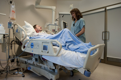
US researchers have created a smart hospital bed that is aware of its surrounding. The intelligent bed will make important decisions regarding the patients' healthcare which will involve both improving and saving precious lives.
A prototype of the smart bed that is being tested at the University of New Hampshire could revolutionise health care In the future


John LaCourse, professor at the University of New Hampshire, is currently negotiating with hospital bed manufacturers

to adopt his prograq~.med algorithm, which could become the basis for computerised hospital beds.

These smart hospital beds would communicate with and respond to medical devices that monitor a patient's condition.

"Perhaps a sleeping patient moves, causing a drop in blood pressure. The blood pressure monitor would communicate

this change to the bed and the bed, in turn, would move up or down until the patients' blood pressure is stabilised," he says.

IMPROVED POST-SURGERY CARE

Post-surgical needs may also be met with this bed. "Procedures such as retinal surgery require exact blood pressure levels," says LaCourse. "A smart hospital bed would adjust itself to maintain these levels for patients."

Even quality-of-life conditions such as bed sores could be addressed. "Instead of requiring hospital staff to move the patient, monitors could send signals to the bed to roll the patient to his left or right to avoid bed sores," says LaCourse.

"Microprocessors installed into the bed can also sense respiration patterns to determine when breathing has ceased, the bed moves in such a way that the breathing resumes," said Jonathan Waters, who is working on modifying the bed for sleep apnoea.

PLUG AND PLAY

The ultimate success of LaCourse's project rests with the plug-and-play component. Plug-and-play means that medical devices – everything from blood pressure monitors to breathing machines- "can talk to each other and share patient information which greatly reduces care errors," explains LaCourse.

To realise plug-and-play capability, however, LaCourse's technology must become the industry standard for hospital bed manufacturers. 1n this way, medical devices could seamlessly connect to and exchange patient data.

LaCourse is hopeful that, within two to three years, his technology may be accepted by most if not all hospital bed companies.

Friday, October 15, 2010

Feelings of Love: Effective Pain Relief


Intense, passionate feelings of love can provide amazingly effective pain relief, similar to painkillers or such illicit drugs as cocaine, according to a new Stanford University School of Medicine study.
Love-induced pain relief was associated with the activation of primitive brain structures that control rewarding experiences, such as the nucleus accumbens – shown here in color. (Credit: Courtesy of Sean Mackey and Jarred Younger)

"When people are in this passionate, all-consuming phase of love, there are significant alterations in their mood that are impacting their experience of pain," said Sean Mackey, MD, PhD, chief of the Division of Pain Management, associate professor of anesthesia and senior author of the study, which will be published online Oct. 13 in PLoS ONE. "We're beginning to tease apart some of these reward systems in the brain and how they influence pain. These are very deep, old systems in our brain that involve dopamine -- a primary neurotransmitter that influences mood, reward and motivation."

Scientists aren't quite yet ready to tell patients with chronic pain to throw out the painkillers and replace them with a passionate love affair; rather, the hope is that a better understanding of these neural-rewards pathways that get triggered by love could lead to new methods for producing pain relief.

"It turns out that the areas of the brain activated by intense love are the same areas that drugs use to reduce pain," said Arthur Aron, PhD, a professor of psychology at State University of New York at Stony Brook and one of the study's authors. Aron has been studying love for 30 years. "When thinking about your beloved, there is intense activation in the reward area of the brain -- the same area that lights up when you take cocaine, the same area that lights up when you win a lot of money."

The concept for the study was sparked several years ago at a neuroscience conference when Aron, an expert in the study of love, met up with Mackey, an expert in the research of pain, and they began talking.

"Art was talking about love," Mackey said. "I was talking about pain. He was talking about the brain systems involved with love. I was talking about the brain systems involved with pain. We realized there was this tremendous overlapping system. We started wondering, 'Is it possible that the two modulate each other?'"

After the conference, Mackey returned to Stanford and collaborated with postdoctoral scholar Jarred Younger, PhD, now an assistant professor of anesthesia, who was also intrigued with the idea. Together the three set up a study that would entail examining the brain images of undergraduates who claimed to be "in that first phase of intense love."

"We posted fliers around Stanford University and within hours we had undergrads banging on our door," Mackey said. The fliers asked for couples who were in the first nine months of a romantic relationship.

"It was clearly the easiest study the pain center at Stanford has ever recruited for," Mackey said. "When you're in love you want to tell everybody about it.

"We intentionally focused on this early phase of passionate love," he added. "We specifically were not looking for longer-lasting, more mature phases of the relationship. We wanted subjects who were feeling euphoric, energetic, obsessively thinking about their beloved, craving their presence.

"When passionate love is described like this, it in some ways sounds like an addiction. We thought, 'Maybe this does involve similar brain systems as those involved in addictions which are heavily dopamine-related.' Dopamine is the neurotransmitter in our brain that is intimately involved with feeling good."

Researchers recruited 15 undergraduates (eight women and seven men) for the study. Each was asked to bring in photos of their beloved and photos of an equally attractive acquaintance. The researchers then successively flashed the pictures before the subjects, while heating up a computer-controlled thermal stimulator placed in the palm of their hand to cause mild pain. At the same time, their brains were scanned in a functional magnetic resonance imaging machine.

The undergraduates were also tested for levels of pain relief while being distracted with word-association tasks such as: "Think of sports that don't involve balls." Scientific evidence has shown in the past that distraction causes pain relief, and researchers wanted to make sure that love was not just working as a distraction from pain.

Results showed that both love and distraction did equally reduce pain, and at much higher levels than by concentrating on the photo of the attractive acquaintance, but interestingly the two methods of pain reduction used very different brain pathways.

"With the distraction test, the brain pathways leading to pain relief were mostly cognitive," Younger said. "The reduction of pain was associated with higher, cortical parts of the brain. Love-induced analgesia is much more associated with the reward centers. It appears to involve more primitive aspects of the brain, activating deep structures that may block pain at a spinal level -- similar to how opioid analgesics work.

"One of the key sites for love-induced analgesia is the nucleus accumbens, a key reward addiction center for opioids, cocaine and other drugs of abuse. The region tells the brain that you really need to keep doing this," Younger said.

"This tells us that you don't have to just rely on drugs for pain relief," Aron said. "People are feeling intense rewards without the side effects of drugs."

Other Stanford contributors include research assistants Sara Parke and Neil Chatterjee.

Funding for the study was received from the Chris Redlich Pain Research Fund.

Editor's Note: This article is not intended to provide medical advice, diagnosis or treatment.