BTemplates.com

Powered by Blogger.

Pageviews past week

Quantum mechanics

Auto News

artificial intelligence

About Me

Recommend us on Google!

Information Technology

Popular Posts

Showing posts with label Communications. Show all posts

Wednesday, June 12, 2013

Brain-Computer Interfaces: Just Wave a Hand


Small electrodes placed on or inside the brain allow patients to interact with computers or control robotic limbs simply by thinking about how to execute those actions. This technology could improve communication and daily life for a person who is paralyzed or has lost the ability to speak from a stroke or neurodegenerative disease.
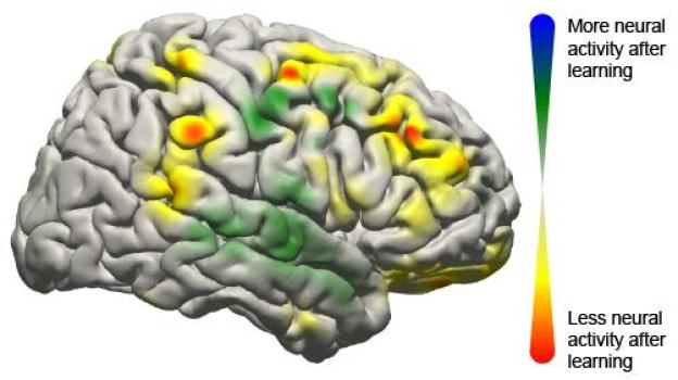
This image shows the changes that took place in the brain for all patients participating in the study using a brain-computer interface. Changes in activity were distributed widely throughout the brain.
This image shows the changes that took place in the brain for all patients participating in the study using a brain-computer interface. Changes in activity were distributed widely throughout the brain. (Credit: Jeremiah Wander, UW)
Now, University of Washington researchers have demonstrated that when humans use this technology -- called a brain-computer interface -- the brain behaves much like it does when completing simple motor skills such as kicking a ball, typing or waving a hand. Learning to control a robotic arm or a prosthetic limb could become second nature for people who are paralyzed.

"What we're seeing is that practice makes perfect with these tasks," said Rajesh Rao, a UW professor of computer science and engineering and a senior researcher involved in the study. "There's a lot of engagement of the brain's cognitive resources at the very beginning, but as you get better at the task, those resources aren't needed anymore and the brain is freed up."

Rao and UW collaborators Jeffrey Ojemann, a professor of neurological surgery, and Jeremiah Wander, a doctoral student in bioengineering, published their results online June 10 in the Proceedings of the National Academy of Sciences.

In this study, seven people with severe epilepsy were hospitalized for a monitoring procedure that tries to identify where in the brain seizures originate. Physicians cut through the scalp, drilled into the skull and placed a thin sheet of electrodes directly on top of the brain. While they were watching for seizure signals, the researchers also conducted this study.

The patients were asked to move a mouse cursor on a computer screen by using only their thoughts to control the cursor's movement. Electrodes on their brains picked up the signals directing the cursor to move, sending them to an amplifier and then a laptop to be analyzed. Within 40 milliseconds, the computer calculated the intentions transmitted through the signal and updated the movement of the cursor on the screen.

Researchers found that when patients started the task, a lot of brain activity was centered in the prefrontal cortex, an area associated with learning a new skill. But after often as little as 10 minutes, frontal brain activity lessened, and the brain signals transitioned to patterns similar to those seen during more automatic actions.

"Now we have a brain marker that shows a patient has actually learned a task," Ojemann said. "Once the signal has turned off, you can assume the person has learned it."

While researchers have demonstrated success in using brain-computer interfaces in monkeys and humans, this is the first study that clearly maps the neurological signals throughout the brain. The researchers were surprised at how many parts of the brain were involved.

"We now have a larger-scale view of what's happening in the brain of a subject as he or she is learning a task," Rao said. "The surprising result is that even though only a very localized population of cells is used in the brain-computer interface, the brain recruits many other areas that aren't directly involved to get the job done."

Several types of brain-computer interfaces are being developed and tested. The least invasive is a device placed on a person's head that can detect weak electrical signatures of brain activity. Basic commercial gaming products are on the market, but this technology isn't very reliable yet because signals from eye blinking and other muscle movements interfere too much.

A more invasive alternative is to surgically place electrodes inside the brain tissue itself to record the activity of individual neurons. Researchers at Brown University and the University of Pittsburgh have demonstrated this in humans as patients, unable to move their arms or legs, have learned to control robotic arms using the signal directly from their brain.

The UW team tested electrodes on the surface of the brain, underneath the skull. This allows researchers to record brain signals at higher frequencies and with less interference than measurements from the scalp. A future wireless device could be built to remain inside a person's head for a longer time to be able to control computer cursors or robotic limbs at home.

"This is one push as to how we can improve the devices and make them more useful to people," Wander said. "If we have an understanding of how someone learns to use these devices, we can build them to respond accordingly."

The research team, along with the National Science Foundation's Engineering Research Center for Sensorimotor Neural Engineering headquartered at the UW, will continue developing these technologies.

This research was funded by the National Institutes of Health, the NSF, the Army Research Office and the Keck Foundation. 


Share this story on Facebook, Twitter, and Google

Thursday, June 14, 2012

Self-Assembling Nanocubes for Lenses


Researchers at the University of California, San Diego Jacobs School of Engineering have developed a technique that enables metallic nanocrystals to self-assemble into larger, complex materials for next-generation antennas and lenses. The metal nanocrystals are cube-shaped and, like bricks or Tetris blocks, spontaneously organize themselves into larger-scale structures with precise orientations relative to one another.

UC San Diego nanoengineers have developed a technique that enables silver nanocubes to self-assemble into larger-scale structures for use in new optical chemical and biological sensors, and optical circuitry. (Credit: Image courtesy of University of California - San Diego)
UC San Diego nanoengineers have developed a 
technique that enables silver nanocubes to 
self-assemble into larger-scale structures for use in 
new optical chemical and biological sensors, and 
optical circuitry. (Credit: Image courtesy of 
University of California - San Diego)

Their findings were published online June 10 in the journal Nature Nanotechnology.

This research is in the new field of nanoplasmonics, where researchers are developing materials that can manipulate light using structures that are smaller than the wavelength of light itself. The nanocubes used in this study were less than 0.1 microns; by comparison, the breadth of a human hair is 100 microns. Precise orientation is necessary so that the cubes can confine light (for a nanoscale antenna) or focus light (for a nanoscale lens) at different wavelengths.

"Our findings could have important implications in developing new optical chemical and biological sensors, where light interacts with molecules, and in optical circuitry, where light can be used to deliver information," said Andrea Tao, a professor in the Department of NanoEngineering at the Jacobs School. Tao collaborated with nanoengineering professor Gaurav Arya and post-doctoral researcher Bo Gao.

To construct objects like antennas and lenses, Tao's team is using chemically synthesized metal nanocrystals. The nanocrystals can be synthesized into different shapes to build these structures; in this study, Tao's team created tiny cubes composed of crystalline silver that can confine light when organized into multi-particle groupings. Confining light into ultra-small volumes could allow optical sensors that are extremely sensitive and that could allow researchers to monitor how a single molecule moves, reacts, and changes with time.

To control how the cubes organize, Tao and her colleagues developed a method to graft polymer chains to the silver cube surfaces that modify how the cubes interact with each other. Normally when objects like cubes stack, they pack side-by-side like Tetris blocks. Using simulations, Tao's team predicted that placing short polymer chains on the cube surface would cause them to stack normally, while placing long polymer chains would cause the cubes to stack edge-to-edge. The approach is simple, robust, and versatile.

In demonstrating their technique, the researchers created macroscopic films of nanocubes with these two different orientations and showed that the films reflected and transmitted different wavelengths of light.

The research was supported by the National Science Foundation, the Hellman Foundation, and Jacobs School of Engineering at UC San Diego.

Wednesday, August 11, 2010

Brains Wiring: More Like the Internet Than a Pyramid?


The brain has been mapped to the smallest fold for at least a century, but still no one knows how all the parts talk to each other.
Image
New research suggests that the distributed network 
of the Internet may be a better model for the human 
brain than a top-down hierarchy. 
(Credit: iStockphoto/Henrik Jonsson)

A study in Proceedings of the National Academy of Sciences answers that question for a small area of the rat brain and in so doing takes a big step toward revealing the brain's wiring.

The network of brain connections was thought too complex to describe, but molecular biology and computing methods have improved to the point that the National Institutes of Health have announced a $30 million plan to map the human "connectome."

The study shows the power of a new method for tracing brain circuits.

USC College neuroscientists Richard H. Thompson and Larry W. Swanson used the method to trace circuits running through a "hedonic hot spot" related to food enjoyment.

The circuits showed up as patterns of circular loops, suggesting that at least in this part of the rat brain, the wiring diagram looks like a distributed network.

Neuroscientists are split between a traditional view that the brain is organized as a hierarchy, with most regions feeding into the "higher" centers of conscious thought, and a more recent model of the brain as a flat network similar to the Internet.

"We started in one place and looked at the connections. It led into a very complicated series of loops and circuits. It's not an organizational chart. There's no top and bottom to it," said Swanson, a member of the National Academy of Sciences and the Milo Don and Lucille Appleman Professor of Biological Sciences at USC College.

The circuit tracing method allows the study of incoming and outgoing signals from any two brain centers. It was invented and refined by Thompson over eight years. Thompson is a research assistant professor of biological sciences at the College.

Most other tracing studies at present focus only on one signal, in one direction, at one location.

"[We] can look at up to four links in a circuit, in the same animal at the same time. That was our technical innovation," Swanson said.

The Internet model would explain the brain's ability to overcome much local damage, Swanson said.

"You can knock out almost any single part of the Internet and the rest of it works."

Likewise, Swanson said, "There are usually alternate pathways through the nervous system. It's very hard to say that any one part is absolutely essential."

Swanson first argued for the distributed model of the brain in his acclaimed book Brain Architecture: Understanding the Basic Plan (Oxford University Press, 2003).

The PNAS study appears to support his view.

"There is an alternate model. It's not proven, but let's rethink the traditional way of regarding how the brain works," he said.

"The part of the brain you think with, the cortex, is very important, but it's certainly not the only part of the nervous system that determines our behavior."

The research described in the PNAS study was supported by the National Institute of Neurological Disorders and Stroke in the National Institutes of Health.

Friday, March 12, 2010

Computer Algorithm Able to 'Read' Memories


Computer programs have been able to predict which of three short films a person is thinking about, just by looking at their brain activity. The research, conducted by scientists at the Wellcome Trust Centre for Neuroimaging at UCL (University College London), provides further insight into how our memories are recorded.
To explore how memories are recorded, researchers showed volunteers three short films and asked them to memorize what they saw. The films were very simple, sharing a number of similar features -- all included a woman carrying out an everyday task in a typical urban street, and each film was the same length, seven seconds long. For example, one film showed a woman posting a letter. (Credit: Wellcome Trust Centre for Neuroimaging at UCL)

Professor Eleanor Maguire led this Wellcome Trust-funded study, an extension of work published last year which showed how spatial memories -- in that case, where a volunteer was standing in a virtual reality room -- are recorded in regular patterns of activity in the hippocampus, the area of the brain responsible for learning and memory.

"In our previous experiment, we were looking at basic memories, at someone's location in an environment," says Professor Maguire. "What is more interesting is to look at 'episodic' memories -- the complex, everyday memories that include much more information on where we are, what we are doing and how we feel."

To explore how such memories are recorded, the researchers showed ten volunteers three short films and asked them to memorise what they saw. The films were very simple, sharing a number of similar features -- all included a woman carrying out an everyday task in a typical urban street, and each film was the same length, seven seconds long. For example, one film showed a woman drinking coffee from a paper cup in the street before discarding the cup in a litter bin; another film showed a (different) woman posting a letter.

The volunteers were then asked to recall each of the films in turn whilst inside an fMRI scanner, which records brain activity by measuring changes in blood flow within the brain.

A computer algorithm then studied the patterns and had to identify which film the volunteer was recalling purely by looking at the pattern of their brain activity. The results are published in the journal Current Biology.

"The algorithm was able to predict correctly which of the three films the volunteer was recalling significantly above what would be expected by chance," explains Martin Chadwick, lead author of the study. "This suggests that our memories are recorded in a regular pattern."

Although a whole network of brain areas support memory, the researchers focused their study on the medial temporal lobe, an area deep within the brain believed to be most heavily involved in episodic memory. It includes the hippocampus -- an area which Professor Maguire and colleagues have studied extensively in the past.

They found that the key areas involved in recording the memories were the hippocampus and its immediate neighbours. However, the computer algorithm performed best when analysing activity in the hippocampus itself, suggesting that this is the most important region for recording episodic memories. In particular, three areas of the hippocampus -- the rear right and the front left and front right areas -- seemed to be involved consistently across all participants. The rear right area had been implicated in the earlier study, further enforcing the idea that this is where spatial information is recorded. However, it is still not clear what role the front two regions play.

"Now that we are developing a clearer picture of how our memories are stored, we hope to examine how they are affected by time, the ageing process and by brain injury," says Professor Maguire.
Reblog this post [with Zemanta]