BTemplates.com

Powered by Blogger.

Pageviews past week

Quantum mechanics

Auto News

artificial intelligence

About Me

Recommend us on Google!

Information Technology

Popular Posts

Showing posts with label Retina. Show all posts

Tuesday, June 7, 2011

Early Light Refines Brain's Circuitry for Vision: Studies Show Importance of Visual Stimulation in Wiring Up Species' Brains to See


Any parent knows that newborns still have a lot of neurological work to do to attain fully acute vision. In a wide variety of nascent animals, genes provide them with only a rough wiring plan and then leave it to the developing nervous system to do its own finish work. Two studies by Brown University researchers provide new evidence of a role for exposure to light in the environment as mouse pups and tadpoles organize and refine the circuitry of their vision systems.
Light and sight: connected at the beginning Because 
the retinal layer of rods and cones is not connected 
early in mice, neuroscientists had no reason to suspect 
that light helps develop neural connections for vision. 
David Berson, right, with Jordan Renna, has shown 
that photosensitive cells he discovered a decade ago are 
connected and do help with neural development. 
(Credit: Mike Cohea/Brown University)

"Through a combination of light-independent and light-dependent processes, the visual system is getting tuned up over time," said David Berson, professor of neuroscience.

His new work, published in advance online June 5 in Nature Neuroscience, offers the surprising result that light exposure can enhance how well mice can organize the nerve endings from their left eye and their right eye in an area of the brain where they start out somewhat jumbled. Neuroscientists had thought that mammals were unable to see at this stage, but a new type of light-sensitive cell that Berson discovered a decade ago turns out to let in the light.

Meanwhile, Berson's colleague Carlos Aizenman, assistant professor of neuroscience, co-authored a paper online May 31 in the Journal of Neuroscience showing that newborn tadpoles depend on light to coordinate and improve the response speed, strength and reliability of a network of neurons in a vision-processing region of their brains.

"This is how activity is allowing visual circuits to refine and sort themselves out," said Aizenman. "Activity is fine-tuning all these connections. It's making the circuit function in a much more efficient, synchronous way."

Not completely blind mice
Berson, postdoctoral scholar Jordan Renna, and former postdoctoral researcher Shijun Weng conducted several experiments in newborn mice to see whether light influences the process by which the mice rewire to distinguish between their eyes.

"For certain functions, the brain wants to keep track of which eye is which," Berson said. Among those functions are the perception of depth and distance.

At a circuit level, the brain keeps signals from the two eyes distinct by segregating their nerve endings into separate regions in the dorsal lateral geniculate nucleus (dLGN), a key waystation on the path to the visual cortex and conscious visual perception. Scientists have long known this sorting-out process depends on waves of activity that spontaneously excite cells in the inner retina. They did not know until now that the waves are influenced by a light-sensitive type of cell called intrinsically photosensitive retinal ganglion cells (ipRGCs).

About a decade ago, a team Berson led at Brown discovered the ipRGCs, which are the first light-sensitive cells to develop in the eye. They reside in the inner retina, the home of retinal cells that send visual information directly to the brain. The outer retina is where the more familiar rods and cones sense light. Early in life, when the brain is segregating nerve endings into distinct regions in the dLGN, the two retinal layers are not connected, so until ipRGCs were discovered there was no reason to believe that light would affect the sorting process.

The new research doesn't say anything definitive about the consequences of light exposure at this stage for eyesight in adults, especially given that some mammals (such as monkeys) experience this developmental stage in utero.

"Whether different animals in nature are exposed to enough light to induce a change in segregation patterns is unclear," Renna said.

But the research shows that light exposure does improve how well the sorting goes, Berson said, and the work advances neuroscientists' understanding of the eye-distinction process, which is widely studied as a model of "activity-driven" neural development.

To assess the effect of light on retinal waves, Renna used electrodes to record the activity of cells in the inner retinas of newborn mice, first recording in the dark, then in the light, and then again in the dark. In every case retinas experienced waves, but when the retinas were exposed to light, the waves lasted about 50 percent longer.

Renna then tested whether the light-sensitive cells were really creating this wave-lengthening effect by repeating the study in "knock-out" mice in which the ability of the ipRGCs to sense light had been genetically abolished. With the cells disabled, exposure to light no longer made any difference in the duration of the waves.

Finally, to assess the effect of light on the left-right sorting process in the dLGN, Renna examined the tissues from normal mice and the mice whose ipRGCs couldn't sense light. In each case he fluorescently labeled the nerve endings from one eye red and the other green. A computer comparison of the tissues showed that the normal mice developed a higher degree of segregation between red and green than the knockout mice. In other words, the ability of ipRGCs to sense light improved sorting out one eye from another in the dLGN.

Twinkling tadpoles

In his study, Aizenman collaborated with Arto Nurmikko, professor of engineering and physics, to investigate the function of in the optic tectum of tadpole brains. They flooded the tectal neurons in live tadpoles with a molecule that makes calcium ions fluoresce. As whole networks of neurons became active, they'd take in the ions and glow. The researchers recorded the tadpoles with a high-resolution, high-speed camera that could capture the millisecond-to-millisecond activity of the neurons.

Led in the lab by engineering graduate student Heng Xu, the lead author, and postdoctoral researcher Arseny Khakhalin, the team reared some young tadpoles under normal conditions of 12 hours of light and 12 hours of darkness during the crucial days of development when the tectum is developing. They reared others in the dark, and still others with a chemical that blocks the activity of NMDA receptors, a subtype of receptor to the neurotransmitter glutamate, that is known to promote neural rewiring.

Then they exposed all the tadpoles, however they were reared, to blue LED light flashes delivered via a fiber optic cable mounted next to the eye.

What they found over the course of several experiments was that the neural networks in the tectums of tadpoles reared under normal conditions developed a faster, more cohesive, and stronger response (in terms of the number of neurons) to light.

The tectal neural networks of tadpoles kept in the dark during development failed to progress at all. Those whose NMDA receptors were blocked occupied a middle ground, showing more progress than dark-reared tadpoles but less than normal tadpoles. Tadpoles, they found, train their brains with the light they see.

Aizenman said he hopes the calcium ion imaging technique will prove useful in a wide variety of other neuroscience experiments, including studying how tadpoles neurally encode behaviors such as fleeing when they see certain stimuli.

In the meantime, his team and Berson's have added to the understanding scientists have been building of how creatures turn the somewhat mushy approximations of their brains at birth into high-functioning animal minds.

"That's what everybody is after," Aizenman said. "How do you get this fine-tuned, finely wired brain in the first place?"

Berson and Renna's work was funded by the National Institutes of Health. Aizemnan and Nurmikko's research received support from the National Science Foundation, the NIH's National Eye Institute, and the Whitehall Foundation.

Tuesday, May 17, 2011

Sections of Retinas Regenerated and Visual Function Increased With Stem Cells from Skin


Scientists from Schepens Eye Research Institute are the first to regenerate large areas of damaged retinas and improve visual function using IPS cells (induced pluripotent stem cells) derived from skin. The results of their study, which is published in PLoS ONE this month, hold great promise for future treatments and cures for diseases such as age-related macular degeneration, retinitis pigmentosa, diabetic retinopathy and other retinal diseases that affect millions worldwide.
Histological staining of a teratoma containing Rho-/- eye
at 21 days post-injection of a heterogeneous population of
SSEA1-containing D33 differentiated cells. (Credit: Tucker
et al.,DOI: 10.1371/journal.pone.0018992)

"We are very excited about these results," says Dr. Budd A. Tucker, the study's first author. "While other researchers have been successful in converting skin cells into induced pluripotent stem cells (iPSCs) and subsequently into retinal neurons, we believe that this is the first time that this degree of retinal reconstruction and restoration of visual function has been detected," he adds. Tucker, who is currently an Assistant Professor of Ophthalmology at the University of Iowa, Carver College of Medicine, completed the study at Schepens Eye Research Institute in collaboration with Dr. Michael J. Young, the principle investigator of the study, who heads the Institute's regenerative medicine center.

Today, diseases such as retinitis pigmentosa (RP) and age-related macular degeneration (AMD) are the leading causes of incurable blindness in the western world. In these diseases, retinal cells, also known as photoreceptors, begin to die and with them the eye's ability to capture light and transmit this information to the brain. Once destroyed, retinal cells, like other cells of the central nervous system have limited capacity for endogenous regeneration.

"Stem cell regeneration of this precious tissue is our best hope for treating and someday curing these disorders," says Young, who has been at the forefront of vision stem cell research for more than a decade.

While Tucker, Young and other scientists were beginning to tap the potential of embryonic and adult stem cells early in the decade, the discovery that skin cells could be transformed into "pluripotent" cells, nearly identical to embryonic cells, stirred excitement in the vision research community. Since 2006 when researchers in Japan first used a set of four "transcription factors" to signal skin cells to become iPSCs, vision scientists have been exploring ways to use this new technology. Like embryonic stem cells, iPSCs have ¬the ability to become any other cell in the body, but are not fraught with the ethical, emotional and political issues associated with the use of tissue from human embryos.

Tucker and Young harvested skin cells from the tails of red fluorescent mice. They used red mice, because the red tissue would be easy to track when transplanted in the eyes of non-fluorescent diseased mice.

By forcing these cells to express the four Yamanaka transcription factors (named for their discoverer) the group generated red fluorescent IPSCs, and, with additional chemical coaxing, precursors of retinal cells. Precursor cells are immature photoreceptors that only mature in their natural habitat -- the eye.

Within 33 days the cells were ready to be transplanted and were introduced into the eyes of a mouse model of retina degenerative disease. Due to a genetic mutation, the retinas of these recipient mice quickly degenerate, the photoreceptor cells die and at the time of transplant electrical activity, as detected by ERG (electroretinography), is absent.

Within four to six weeks, the researchers observed that the transplanted "red" cells had taken up residence in the appropriate retinal area (photoreceptor layer) of the eye and had begun to integrate and assemble into healthily looking retinal tissue.

The team then retested the mice with ERG and found a significant increase in electrical activity in the newly reconstructed retinal tissue. In fact, the amount of electrical activity was approximately half of what would be expected in a normal retina. They also conducted a dark adaption test to see if connections were being made between the new photoreceptor cells and the rest of the retina. In brief, the group found that by stimulating the newly integrated photoreceptor cells with light they could detect a signal in the downstream neurons, which was absent in the other untreated eye.

Based on the results of their study, Tucker and Young believe that harvesting skin cells for use in retinal regeneration is and will continue to be a promising resource for the future.

The two scientists say their next step will be to take this technology into large animal models of retinal degenerative disease and eventually toward human clinical trials.

Other scientists involved in the PLoS ONE study include In-Hyun Park, Sara D. Qi, Henry J. Klassen, Caihui Jiang, Jing Yao, Stephen Redenti, and George Q. Daley.

Friday, July 16, 2010

Blind Mice Can 'See' Thanks to Special Retinal Cells


It would make the perfect question for the popular television show "Are You Smarter than a 5th Grader:" What parts of the eye allow us to see?
Image
Mice without rods and cones function can still see -- 
and not just light, but also patterns and images -- 
thanks to a third kind of photosensitive cell in the 
retina, according to new research. 
(Credit: iStockphoto/Steven Maltby)

The conventional wisdom: rods and cones. The human retina contains about 120 million rods, which detect light and darkness, shape and movement, and about 7 million cones, which in addition detect color. Without them, or so we are taught, our eyesight simply would not exist.

But that might not be true, according to a study -- published July 15 in the journal Neuron -- that provides new hope to people who have severe vision impairments or who are blind.

A team led by biologist Samer Hattar of The Johns Hopkins University's Krieger School of Arts and Sciences found that mice that didn't have any rods and cones function could still see -- and not just light, but also patterns and images -- courtesy of special photosensitive cells in the rodents' retinas. Until now, it was presumed that those cells, called intrinsically photosensitive Retinal Ganglion Cells, (or ipRGCs), didn't play a role in image formation, but instead served other functions, such as dictating when the animals went to sleep or woke up. (All mammals, including humans, have ipRGCs, as well as rods and cones.)

"Up until now, it was assumed that rods and cones were the only cells capable of detecting light to allow us to form images," said Hattar, who as an assistant professor in the Department of Biology, studies mammals' sleep-wake cycles, also called "circadian rhythms." "But our study shows that even mice which were blind could form low-acuity yet measurable images, using ipRGCs. The exciting thing is that, in theory at least, this means that a blind person could be trained to use his or her ipRGCs to perform simple tasks that require low visual acuity."

"Visual acuity" refers to the sharpness or clarity of a person's (or animal's) vision. Someone with so-called "20/20 vision" can see clearly at a distance of 20 feet what the "average" human being can see at that distance. In contrast, a person with "20/100" vision would have to stand 20 feet away from, for instance, an eye chart that the average person could read from 100 feet away. People with very low visual acuity (worse than "20/100" with corrective lenses) are considered "legally blind."

In addition to providing hope for people with serious vision problems, Hattar's findings hint that, in the past, mammals may have used their ipRGCs for sight/image formation, but during the course of evolution, that function was somehow taken over by rods and cones.

The study also concludes that, far from being homogenous, ipRGCs come in five different subtypes, with the possibility that each may have different light-detecting physiological functions.

To conduct the study, the team used a special system to genetically label cells and then "trace" them to the rodents' brains before subjecting the mice to a number of vision tests. In one, mice followed the movements of a rotating drum, a test that assessed the animals' ability to track moving objects. In another, the rodents were placed within a "Y"-shaped maze and challenged to escape by selecting the lever that would let them out. That lever was associated with a certain visual pattern. The mice that were blind -- they lacked rods, cones and ipRGCs -- couldn't find that lever. But those with only ipRGCs could.

"These studies are extremely exciting to me, because they show that even a simple light-detecting system like ipRGCs has incredible diversity and may support low-acuity vision, allowing us to peer into evolution to understand how simple vision may have originally evolved before the introduction of the fancy photoreceptors rods and cones," Hattar said.

Hattar's team worked on this study in collaboration with groups led by David Berson of Brown University and Glen Prusky of Weill Cornell Medical College. It was supported by grants from the National Institutes of Health, the David and Lucile Packard Foundation and the Alfred P. Sloan Foundation.

Thursday, April 1, 2010

Toward Making the Blind See: Gene Therapy Restores Vision in Mice


Scientists from Buffalo, Cleveland, and Oklahoma City made a huge step toward making the blind see, and they did it by using a form of gene therapy that does not involve the use of modified viruses. In a research report published in the April 2010 print issue of The FASEB Journal, scientists describe how they used a non-viral, synthetic nanoparticle carrier to improve and save the sight of mice with retinitis pigmentosa, an inherited disease characterized by progressive vision loss and eventual blindness.
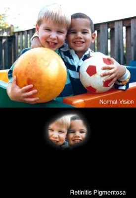
Gene Therapy
Top: Normal vision. Bottom: The same scene as it might be viewed by a person with retinitis pigmentosa. (Credit: National Eye Institute, National Institutes of Health)
"We hope the results of our study will be instrumental in generating a cure for the debilitating blindness associated with retinitis pigmentosa and other inherited and acquired retinal diseases," said Muna I. Naash, Ph.D., a researcher involved in the work from the Department of Cell Biology at the University of Oklahoma Health Sciences Center in Oklahoma City. "Compacted DNA nanoparticles are an exciting treatment strategy for these diseases and we look forward to exciting new developments."

To make this discovery, Naash and colleagues used groups of mice with the retinal degeneration slow (Rds) gene, which causes retinitis pigmentosa. The mice received one of three types of "treatments:" nanoparticles containing the normal copy of the Rds gene, the normal gene alone, or saline solution. After these treatments were delivered to the mice, the structure and function of the retina were analyzed by comparing them to untreated mice with retinitis pigmentosa and healthy mice with the normal Rds gene. Researchers also measured the level and pattern of Rds gene expression, as well as functional, structural and biochemical improvements in disease symptoms. They discovered that mice receiving the nanoparticle gene therapy show significant signs of healing. These mice had structural improvement in their retinas, as well as functional vision improvements, which lasted throughout the duration of the study. The mice that received the gene alone or saline continued to lose their vision. The nanoparticles were safe and well-tolerated with no adverse effects.

"Making the blind see was once called a miracle," said Gerald Weissmann, M.D., Editor-in-Chief of The FASEB Journal. "As we have expanded our understanding of evolution, genetics, and nanotechnology, chances are that "miraculous" cures will become as commonplace as those claimed by faith-healers past and present."

According to the National Institutes of Health Office of Rare Diseases Research, retinitis pigmentosa is a group of inherited eye diseases that affect the retina. Retinitis pigmentosa causes cells in the retina to die prematurely, eventually leading to vision loss. There is no cure.