BTemplates.com

Powered by Blogger.

Pageviews past week

Quantum mechanics

Auto News

artificial intelligence

About Me

Recommend us on Google!

Information Technology

Popular Posts

Showing posts with label Nature Medicine. Show all posts

Monday, June 6, 2011

Neuroscientists Map a New Target to Wipe Pain Away


Researchers at the Indiana University School of Medicine have discovered a peptide that short circuits a pathway for chronic pain. Unlike current treatments this peptide does not exhibit deleterious side effects such as reduced motor coordination, memory loss, or depression, according to an article in Nature Medicine posted online June 5, 2011.
A newly discovered peptide short circuits a 
pathway for chronic pain. (Credit: 
iStockphoto/Sebastian Meckelmann)

The peptide, CBD3, has been shown in mice to interfere with signals that navigate calcium channels to produce pain. Unlike other substances that block pain signals, CBD3 does not directly inhibit the influx of calcium. This is important as influx of calcium regulates heart rhythm and vital functions in other organs.

Rajesh Khanna, Ph.D., assistant professor of pharmacology and toxicology at the Indiana University School of Medicine, said the peptide discovered by him and his colleagues is potentially safer to use than addictive opioids or cone snail toxin Prialt® -- a recognized analgesic that is injected into the spinal column, both of which can cause respiratory distress, cardiac irregularities and other problems.

"After opioids-the gold standard for pain control -- the next target is calcium channels," said Dr. Khanna. "Along the pain pathway in the spinal cord, there are pain-sensing neurons called nociceptors that have an abundance of calcium channels."

Earlier international research has shown that the calcium channel is a key player within the pathway for pain signals. Based on work from Dr. Khanna's laboratory, it is also accepted that an axonal protein, CRMP-2, binds to the calcium channel "acting like a remote control" to modulate transmission of excitability and pain signals, Dr. Khanna explained.

He and his colleagues discovered the CBD3 peptide, a portion of the CRMP-2 protein, realizing that its smaller size would be beneficial in producing a synthetic version for drug development.


CBD3 can be given systemically and blocks pain in a variety of acute as well as chronic pain models, he said. The novel peptide binds to the calcium channel and reduces the number of excitability signals without disrupting the beneficial global calcium flow. Upon reaching the brain, these signals are interpreted as the sensation of pain.

"Since our approach does not directly inhibit calcium entry through voltage-gated channels, we expect that this molecule will be more specific and have fewer side effects than currently available analgesics," said Dr. Khanna. "We anticipate that this peptide will serve as a novel pharmacological therapeutic for the relief of chronic pain."

Dr. Khanna is a primary investigator in the Paul and Carole Stark Neurosciences Research Institute and the Indiana Spinal Cord and Brain Injury Research Group. His Stark Neuroscience Institute colleagues involved in the research are first author Joel M. Brittain and second author Sarah M. Wilson, both PhD students in his laboratory, and co-first-author Djane B. Duarte, Ph.D., a post-doctoral fellow. Members of the Harvard University Department of Anesthesiology also assisted with the research.

Funding for the research was provided in part by a American Heart Association National Scientist Development Grant, the Ralph W. and Grace M. Showalter Research Trust Fund and the Indiana Genomics Initiative.
Wednesday, June 16, 2010

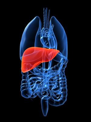
Discarded Livers: Replacement Organs?


A team led by researchers from the Center for Engineering in Medicine at Massachusetts General Hospital (MGH) has developed a technique that someday may allow growth of transplantable replacement livers. In a study appearing in Nature Medicine, the investigators describe using the structural tissue of rat livers as scaffolding for the growth of tissue regenerated from liver cells introduced through a novel reseeding process.
Image
Researchers have developed a technique that someday 
may allow growth of transplantable replacement livers. 
(Credit: iStockphoto/Sebastian Kaulitzki)

"Having the detailed microvasculature of the liver within a biocompatible, natural scaffold is a major advantage to growing liver tissue in a synthetic environment," says Basak Uygun, PhD, research associate at the MGH Center for Engineering in Medicine (MGH-CEM) and the paper's lead author. "Our technique of 'decellularizing' organs leaves the vascular system intact, which facilitates repopulation of the structural matrix and the subsequent survival and function of the introduced liver cells."

Liver transplantation is the only effective treatment for liver failure but is greatly limited by the shortage of donor organs. Each year 4,000 individuals who might have survived with a liver transplant die in the U.S. The shortage of donor livers and other organs is a major force behind the emerging field of tissue engineering and regenerative medicine. Efforts to build tissues from the ground up have not yet approached the goal of transplantable replacement organs, and replacing the liver -- in which each cell is a metabolic factory requiring constant, direct contact with the vascular system -- has been particularly challenging.

The current report describes a refinement of an approach to re-engineering replacement rat hearts that was reported in 2008 by University of Minnesota researchers. Since liver tissue is much more delicate than the muscular structure of the heart, the MGH-CEM team developed a gentler way of flushing living cells out of the liver's structural matrix, which is primarily made of connective tissue like collagen. After the cells were removed, the lobular structure of the liver and its extracellular matrix remained. Containing specific biochemical signals and cues that would direct liver cells to travel to the correct location and resume function -- something quite difficult to replicate using synthetic methods -- the matrix also maintained the organ's intricate network of blood vessels.

Another novel technique was used to reintroduce hepatocytes, the cells that carry out most of the liver's primary functions, into the decellularized matrix. The MGH-CEM approach actually caused cells to penetrate the vascular network and become embedded in the matrix, leaving major vessels clear to carry the essential blood supply. The repopulated matrix displayed normal liver function for up to 10 days in culture, and recellularized grafts were successfully connected to the circulation of live rats with minimal cellular damage and normal hepatocyte function.

"As far as we know, a transplantable liver graft has never been constructed in a laboratory setting before," explains Korkut Uygun, PhD, of the MGH-CEM, the paper's senior author. "Even though this is very exciting and promising, it is a proof-of-concept study only. Much more work will be required to make long-term functional liver grafts that can actually be transplanted into humans. We haven't been able to go beyond several hours in the rats, but it's a great start."

Martin Yarmush, MD, PhD, director of the MGH-CEM and a co-author of the Nature Medicine study, explains that the quarter of a million donor livers discarded each year because they are not suitable for transplantation would be an obvious source of supply for the creation of whole-organ scaffolds. "There is great potential for constructing full-fledged liver lobes containing animal or human cells, but several thorny issues must first be tackled, including formation of a layer of endothelial cells to line graft blood vessels," he says. "Given enough careful work, this approach could ultimately revolutionize tissue engineering and provide real working grafts for the liver and other complex tissues." Yarmush and Korkut Uygun both have faculty appointments at Harvard Medical School.

Additional co-authors of the Nature Medicine report are Alejandro Soto-Gutierrez, MD, PhD, Hiroshi Yagi, MD, Maria-Louisa Izamis, Maria Guzzardi, Carley Shulman, Jack Milwid, Arno Tilles, MD, Francois Berthiaume, PhD, and Yaahov Nahmias, PhD, MGH Center for Engineering in Medicine; Martin Hertl, MD, MGH Surgery; and Naoya Kobyashi, MD, PhD, Okayama University School of Medicine and Dentistry, Japan. The study was partially supported by grants from the National Institutes of Health, National Science Foundation and Shriners Hospitals for Children.
Enhanced by Zemanta