BTemplates.com

Powered by Blogger.

Pageviews past week

Quantum mechanics

Auto News

artificial intelligence

About Me

Recommend us on Google!

Information Technology

Popular Posts

Showing posts with label Gene. Show all posts

Thursday, July 7, 2011

The turn of the corkscrew : Structural analysis uncovers mechanisms of gene expression


The diverse functions of living cells are all based on the information encoded in the structure of the hereditary material DNA. Gene expression must therefore be tightly controlled, and this task is accomplished by the binding of regulatory proteins to, and their removal from, specific DNA sequences. One class of large molecular machines known as Swi2/Snf2 remodelers plays a central role in modulating these processes. However, until now, it was not clear how Swi2/Snf2 remodelers actually work. A team led by Professor Karl-Peter Hopfner at the Gene Center at Ludwig-Maximilians-Universität (LMU) in Munich has clarified the structure and function of the remodeler Mot1 (Modifier of Transcription 1), which binds directly to DNA. It turns out that Mot1 acts like a molecular corkscrew that migrates along the DNA, following its helical contour. During its progress, Mot1 displaces a crucial transcription factor called TBP (for “TATA Box Binding Protein”) from the DNA. Removal of TBP from a TATA box represses transcription of the adjacent gene, and the protein encoded by that gene is no longer synthesized. At the same time, TBP is stabilized and its binding specificity is changed, which facilitates the expression of genes that lack TATA boxes and code for other proteins. (Nature 6 July 2011)

The DNA in the cells of higher organisms is tightly wrapped around protein complexes called nucleosomes. This type of structural organization not only makes it possible to package the long DNA molecules in a highly compact form, it also provides the basis for the controlled expression of genetic information. Densely packed sections of the molecule are effectively in a repressed state, and genes located in these DNA segments cannot be transcribed. Activation of repressed genes depends on the intervention of complex molecular machines, so-called Swi2/Snf2 remodelers, which reorganize condensed stretches of DNA so as to make them accessible for transcription. The precise mode of action of remodelers has so far been unclear, mainly because most of them are made up of several components and the active complexes are difficult to study. This is why Hopfner chose to study Mot1, which is a comparatively simple representative of the family that functions as an Swi2/Snf2 remodeler on its own, and can serve as a guide to understanding the more complicated members of the class. Mot1 is known to participate in the control of gene expression, but how exactly it does so is not well understood.

The first stage of the process that leads to the synthesis of a given protein is the transcription of the specific segment of DNA that codes for it into molecules of messenger RNA. This initial step requires the action of so-called transcription factors. One of the most important of these is TBP, which binds preferentially to DNA sequences called TATA boxes that are located near the beginnings of many genes. Binding of TBP introduces a kink into the DNA, and this landmark serves as a platform for the binding of further proteins, ultimately leading to the assembly of the complex necessary for the initiation of transcription. Mot1 regulates transcription by actively removing TBP from the DNA, using ATP as a source of energy. “How Mot1 dissociates the TBP-DNA complex was completely unclear up to now,” says Hopfner. With the aid of so-called hybrid methods - in which data obtained from high-resolution X-ray diffraction analysis of the crystallized protein complex with images of the same molecular complex taken with the electron microscope were combined - Hopfner’s team was able to define the three-dimensional structure of the Mot1-TBP complex for the first time. This revealed how Mot1 recognizes the surface of the DNA-bound TBP. “Once Mot1 has recognized TBP, it binds to the adjacent DNA and begins to migrate along the DNA strand, using the energy released by the hydrolysis of ATP to power its movement. This helical movement, which is reminiscent of the insertion of a corkscrew, causes TBP to detach from the DNA,” explains Dr Petra Wollmann, who is first author on the new study. The researchers were surprised to find that Mot1 contains a strikingly extended loop. After TBP has dissociated from the DNA, this loop masks TBP’s DNA binding site and prevents the protein from reoccupying it.


Previous studies had reported what appeared to be paradoxical observations, which indicated that Mot1 inhibits transcription of TATA box-containing genes while facilitating the expression of genes that lack canonical TATA boxes. “Our results suggest that Mot1 also stabilizes the DNA-free conformation of TBP, increasing the probability that it can reach, bind to and activate genes that lack TATA boxes,” explains Hopfner. In other words, Mot1 is also a redistribution factor, which enables TBP to bind to different sequences and thus controls its association with other cellular components. This combination of detachment and redistribution functions may be a common feature of remodeling complexes, and would help to explain how they mediate the large-scale redistribution of DNA-binding regulatory proteins. (göd/PH)

The project was carried out under the auspices of two Clusters of Excellence - the Center for Integrated Protein Science Munich (CiPSM) and the Munich Centre for Advanced Photonics (MAP). The work was also supported by the German Research Foundation (DFG) as part of the Collaborate Research Centers (SFB) 646 and TR5, and by the LMUexcellent Investment Fund.

Friday, April 8, 2011

Coffee Drinking in Your Genes? Genetic Variants in Two Genes Linked With Caffeine Intake


Two genes in which variation affects intake of caffeine, the most widely consumed stimulant in the world, have been discovered. A team of investigators from the National Cancer Institute, Harvard School of Public Health, Brigham and Women's Hospital, and the University of North Carolina at Chapel Hill examined genetic variation across the entire genome of more than 47,000 individuals from the U.S., as described in the open-access journal PLoS Genetics.
Researchers have discovered two genes in which variation 
affects intake of caffeine, the most widely consumed stimulant
in the world. (Credit: iStockphoto/Sergey Galushko)

The genes identified were CYP1A2, which has previously been implicated in the metabolism of caffeine, and AHR, involved in the regulation of CYP1A2. Individuals with the highest-consumption genotype for either gene consumed ~40 mg more caffeine than those with the lowest-consumption genotype, equivalent to the amount of 1/3 cup of caffeinated coffee, or 1 can of cola.

Caffeine is implicated in numerous physiological and medical conditions; it affects sleep patterns, energy levels, mood, and mental and physical performance. The identification of genes that have an impact on daily consumption offers opportunities to better understand these conditions. Further exploration of the identified genetic variants may provide insight into the speed of caffeine metabolism, how long caffeine circulates in the blood, or how strong the physiological effects of consuming a given amount of caffeine are.

Apart from smoking, genetic determinants of lifestyle behaviors have generally not been consistently described. This study is among the first to examine the entire genome for a relationship between genetics and caffeine intake, a lifestyle behavior relevant to over 90% of U.S. adults. The study's success also suggests that additional genetic determinants of dietary and lifestyle behaviors may be identified in the future using a similar genome-based research strategy.


Wednesday, January 19, 2011

Birds of a feather? Your genes help you select friends


Vices attract while virtues repel people, according to a new study, which has found that genetic make-up plays a part in who one chooses as friends.

Researchers have found that people partly choose their friends by their genes especially those that have a big impact on their behaviour sometimes it can be a case of "opposites attract'' while sometimes "birds of a feather flock together". To research the conclusion, a team, led by James Fowler at University of California, used data from two large health studies to search for genetic links to friendship.
OF HUMAN BONDAGE

The researchers identified genetic markers, or genotypes, in six specific genes and looked at how often they occurred among friends. They found two clear examples where "birds of a feather flock together" and "opposites attract". The first involved a variant of the gene DRD2 which has been associated with alcoholism. People who carried the DRD2 genotype tended to befriend others with the same marker. Those who lacked the marker were also more likely to be friends with one another.

A less obvious opposite association was seen between people with a version of the gene CYP2A6linked to having an "open" personality. In this case, people with the genetic marker gravitated towards individuals who did not have it.

The findings, published in the 'Proceedings of the National Academy of Sciences' journal, remained significant after taking account of people's tendency to form "local" friendships within the same geographical area.

"An important implication of these results is that genetic structure in human populations may result not only from the formation of reproductive unions, but also from the formation of friendship unions within• a population," said the researchers in a statement on Tuesday.

Human evolution may to some extent have been shaped by interactions between genes and friendship choices." The human evolutionary environment is not limited to the physical and biological environment, but also includes the social environment, which may itself be an evolutionary force," they Said.

Tuesday, September 21, 2010

Study Reveals Structure of Cell Division’s Key Molecule At the Crossroads of Chromosomes


On average, one hundred billion cells in the human body divide over the course of a day. Most of the time the body gets it right but sometimes, problems in cell replication can lead to abnormalities in chromosomes resulting in many types of disorders, from cancer to Down Syndrome.
Human chromosome, with conventional nucleosomes 
containing the major form of the histones (green), and 
localization of the centromere histone H3 variant, 
CENP-A (red). (Credit: Ben E. Black, University 
of Pennsylvania School of Medicine)

Now, researchers at the University of Pennsylvania's School of Medicine have defined the structure of a key molecule that plays a central role in how DNA is duplicated and then moved correctly and equally into two daughter cells to produce two exact copies of the mother cell. Without this molecule, entire chromosomes could be lost during cell division.

Ben Black, PhD, assistant professor of Biochemistry and Biophysics, and Nikolina Sekulic, PhD, a postdoctoral fellow in the Black lab, report in the Sept. 16 issue of Nature the structure of the CENP-A molecule, which defines a part of the chromosome called the centromere. This is a constricted area to which specialized molecules called spindle fibers attach that help pull daughter cells apart during cell division.

"Our work gives us the first high-resolution view of the molecules that control genetic inheritance at cell division," says Black. "This is a big step forward in a puzzle that biologists have been chipping away at for over 150 years."

Investigators have known for the last 15 years that part of cell division is controlled by epigenetic processes, the series of actions that affect the protein spools around which DNA is tightly bound, rather than encoded in the DNA sequence itself. Those spools are built of histone proteins, and chemical changes to these spool proteins can either loosen or tighten their interaction with DNA. Epigenetics alter the readout of the genetic code, in some cases ramping a gene's expression up or down. In the case of the centromere, it marks the site where spindle fibers attach independently of the underlying DNA sequence. CENP-A has been suspected to be the key epigenetic marker protein.

However, what hasn't been known is how CENP-A epigenetically marks the centromere to direct inheritance. The Black team found the structural features that confer CENP-A the ability to mark centromere location on each chromosome. This is important because without CENP-A or the centromere mark it creates, the entire chromosome -- and all of the genes it houses -- are lost at cell division.

In this study, Black solved CENP-A's structure to determine how it specifically marks the centromere on each chromosome and surmise from that how the epigenetic mark is copied correctly in each cell division. They found that CENP-A changes the shape of the nucleosome of which it's a part, also making it more rigid than other nucleosomes without CENP-A. The nucleosome is the combination of DNA wound around a histone protein core --the DNA thread wrapped around the histone spool. The CENP-A nucleosome is copied several times to create a unique epigenetic area, different from the rest of the chromosome. CENP-A replaces histone H3 in the nucleosomes located at the centromere.

This CENP-A centromere identifier attracts other proteins, and in cell division builds a massive structure, the kinetochore, for pulling the duplicated chromosomes apart during cell division.

Besides the major advance in the understanding of the molecules driving human inheritance, this work also brings about the exciting prospect that the key epigenetic components are now in hand to engineer clinically useful artificial chromosomes that will be inherited alongside our own natural chromosomes -- and with the same high fidelity, says Black.

Co-authors are graduate student Emily A. Bassett and research specialist Danielle J. Rogers. The work was funded by National Institute for General Medical Sciences, the Burroughs Wellcome Fund, the Rita Allen Foundation, the American Cancer Society, and the American Heart Association.

Tuesday, September 14, 2010

Ancient Viral Invasion Shaped Human Genome


Scientists at the Genome Institute of Singapore (GIS), a biomedical research institute of the Agency for Science, Technology and Research (A*STAR), and their colleagues from the National University of Singapore, Nanyang Technological University, Duke-NUS Graduate Medical School and Princeton University have recently discovered that viruses that 'invaded' the human genome millions of years ago have changed the way genes get turned on and off in human embryonic stem (ES) cells.
Scientists have discovered that viruses that "invaded" 
the human genome millions of years ago have changed the 
way genes get turned on and off in human embryonic
stem cells. (Credit: iStockphoto/Martin McCarthy)

The study provides definitive proof of a theory that was first proposed in the 1950s by Nobel Laureate in physiology and medicine, Barbara McClintock, who hypothesized that transposable elements, mobile pieces of the genetic material (DNA), such as viral sequences, could be "control elements" that affect gene regulation once inserted in the genome.

This finding is an important contribution to the advancement of stem cell research and to its potential for regenerative medicine. Led by GIS Senior Group Leader Dr Guillaume Bourque, the study was published in Nature Genetics on June 6, 2010.

Through the use of new sequencing technologies, the scientists studied the genomic locations of three regulatory proteins (OCT4, NANOG and CTCF) in human and mouse embryonic stem (ES) cells. Interestingly, while the scientists found a lot of similarities, they also found many differences in the methods and the types of genes that are being regulated in humans. In particular, it was discovered that specific types of viruses that inserted themselves in the human genomes millions of years ago have dramatically changed the gene regulatory network in human stem cells.

"This study is a computational and experimental tour de force. It provides undeniable evidence that some transposable elements, which are too often dismissed as merely junk DNA, are key components of a regulatory code underlying human development," said Dr Cedric Feschotte, Associate Professor of the University of Texas Arlington.

The comparisons between the human and mouse model system in the study of gene regulatory networks help to advance the understanding of how stem cells differentiate into various cell types of the body. "This understanding is crucial in the improved development of regenerative medicine for diseases such as Parkinson's disease and leukaemia," said Dr Bourque. "Despite the advantages of using mouse ES cells in the study of gene regulatory networks, further research must focus more directly on human stem cells. This is due to the inherent challenges of converting the results of studies done from one species to that of the next. More research will need to be done in both human and non-human primate stem cells for findings on stem cells to be used in clinical application."

Prof Raymond L. White, PhD, Rudi Schmid Distinguished Professor of Neurology, University of California said, "The paper reports very exciting new findings that establish a new and fundamentally distinct mechanism for the regulation of gene expression. By comparing the genomes of mouse with human, the scientists were able to show that the binding sites for gene regulatory factors are very often not in the same place between the two species. This by itself would be very surprising, but the investigators go further and demonstrate that many of the sites are imbedded within a class of DNA sequences called "transposable" elements because of their ability to move to new places in the genome. There are a number of such elements believed to be the evolutionary remnants of viral genomes, but it was very surprising to learn that they were carrying binding sites for regulatory elements to new locations. These changes in regulation would be expected to create major changes in the organisms which carry them. Indeed, many think that regulatory changes are at the heart of speciation and may have played a large role in the evolution of humans from their predecessors. This is likely to be a landmark paper in the field."

Dr Eddy Rubin, Director of the U.S. Department of Energy Joint Genome Institute and Director of the Genomics Division at Lawrence Berkeley National Laboratory in Berkeley added, "This study using a comparative genomics strategy discovered important human specific properties of the regulatory network in human ES cells. This information is significant and should contribute to helping move the regenerative medicine field forward."

Saturday, September 4, 2010

Brainy Worms: Scientists Uncover Counterpart of Cerebral Cortex in Marine Worms


Our cerebral cortex, or pallium, is a big part of what makes us human: art, literature and science would not exist had this most fascinating part of our brain not emerged in some less intelligent ancestor in prehistoric times. But when did this occur and what were these ancestors? Unexpectedly, scientists at the European Molecular Biology Laboratory (EMBL) in Heidelberg, Germany, have now discovered a true counterpart of the cerebral cortex in an invertebrate, a marine worm.
A virtual Platynereis brain (left), created by averaging microscopy images of the brains of 36 different individuals, onto which scientists mapped gene activity (right). Perspective shows the brain as viewed from inside a Platynereis larvae, at 48 hours' old. (Credit: EMBL/R.Tomer)


Their findings are published in Cell, and give an idea of what the most ancient higher brain centres looked like, and what our distant ancestors used them for.

It has long been clear that, in evolutionary terms, we share our pallium with other vertebrates, but beyond that was mystery. This is because even invertebrates that are clearly related to us -- such as the fish-like amphioxus -- appear to have no similar brain structures, nothing that points to a shared evolutionary past. But EMBL scientists have now found brain structures related to the vertebrate pallium in a very distant cousin -- the marine ragworm Platynereis dumerilii, a relative of the earthworm -- which last shared an ancestor with us around 600 million years ago.

"Two stunning conclusions emerge from this finding," explains Detlev Arendt, who headed the study: "First, the pallium is much older than anyone would have assumed, probably as old as higher animals themselves. Second, we learn that it came out of 'the blue' -- as an adaptation to early marine life in Precambrian oceans."

To uncover the evolutionary origins of our brain, EMBL scientist Raju Tomer, who designed and conducted the work, took an unprecedentedly deep look at the regions of Platynereis dumerilii's brain responsible for processing olfactory information -- the mushroom-bodies. He developed a new technique, called cellular profiling by image registration (PrImR), which is the first to enable scientists to investigate a large number of genes in a compact brain and determine which are turned on simultaneously. This technique enabled Tomer to determine each cell's molecular fingerprint, defining cell types according to the genes they express, rather than just based on their shape and location as was done before.

"Comparing the molecular fingerprints of the developing ragworms' mushroom-bodies to existing information on the vertebrate pallium," Arendt says, " it became clear that they are too similar to be of independent origin and must share a common evolutionary precursor."

This ancestral structure was likely a group of densely packed cells, which received and processed information about smell and directly controlled locomotion. It may have enabled our ancestors crawling over the sea floor to identify food sources, move towards them, and integrate previous experiences into some sort of learning.

"Most people thought that invertebrate mushroom-bodies and vertebrate pallium had arisen independently during the course of evolution, but we have proven this was most probably not the case," says Tomer. Arendt concludes: "The evolutionary history of our cerebral cortex has to be rewritten."

Wednesday, August 18, 2010

Mitochondrial Eve: Mother of All Humans Lived 200,000 Years Ago


The most robust statistical examination to date of our species' genetic links to "mitochondrial Eve" -- the maternal ancestor of all living humans -- confirms that she lived about 200,000 years ago. The Rice University study was based on a side-by-side comparison of 10 human genetic models that each aim to determine when Eve lived using a very different set of assumptions about the way humans migrated, expanded and spread across Earth.
Image
Artist's cross section of a mitochondrion. (Credit: iStockphoto/David Marchal)

The research is available online in the journal Theoretical Population Biology.

"Our findings underscore the importance of taking into account the random nature of population processes like growth and extinction," said study co-author Marek Kimmel, professor of statistics at Rice. "Classical, deterministic models, including several that have previously been applied to the dating of mitochondrial Eve, do not fully account for these random processes."

The quest to date mitochondrial Eve (mtEve) is an example of the way scientists probe the genetic past to learn more about mutation, selection and other genetic processes that play key roles in disease.

"This is why we are interested in patterns of genetic variability in general," Kimmel said. "They are very important for medicine."

For example, the way scientists attempt to date mtEve relies on modern genetic techniques. Genetic profiles of random blood donors are compared, and based upon the likenesses and differences between particular genes, scientists can assign a number that describes the degree to which any two donors are related to one another.

Using mitochondrial genomes to gauge relatedness is a way for geneticists to simplify the task of finding common ancestors that lived long ago. That is because the entire human genome contains more than 20,000 genes, and comparing the differences among so many genes for distant relatives is problematic, even with today's largest and fastest supercomputers.

But mitochondria -- the tiny organelles that serve as energy factories inside all human cells -- have their own genome. Besides containing 37 genes that rarely change, they contain a "hypervariable" region, which changes fast enough to provide a molecular clock calibrated to times comparable to the age of modern humanity. Because each person's mitochondrial genome is inherited from his or her mother, all mitochondrial lineages are maternal.

To infer mtEve's age, scientists must convert the measures of relatedness between random blood donors into a measure of time.

"You have to translate the differences between gene sequences into how they evolved in time," said co-author Krzysztof Cyran, vice head of the Institute of Informatics at Silesian University of Technology in Gliwice, Poland. "And how they evolved in time depends upon the model of evolution that you use. So, for instance, what is the rate of genetic mutation, and is that rate of change uniform in time? And what about the process of random loss of genetic variants, which we call genetic drift?"

Within each model, the answers to these questions take the form of coefficients -- numeric constants that are plugged into the equation that returns the answer for when mtEve lived.

Each model has its own assumptions, and each assumption has mathematical implications. To further complicate matters, some of the assumptions are not valid for human populations. For example, some models assume that population size never changes. That is not true for humans, whose population has grown exponentially for at least several thousand generations. Other models assume perfect mixing of genes, meaning that any two humans anywhere in the world have an equal chance of producing offspring.

Cyran said human genetic models have become more complex over the past couple of decades as theorists have tried to correct for invalid assumptions. But some of the corrections -- like adding branching processes that attempt to capture the dynamics of population growth in early human migrations -- are extremely complex. Which raises the question of whether less complex models might do equally well in capturing what's occurring.

"We wanted to see how sensitive the estimates were to the assumptions of the models," Kimmel said. "We found that all of the models that accounted for random population size -- such as different branching processes -- gave similar estimates. This is reassuring, because it shows that refining the assumptions of the model, beyond a certain point, may not be that important in the big picture."

The research was supported by grants from the Polish Ministry of Science and Higher Education and the Cancer Prevention and Research Institute of Texas. It has resulted from a standing collaboration between Rice University and Silesian University of Technology.

Monday, August 9, 2010

Protein That Shuttles RNA Into Cell Mitochondria Discovered


Researchers at UCLA's Jonsson Comprehensive Cancer Center and the departments of Chemistry and Biochemistry and Pathology and Laboratory Medicine have uncovered a role for an essential cell protein in shuttling RNA into the mitochondria, the energy-producing "power plant" of the cell.
Image
Researchers at UCLA's Jonsson Comprehensive Cancer Center and the departments of Chemistry and Biochemistry and Pathology and Laboratory Medicine have uncovered a role for an essential cell protein in shuttling RNA into the mitochondria, the energy-producing "power plant" of the cell. (Credit: Maureen Heaster)

The import of nucleus-encoded small RNAs into mitochondria is essential for the replication, transcription and translation of the mitochondrial genome, but the mechanisms that deliver RNA into mitochondria remain poorly understood.

In the current study, UCLA scientists show a new role for a protein called polynucleotide phosphorylase (PNPASE) in regulating the import of RNA into mitochondria. Reducing the expression of PNPASE decreased RNA import, which impaired the processing of mitochondrial genome-encoded RNAs. Reduced RNA processing inhibited the translation of proteins required to maintain the electron transport chain that handles oxygen to produce energy in the form of adenosine triphosphate, the energy currency of a cell. With reduced PNPASE, unprocessed mitochondrial RNAs accumulated, protein translation was inhibited and energy production was compromised, leading to stalled cell growth.

The study appears Aug. 5, 2010, in the peer-reviewed journal Cell.

"This discovery tells us that PNPASE regulates the energy producing function of mitochondria by mediating cytoplasmic RNA import," said Dr. Michael Teitell, a professor of pathology and laboratory medicine, a Jonsson Cancer Center researcher and co-senior author of the study. "The study yields new insight for how cells function at a very fundamental level. This information provides a potential new pathway to control mitochondrial energy production and possibly impact the growth of cells, including certain types of cancer cells."

Mitochondria are described as cellular power plants because they generate most of the energy supply of the cell. In addition to supplying energy, mitochondria also are involved in a broad range of other cellular processes, such as signaling, differentiation, death, control of the cell cycle and growth.

The study could have implications for studying and treating certain cancers, which rely on cellular energy to grow and spread, as well as mitochondrial disorders such as neuromuscular diseases. The study could also result in new ways to think about attacking neurodegenerative disorders, such as Parkinson and Alzheimer diseases, which have recently been linked to the function of mitochondria.

"When we're talking about looking for ways to cure cancer, we fundamentally need to understand what makes cells grow and die and the mitochondrion is right at the heart of these issues," said Carla Koehler, a professor of chemistry and biochemistry, Jonsson Cancer Center researcher and co-senior author of the study. "This new and novel pathway for transporting RNA into the mitochondria is shedding new light on the evolving role and importance of mitochondria function in normal physiology and a wide variety of diseases. If we can understand how this pathway functions in healthy cells we could potentially uncover defects that help in transforming normal cells into cancer cells."

PNPASE was identified in 2004 by Teitell and his team as they attempted to find proteins that interact with TCL1, a human lymphoma-promoting cancer gene that has been used to generate genetic models of lymphocyte cancer. Mass spectrometry uncovered PNPASE, which had a signature sequence that suggested that it trafficked into and localized within the mitochondria of cells.

Once localized, Teitell, Koehler and post-doctoral fellow Geng Wang turned their attention to the function of PNPASE, which generated the unexpected results reported in this study. Prior to their discovery, it was not known what pathway was used to get RNA into the mitochondria. PNPASE mediates the movement of RNA from the cell cytoplasm, the area of the cell enclosed by the cell membrane, into the matrix of mitochondria, where the mitochondrial genome is located. The protein acts as receptor and binds to cytoplasmic RNAs that have a particular stem-loop signature sequence, mediating import, Teitell said.

Without this RNA import, the cell lacks the machinery to assemble the mitochondria's energy source, Koehler said.

"The cell would lose most of its ability to make energy," she said. "It would be crippled. Mitochondria are fantastically complex and our study reveals another cellular pathway in which these tiny but important powerhouses participate in essential cell activities, such as the generation of energy essential for life."

The study was funded by the National Institutes of Health, the California Institute for Regenerative Medicine, the American Heart Association, the Leukemia & Lymphoma Society and a NIH Nanomedicine Roadmap Grant.

Saturday, July 17, 2010

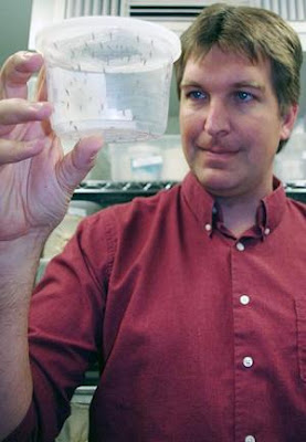
First Malaria-Proof Mosquito: Genetic Manipulation Renders Them Completely Immune to the Parasite


For years, researchers worldwide have attempted to create genetically altered mosquitoes that cannot infect humans with malaria. Those efforts fell short because the mosquitoes still were capable of transmitting the disease-causing pathogen, only in lower numbers.
Image
Michael Riehle, holding genetically altered 
mosquitoes, and his team work in a highly secure 
lab environment to prevent genetically altered 
mosquitoes from escaping. 
(Credit: Riehle Lab, University of Arizona)

Now for the first time, University of Arizona entomologists have succeeded in genetically altering mosquitoes in a way that renders them completely immune to the parasite, a single-celled organism called Plasmodium. Someday researchers hope to replace wild mosquitoes with lab-bred populations unable to act as vectors, i.e. transmit the malaria-causing parasite.

"If you want to effectively stop the spreading of the malaria parasite, you need mosquitoes that are no less than 100 percent resistant to it. If a single parasite slips through and infects a human, the whole approach will be doomed to fail," said Michael Riehle, who led the research effort, the results of which will be published July 15 in the journal Public Library of Science Pathogens. Riehle is a professor of entomology in the UA's College of Agriculture and Life Sciences and is a member of the BIO5 Institute.

Riehle's team used molecular biology techniques to design a piece of genetic information capable of inserting itself into a mosquito's genome. This construct was then injected into the eggs of the mosquitoes. The emerging generation carries the altered genetic information and passes it on to future generations. For their experiments, the scientists used Anopheles stephensi, a mosquito species that is an important malaria vector throughout the Indian subcontinent.

The researchers targeted one of the many biochemical pathways inside the mosquito's cells. Specifically, they engineered a piece of genetic code acting as a molecular switch in the complex control of metabolic functions inside the cell. The genetic construct acts like a switch that is always set to "on," leading to the permanent activity of a signaling enzyme called Akt. Akt functions as a messenger molecule in several metabolic functions, including larval development, immune response and lifespan.

When Riehle and his co-workers studied the genetically modified mosquitoes after feeding them malaria-infested blood, they noticed that the Plasmodium parasites did not infect a single study animal.

"We were surprised how well this works," said Riehle. "We were just hoping to see some effect on the mosquitoes' growth rate, lifespan or their susceptibility to the parasite, but it was great to see that our construct blocked the infection process completely."

Of the estimated 250 million people who contract malaria each year, 1 million -- mostly children -- do not survive. Ninety percent of the number of fatalities, which Riehle suspects to be underreported, occur in Sub-Saharan Africa.

Each new malaria case starts with a bite from a vector -- a mosquito belonging to the genus Anopheles. About 25 species of Anopheles are significant vectors of the disease.

Only the female Anopheles mosquitoes feed on blood, which they need to produce eggs. When they bite an infected human or animal, they ingest the malaria parasite.

Once the Plasmodium cells find themselves in the insect's midgut, they spring into action. They leave the insect's digestive tract by squeezing through the midgut lining. The vast majority of Plasmodium cells do not survive this journey and are eliminated by the mosquito's immune cells. A tiny fraction of parasite cells, usually not more than a handful, make it and attach themselves on the outside of the midgut wall where they develop into brooding cells called oocysts.

Within 10-12 days, thousands of new Plasmodium cells, so-called sporozoites, sprout inside the oocyst. After hatching from the oocyst, the sporozoites make their way into the insect's salivary glands where they lie in wait until the mosquito finds a victim for a blood meal. When the mosquito bites, some sporozoites are flushed into the victim's bloodstream.

"The average mosquito transmits about 40 sporozoites when it bites," said Riehle, "but it takes only one to infect a human and make a new malaria victim."

Several species of Plasmodium exist in different parts of the world, all of which are microscopically small single-celled organisms that live in their hosts' red blood cells. Each time the parasites undergo a round of multiplication, their host cells burst and release the progeny into the bloodstream, causing the painful bouts of fever that malaria is known and feared for.

Malaria killed more soldiers in the Civil War than the fighting, according to Riehle. In fact, malaria was prevalent in most parts of the U.S. until the late 1940s and early 1950, when DDT spraying campaigns wiped the vectors off the map. Today, a new case of malaria occurs in the U.S. only on rare occasions.

The severity of the disease depends very largely on the species of the Plasmodium parasite the patient happens to contract.

"Only two species of Plasmodium cause the dreaded relapses of the disease," said Riehle. "One of them, Plasmodium vivax, can lie dormant in the liver for 10 to 15 years, but now drugs have become available that target the parasites in the liver as well as those in the blood cells."

That said, there are no effective or approved malaria vaccines. A few vaccine candidates have gone to clinical trials but they were shown to either be ineffective or provide only short-term protection. If an effective vaccine were to be developed, distribution would be a major problem, Riehle said.

Researchers and health officials put higher hopes into eradication programs, which aim at the disease-transmitting mosquitoes rather than the pathogens that cause it.

"The question is 'What can we do to turn a good vector into a bad vector?'" Riehle said.

"The eradication scenario requires three things: A gene that disrupts the development of the parasite inside the mosquito, a genetic technique to bring that gene into the mosquito genome and a mechanism that gives the modified mosquito an edge over the natural populations so they can displace them over time."

"The third requirement is going to be the most difficult of the three to realize," he added, which is why his team decided to tackle the other two first.

"It was known that the Akt enzyme is involved in the mosquito's growth rate and immune response, among other things," Riehle said. "So we went ahead with this genetic construct to see if we can ramp up Akt function and help the insects' immune system fight off the malaria parasite."

The second rationale behind this approach was to use Akt signaling to stunt the mosquitoes' growth and cut down on its lifespan.

"In the wild, a mosquito lives for an average of two weeks," Riehle explained. "Only the oldest mosquitoes are able to transmit the parasite. If we can reduce the lifespan of the mosquitoes, we can reduce the number of infections."

His research team discovered that mosquitoes carrying two copies of the altered gene had lost their ability to act as malaria vectors altogether.

"In that group of mosquitoes, not a single Plasmodium oocyst managed to form."

At this point, the modified mosquitoes exist in a highly secured lab environment with no chance of escape. Once researchers find a way to replace wild mosquito populations with lab-bred ones, breakthroughs like the one achieved by Riehle's group could pave the way toward a world in which malaria is all but history.

This study was funded by the National Institutes of Health.

Saturday, July 3, 2010

Scientists Find Direct Line from Development to Growth


It may seem intuitive that growth and development somehow go together so that plants and animals end up with the right number of cells in all the right places. But it is only now that scientists at the Duke Institute for Genome Sciences & Policy have gotten some of the first insights into how this critical coordination actually works in a plant.
Arabidopsis thaliana.
Arabidopsis thaliana. (Credit: iStockphoto)

The answer is surprisingly simple.

A well-known developmental protein called Short-root has been found to directly control the activity, in both time and space, of other well-known genes involved in cell division.

"It's a remarkably straightforward answer," said Philip Benfey, director of the IGSP's Center for Systems Biology. "Considering the level of complexity that is so often found in biology, this is simplicity itself."

The researchers report their findings on July 1 in the journal Nature.

Benfey's group and others have studied the molecular-level events that determine what particular cells in Arabidopsis plants will become in considerable detail. Those events involve genetic partners Short-root and Scarecrow along with a couple of microRNAs.

Researchers also have a pretty good understanding of the intrinsic factors that allow cells to go through their cycle and divide into two daughter cells. "What was missing was a connection between the two," said Rosangela Sozzani, a postdoctoral researcher in Benfey's lab and first author of the new study.

To shed light on that connection, Sozzani and her collaborators combined a number of experimental techniques and technologies to produce a dynamic, genome-wide view of the genetic events that Short-root and its partner Scarecrow set into motion within a single type of cell. At the very same time that cells divide, Short-root and Scarecrow switch on the gene cyclin D6, they report. Cyclin D6 is one of a family of genes that govern cell growth and division.

Benfey says the discovery in plants has immediate practical relevance given the central role of plants to human life, in the form of "food, feed, fuel and fiber." It's also likely that the "logic" behind plants' growth and development will carry over to other species, perhaps even our own. In fact, he and Sozzani note, animals including humans have cyclin D6 too.

"It's not just molecules," Benfey said. "There are evolutionary relationships. Once these fundamental processes got worked out, they are likely to have been kept around."

Jim Murray from the University of Cardiff was a key collaborator on the study. The work was funded by grants from the National Institutes of Health and the National Science Foundation.

Thursday, June 10, 2010

Genetic 'Parts' List Now Available for Hypothalamus


A Johns Hopkins and Japanese research team has generated the first comprehensive genetic "parts" list of a mouse hypothalamus, an enigmatic region of the brain -- roughly cherry-sized, in humans -- that controls hunger, thirst, fatigue, body temperature, wake-sleep cycles and links the central nervous system to control of hormone levels.
Image
A. A diagram of the developing mouse brain at 12 
days gestation. The hypothalamus is outlined in 
red, while cells that express the Shh gene, which 
was used as a landmark in this study, are shown in 
brown. B. Various different genes that are turned on 
in different parts of the hypothalamus are shown in 
purple, while Shh is shown in brown. (Credit: Image 
courtesy of Johns Hopkins Medical Institutions)

Flaws in hypothalamus development may underlie both inborn and acquired metabolic balance problems that can lead to obesity, diabetes, mood disorders and high blood pressure, according to a report on the study published May 2 in the advance online publication of Nature Neuroscience.

"Knowing how cells develop in this part of the brain will help us understand how they regulate behavior, mood and metabolism," says Seth Blackshaw, Ph.D., an assistant professor in the Solomon H. Snyder Department of Neuroscience, Johns Hopkins University School of Medicine.

The hypothalamus is one of the most diverse and complex parts of the brain, and having an index of the genes involved in producing its many cell types is a toolbox that researchers can use to manipulate the activity of brain cells by turning them on and off, or tracing their connections. This may ultimately lead to better diagnostic and treatment options for a variety of disorders.

"The study of hypothalamic development, particularly of cell specification, will help us to understand how hypothalamic neurons function to regulate behavior and physiology," says Blackshaw. "Because of when and where we saw certain genes turn on, we now have identified a set of candidate players that guide the assembly of the different parts of the hypothalamus and that specify the many individual cell types within it."

The hypothalamus is composed of at least dozens of types of neurons -- and more likely hundreds -- each of which corresponds to a gene that has remained unidentified until now. Its cellular arrangement is more akin to a bowl of spaghetti than a neatly organized club sandwich, according to Blackshaw. The catalog of molecular markers identified here helps unravel this complexity.

The team's first challenge was to dissect away, at the very start of neural development, the part of the mouse brain which develops into the hypothalamus, and then cut tiny slices of this region for use in microarray analysis, a technology that reveals multiple gene activity. By analyzing all the roughly 20,000 genes in the mouse genome, the team identified 1200 as strongly activated in developing hypothalamus and characterized the cells within the hypothalamus in which they were activated. The team then characterized the expression of the most interesting 350 genes in detail using another gene called Shh, for sonic hedgehog, as a landmark to identify the precise region of the hypothalamus in which these genes were turned on. This involved processing close to 20,000 tissue sections -- painstakingly sliced at one-fiftieth of a millimeter thickness and then individually examined.

"We were able to use this data to find genes whose expression matched every individual hypothalamic nucleus and essentially assemble a jigsaw puzzle of gene expression patterns that completely covered the developing hypothalamus," Blackshaw says. "Now that we have a complete set of molecular landmarks, along with an extensive molecular parts list, we can begin to learn how all these parts fit together to create this essential and highly complex brain region."

Authors of the paper, in addition to Blackshaw, are Daniel A. Lee, Ana Miranda-Angulo, Yangqin Yang, Aya C. Yoshida, Hong Wang, Hiromi Mashiko, Lizhi Jiang, Marina Avetisyan, Lixin Qi, and Jiang Qian, all of Johns Hopkins; Ayane Kataoka and Tomomi Shimogori of RIKEN-BSI, 2-1 Hirosawa, Wako-shi, Saitama, Japan.

This research was supported by March of Dimes, the Klingenstein Fund, the W.M. Keck Foundation, and the Japan Society for Promotion of Science.

Monday, April 12, 2010

Gene That Changes the Brain’s Response to Stress Identified


Stress can literally warp your brain, reshaping some brain structures that help cope with life's pressures. In the short term, the stress response can be helpful -- i.e., fight or flight -- but over time it leads to a wear and tear that can cause disease in both the brain and other parts of the body. Digging deeper into what underlies these potentially harmful changes, new research has identified a key protein involved in remodeling the brain under stress. Experiments have found that the brains of mice with an inadequate amount of this protein, called brain-derived neurotrophic factor (BDNF), look similar to those of normal mice that have been under stress for long periods.
BDNF
Reduced reach. Tracings of neurons from mice 
with different levels of BDNF show that a short 
supply of the protein causes relatively shrunken 
neurons (right) in some parts of the hippocampus. 
(Credit: Image courtesy of Rockefeller University)

The experiments homed in on the gene for a protein that, among other things, enhances the adaptability of neurons in the hippocampus, a brain region that plays a key role in mood, cognition and memory. When normal mice are exposed to chronic stress (simulated by confinement in a wire mesh restraint), there is a significant retraction in the projections, or dendrites, of some of the neurons in the hippocampus, which shrinks in overall volume as well. The new experiments, reported recently in Hippocampus, looked at mice that had only one instead of the usual two copies of the gene that produces BDNF. The researchers, from Rockefeller University and Weill Cornell Medical College, found that these mice had brains resembling those of normal mice after extended stress. In other words, stress did not have any effect on the experimental mice.

"The findings suggest that BDNF is one of the proteins that play a role in mediating the brain's plasticity," says Bruce S. McEwen, head of Rockefeller's Harold and Margaret Milliken Hatch Laboratory of Neuroendocrinology.

If researchers can find a way to deplete or supplement BDNF in adult mice, they may be able to answer the question of when in development, or even in adult life, it has the greatest impact. "What we're seeing is that there may be a developmental window for BDNF's role, or also that there may be a floor and a ceiling for the right amount of the protein that helps enable adaptive plasticity," McEwen says.

The new work adds to the understanding of BDNF's interaction with hormones in the brain. McEwen's lab recently discovered that a variant of the BDNF gene is a likely contender for a role in premenstrual disorders, changing mice's performance on certain memory tasks according to their stage of the estrous cycle.
Reblog this post [with Zemanta]

Saturday, March 20, 2010

What Makes You Unique? Not Genes So Much as Surrounding Sequences, Study Finds


The key to human individuality may lie not in our genes, but in the sequences that surround and control them, according to new research by scientists at the Stanford University School of Medicine and Yale University. The interaction of those sequences with a class of key proteins, called transcription factors, can vary significantly between two people and are likely to affect our appearance, our development and even our predisposition to certain diseases, the study found.

Me
Researchers have found that the unique, specific changes among individuals in the sequence of DNA affect the ability of "control proteins" called transcription factors to bind to the regions that control gene expression. (Credit: iStockphoto/Andrey Prokhorov)

The discovery suggests that researchers focusing exclusively on genes to learn what makes people different from one another have been looking in the wrong place.

"We are rapidly entering a time when nearly anyone can have his or her genome sequenced," said Michael Snyder, PhD, professor and chair of genetics at Stanford. "However, the bulk of the differences among individuals are not found in the genes themselves, but in regions we know relatively little about. Now we see that these differences profoundly impact protein binding and gene expression."

Snyder is the senior author of two papers -- one in Science Express and one in Nature -- exploring these protein-binding differences in humans, chimpanzees and yeast. Snyder, the Stanford W. Ascherman, MD, FACS, Professor in Genetics, came to Stanford in July 2009 from Yale, where much of the work was conducted.

Genes, which carry the specific instructions necessary to make proteins do the work of the cell, vary by only about 0.025 percent across all humans. Scientists have spent decades trying to understand how these tiny differences affect who we are and what we become. In contrast, non-coding regions of the genome, which account for approximately 98 percent of our DNA, vary in their sequence by about 1 to 4 percent. But until recently, scientists had little, if any, idea what these regions do and how they contribute to the "special sauce" that makes me, me, and you, you.

Now Snyder and his colleagues have found that the unique, specific changes among individuals in the sequence of DNA affect the ability of "control proteins" called transcription factors to bind to the regions that control gene expression. As a result, the subsequent expression of nearby genes can vary significantly.

"People have done a lot of work over the years to characterize differences in gene expression among individuals," said Snyder. "We're the first to look at differences in transcription-factor binding from person to person." What's more, by selectively breeding, or crossing, yeast strains, Snyder and his colleagues found that many, but not all, of these differences in binding and expression levels are heritable.

In the Science Express paper, which will be published online March 18, Snyder and his colleagues compared the binding patterns of two transcription factors in 10 people and one chimpanzee. They identified more than 15,000 binding sites across the genome for the transcription factor called NF-kB and more than 19,000 sites for another factor called RNA PolII. They then looked to see if every site was bound equally strongly by the proteins, or if there were variations among individuals.

They found that about 25 percent of the PolII sites and 7.5 percent of the NF-kB sites exhibited significant binding differences among individuals -- in some cases greater than two orders of magnitude from one person to another. (For comparison, the binding differences between the humans and the chimpanzee were about 32 percent.) Many of these binding differences could be traced to differences in sequences or structure in the protein binding sites, and several were directly correlated to changes in gene expression levels.

"These binding regions, or chunks, vary among individuals," said Snyder, "and they have a profound impact on gene expression." In particular, the researchers found that several of the variable binding regions were near genes involved in such diseases as type-1 diabetes, lupus, leukemia and schizophrenia.

The researchers confirmed and extended their findings in the Nature paper, which will be published online March 17. In this study, they used yeast to determine that many of the binding differences and variations in gene expression levels in individuals are passed from parent to progeny, and they identify several control proteins that vary -- a study that would have been impossible to perform in humans.

"We conducted the two studies in parallel," said Snyder, "and found the same thing. Many of the binding sites differed. When we mapped the areas of difference, we found that they were associated with key regulators of variation in the population. Together these two studies tell us a lot about the so-called regulatory code that controls variation among individuals."

The research in the Science Express study was supported by the National Institutes of Health, the European Molecular Biology Laboratory and the Howard Hughes Medical Institute's Medical Fellows Program. The research in the Nature study was supported by the National Institutes of Health. In addition to Snyder, other Stanford researchers involved in the two studies include postdoctoral scholars Fabian Grubert, PhD; Minyi Shi, PhD; and Manoj Hariharan, PhD; and graduate student Konrad Karczewski.


Reblog this post [with Zemanta]