BTemplates.com

Powered by Blogger.

Pageviews past week

Quantum mechanics

Auto News

artificial intelligence

About Me

Recommend us on Google!

Information Technology

Popular Posts

Tuesday, January 11, 2011

Why it's not good to forgive partner for all offences


Everyone may advice you to forgive your partner's bad behaviour, but in some cases, it may be better to hold a grudge against your transgressing spouse, a new study has suggested.
Newlyweds who forgive their partner's bad 
behaviour are more likely to face additional 
bad behavior next day, says a study

Newlyweds who forgave their partner's bad behaviour were more likely to face additional bad behaviour the next day compared with those who stayed mad, a study of newlyweds the University of Tennessee. Hearing "it's okay, honey," may be just the fuel the transgressing spouse needs for more lapses of judgement, study author James McNulty said, adding the benefits of forgiveness may need to be weighed against the risks.

"You may feel better if you forgive me," McNulty told Live Science. "But the question is, what happens down the road?" So it's likely that the unforgiven spouses behaved better in an attempt to get out of the dog house, he said.

For his research McNulty asked 135 newlywed couples to fill out individual relationship diaries every day for a week. The diaries included a questionnaire about whether the person's spouse had done something to upset them, and whether they'd forgiven their spouse for the transgression.

Overall, spouses who forgave their partners were almost twice as likely to report that their partner misbehaved the next day as those who held a grudge, McNulty found. However, in a second study that has not yet published, McNulty followed couples for four years and the results showed a similar pattern. However, McNulty said the fmdings don't suggest that forgiveness is always bad, nor is it a given that forgiving someone will turn you into a doormat.


Monday, January 10, 2011

Emotional Signals Are Chemically Encoded in Tears, Researchers Find


Emotional crying is a universal, uniquely human behavior. When we cry, we clearly send all sorts of emotional signals. In a paper published online January 6 in Science Express, scientists at the Weizmann Institute have demonstrated that some of these signals are chemically encoded in the tears themselves. Specifically, they found that merely sniffing a woman's tears -- even when the crying woman is not present -- reduces sexual arousal in men.
Scientists have found that certain emotional signals are chemically encoded in tears. (Credit: iStockphoto/Arpad Nagy-Bagoly)

Humans, like most animals, expel various compounds in body fluids that give off subtle messages to other members of the species. A number of studies in recent years, for instance, have found that substances in human sweat can carry a surprising range of emotional and other signals to those who smell them.

But tears are odorless. In fact, in a first experiment led by Shani Gelstein, Yaara Yeshurun and their colleagues in the lab of Prof. Noam Sobel in the Weizmann Institute's Neurobiology Department, the researchers first obtained emotional tears from female volunteers watching sad movies in a secluded room and then tested whether men could discriminate the smell of these tears from that of saline. The men could not.

In a second experiment, male volunteers sniffed either tears or a control saline solution, and then had these applied under their nostrils on a pad while they made various judgments regarding images of women's faces on a computer screen. The next day, the test was repeated -- the men who were previously exposed to tears getting saline and vice versa. The tests were double blinded, meaning neither the men nor the researchers performing the trials knew what was on the pads. The researchers found that sniffing tears did not influence the men's estimates of sadness or empathy expressed in the faces. To their surprise, however, sniffing tears negatively affected the sex appeal attributed to the faces.

To further explore the finding, male volunteers watched emotional movies after similarly sniffing tears or saline. Throughout the movies, participants were asked to provide self-ratings of mood as they were being monitored for such physiological measures of arousal as skin temperature, heart rate, etc. Self-ratings showed that the subjects' emotional responses to sad movies were no more negative when exposed to women's tears, and the men "smelling" tears showed no more empathy. They did, however, rate their sexual arousal a bit lower. The physiological measures, however, told a clearer story. These revealed a pronounced tear-induced drop in physiological measures of arousal, including a significant dip in testosterone -- a hormone related to sexual arousal.

Finally, in a fourth trial, Sobel and his team repeated the previous experiment within an fMRI machine that allowed them to measure brain activity. The scans revealed a significant reduction in activity levels in brain areas associated with sexual arousal after the subjects had sniffed tears.

Sobel said, "This study raises many interesting questions. What is the chemical involved? Do different kinds of emotional situations send different tear-encoded signals? Are women's tears different from, say, men's tears? Children's tears? This study reinforces the idea that human chemical signals -- even ones we're not conscious of -- affect the behavior of others."

Human emotional crying was especially puzzling to Charles Darwin, who identified functional antecedents to most emotional displays -- for example, the tightening of the mouth in disgust, which he thought originated as a response to tasting spoiled food. But the original purpose of emotional tears eluded him. The current study has offered an answer to this riddle: Tears may serve as a chemosignal. Sobel points out that some rodent tears are known to contain such chemical signals. "The uniquely human behavior of emotional tearing may not be so uniquely human after all," he says.

The work was authored by Shani Gelstein, Yaara Yeshurun, Liron Rozenkrantz, Sagit Shushan, Idan Frumin, Yehudah Roth and Noam Sobel, was conducted in collaboration with the Edith Wolfson Medical Center, Holon.

Prof. Noam Sobel's research is supported by the James S. McDonnell Foundation 21st Century Science Scholar in Understanding Human Cognition Program; the Minerva Foundation; the European Research Council; and Regina Wachter, NY.

Wednesday, January 5, 2011

Resurrecting the So-Called 'Depression Gene'::Genes May Play Role in Response to Adversity


University of Michigan Health System researchers have found new evidence that our genes help determine our susceptibility to depression.
Our genes help determine our susceptibility to depression, 
new research suggests. (Credit: iStockphoto/
Nicholas Belton)

Their findings, published online in the Archives of General Psychiatry, challenge a 2009 study that called the genetic link into question and add new support to earlier research hailed as a medical breakthrough.

In the summer of 2003, scientists announced they had discovered a connection between a gene that regulates the neurotransmitter serotonin and an individual's ability to rebound from serious emotional trauma, such as childhood physical or sexual abuse.

The journal Science ranked the findings among the top discoveries of the year and the director of the National Institute of Mental Health proclaimed, "It is a very important discovery and a real advance for the field."

That excitement was dampened in 2009, however, after the research was called into question by a study published in the Journal of the American Medical Association. The New York Times reported that analysis, which examined results from 14 different studies, showed the initial findings had "not held up to scientific scrutiny."

Srijan Sen, M.D., Ph.D, an assistant professor of psychiatry at the University of Michigan Medical School, and his colleagues are presenting a new, broader analysis of the follow-up studies to date. The U-M team examined 54 studies dating from 2001 to 2010 and encompassing nearly 41,000 participants -- making it the largest analysis of the serotonin gene's relationship to depression.

"When we included all the relevant studies, we found that an individual's genetic make-up does make a difference in how he or she responds to stress," says Sen.

The U-M analysis supports previous findings that individuals who had a short allele on a particular area the serotonin gene had a harder time bouncing back from trauma than those with long alleles.

Rudolf Uher, Ph.D., a clinical lecturer at the Institute of Psychiatry in London, says the U-M research will help cut through the debate about the genetic connection and refocus the field on making new advances to help those affected by mental illness.

"The major strength of the analysis is that it is the first such work that included all studies that were available on the topic," Uher says. "And it gives a very clear answer: the 'short' variant of the serotonin transporter does make people more sensitive to the effects of adversity."

The authors of the initial study from 2003 were also excited by the U-M team's results.

"Their careful and systematic approach reveals why the JAMA meta-analysis got it wrong," says Terrie Moffitt, Ph.D., a professor at Duke University and one of the authors of the 2003 study. "We hope that the same journalists who were so hasty to publish a simplistic claim in 2009 will cover this more thoughtful new analysis."

When the U-M team restricted their analysis to the 14 studies included in the 2009 JAMA paper, they also failed to find a genetic link, suggesting to Sen that the scope of the analysis, not the methodology, was responsible for the new findings.

The U-M analysis found robust support for the link between sensitivity to stress and a short allele in those who had been mistreated as children and in people suffering with specific, severe medical conditions. Only a marginal relationship was found in those who had undergone stressful life events.

But that's also common sense. Different stressful life events may have very different effects, Sen says. For instance, there is no reason to think that the effects of divorce, at a biological level, would be similar to the effects of losing your home or being physically assaulted.

Still, the study results don't mean that everyone should run out and get a genetic test; additional susceptibility from having a short allele is only one factor among many that determine how an individual responds to stress, Sen says.

Additional research will help to map an individual's genetic profile for depression.

"This brings us one step closer to being able to identify individuals who might benefit from early interventions or to tailor treatments to specific individuals," Sen says.

Funding: The research was supported by grants from the National Institutes of Health, University of Michigan Depression Center and Studienstiftung des Deutschen Volkes.

Additional U-M Authors: Margit Burmeister, Ph.D., Kerby Shedden, Ph.D., former graduate student Katja Karg

Sunday, January 2, 2011

Your Genome in Minutes: New Technology Could Slash Sequencing Time


Scientists from Imperial College London are developing technology that could ultimately sequence a person's genome in mere minutes, at a fraction of the cost of current commercial techniques.
Dr Joshua Edel shows the prototype chip,
and an array of the chips prior to use.
(Credit: Image courtesy of Imperial College London)

The researchers have patented an early prototype technology that they believe could lead to an ultrafast commercial DNA sequencing tool within ten years. Their work is described in a study published this month in the journal Nano Letters.

The research suggests that scientists could eventually sequence an entire genome in a single lab procedure, whereas at present it can only be sequenced after being broken into pieces in a highly complex and time-consuming process. Fast and inexpensive genome sequencing could allow ordinary people to unlock the secrets of their own DNA, revealing their personal susceptibility to diseases such as Alzheimer's, diabetes and cancer. Medical professionals are already using genome sequencing to understand population-wide health issues and research ways to tailor individualised treatments or preventions.

Dr Joshua Edel, one of the authors on the study from the Department of Chemistry at Imperial College London, said: "Compared with current technology, this device could lead to much cheaper sequencing: just a few dollars, compared with $1m to sequence an entire genome in 2007. We haven't tried it on a whole genome yet but our initial experiments suggest that you could theoretically do a complete scan of the 3,165 million bases in the human genome within minutes, providing huge benefits for medical tests, or DNA profiles for police and security work. It should be significantly faster and more reliable, and would be easy to scale up to create a device with the capacity to read up to 10 million bases per second, versus the typical 10 bases per second you get with the present day single molecule real-time techniques."

In the new study, the researchers demonstrated that it is possible to propel a DNA strand at high speed through a tiny 50 nanometre (nm) hole -- or nanopore -- cut in a silicon chip, using an electrical charge. As the strand emerges from the back of the chip, its coding sequence (bases A, C, T or G) is read by a 'tunnelling electrode junction'. This 2 nm gap between two wires supports an electrical current that interacts with the distinct electrical signal from each base code. A powerful computer can then interpret the base code's signal to construct the genome sequence, making it possible to combine all these well-documented techniques for the first time.

Sequencing using nanopores has long been considered the next big development for DNA technology, thanks to its potential for high speed and high-capacity sequencing. However, designs for an accurate and fast reader have not been demonstrated until now.

Co-author Dr Emanuele Instuli, from the Department of Chemistry at Imperial College London, explained the challenges they faced in this research: "Getting the DNA strand through the nanopore is a bit like sucking up spaghetti. Until now it has been difficult to precisely align the junction and the nanopore. Furthermore, engineering the electrode wires with such dimensions approaches the atomic scale and is effectively at the limit of existing instrumentation. However in this experiment we were able to make two tiny platinum wires into an electrode junction with a gap sufficiently small to allow the electron current to flow between them."

This technology would have several distinct advantages over current techniques, according to co-author, Aleksandar Ivanov from the Department of Chemistry at Imperial College London: "Nanopore sequencing would be a fast, simple procedure, unlike available commercial methods, which require time-consuming and destructive chemical processes to break down and replicate small sections of the DNA molecules to determine their sequence. Additionally, these silicon chips are incredibly durable compared with some of the more delicate materials currently used. They can be handled, washed and reused many times over without degrading their performance."

Dr Tim Albrecht, another author on the study, from the Department of Chemistry at Imperial College London, says: "The next step will be to differentiate between different DNA samples and, ultimately, between individual bases within the DNA strand (ie true sequencing). I think we know the way forward, but it is a challenging project and we have to make many more incremental steps before our vision can be realised."

This work was supported by the Wellcome Trust Translational Award and the Corrigan Foundation.

Friday, December 17, 2010

Robot Arm Improves Performance of Brain-Controlled Device


The performance of a brain-machine interface designed to help paralyzed subjects move objects with their thoughts is improved with the addition of a robotic arm providing sensory feedback, a new study from the University of Chicago finds.
During the experiment, monkeys used their brain signals to move a computer cursor (red circle) to randomly placed targets (squares). When visual and proprioceptive feedback were included, the monkey's hand was moved by a robotic exoskeleton. The additional sensory information resulted in the cursor hitting the target faster and more directly. (Credit: Courtesy, with permission: Hatsopoulos, et al. The Journal of Neuroscience 2010.)

Devices that translate brain activity into the movement of a computer cursor or an external robotic arm have already proven successful in humans. But in these early systems, vision was the only tool a subject could use to help control the motion.

Adding a robot arm that provided kinesthetic information about movement and position in space improved the performance of monkeys using a brain-machine interface in a study published December 14 in The Journal of Neuroscience. Incorporating this sense may improve the design of "wearable robots" to help patients with spinal cord injuries, researchers said.

"A lot of patients that are motor-disabled might have partial sensory feedback," said Nicholas Hatsopoulos, PhD, Associate Professor and Chair of Computational Neuroscience at the University of Chicago. "That got us thinking that maybe we could use this natural form of feedback with wearable robots to provide that kind of feedback."

In the experiments, monkeys controlled a cursor without actively moving their arm via a device that translated activity in the primary motor cortex of their brain into cursor motion. While wearing a sleeve-like robotic exoskeleton that moved their arm in tandem with the cursor, the monkey's control of the cursor improved, hitting targets faster and via straighter paths than without the exoskeleton.

"We saw a 40 percent improvement in cursor control when the robotic exoskeleton passively moved the monkeys' arm," Hatsopoulos said. "This could be quite significant for daily activities being performed by a paralyzed patient that was equipped with such a system."

When a person moves their arm or hand, they use sensory feedback called proprioception to control that motion. For example, if one reaches out to grab a coffee mug, sensory neurons in the arm and hand send information back to the brain about where one's limbs are positioned and moving. Proprioception tells a person where their arm is positioned, even if their eyes are closed.

But in patients with conditions where sensory neurons die out, executing basic motor tasks such as buttoning a shirt or even walking becomes exceptionally difficult. Paraplegic subjects in the early clinical trials of brain-machine interfaces faced similar difficulty in attempting to move a computer cursor or robot arm using only visual cues. Those troubles helped researchers realize the importance of proprioception feedback, Hatsopoulos said.

"In the early days when we were doing this, we didn't even consider sensory feedback as an important component of the system," Hatsopoulos said. "We really thought it was just one-way: signals were coming from the brain, and then out to control the limb. It's only more recently that the community has really realized that there is this loop with feedback coming back."

Reflecting this loop, the researchers on the new study also observed changes in the brain activity recorded from the monkeys when sensory feedback was added to the set-up. With proprioception feedback, the information in the cell firing patterns of the primary motor cortex contained more information than in trials with only visual feedback, Hatsopoulos said, reflecting an improved signal-to-noise ratio.

The improvement seen from adding proprioception feedback may inform the next generation of brain-machine interface devices, Hatsopoulos said. Already, scientists are developing different types of "wearable robots" to augment a person's natural abilities. Combining a decoder of cortical activity with a robotic exoskeleton for the arm or hand can serve a dual purpose: allowing a paralyzed subject to move the limb, while also providing sensory feedback.

To benefit from this solution, a paralyzed patient must have retained some residual sensory information from the limbs despite the loss of motor function -- a common occurrence, Hatsopoulos said, particularly in patients with ALS, locked-in syndrome, or incomplete spinal cord injury. For patients without both motor and sensory function, direct stimulation of sensory cortex may be able to simulate the sensation of limb movement. Further research in that direction is currently underway, Hatsopoulos said.

"I think all the components are there; there's nothing here that's holding us back conceptually," Hatsopoulos said. "I think using these wearable robots and controlling them with the brain is, in my opinion, probably the most promising approach to take in helping paralyzed individuals regain the ability to move."

Funding for the research was provided by the National Institute of Neurological Disorders and Stroke and the Paralyzed Veterans of America Research Foundation.

Disclaimer: This article is not intended to provide medical advice, diagnosis or treatment. Views expressed here do not necessarily reflect to us.

Wednesday, December 15, 2010

Bioengineers Discover How Particles Self-Assemble in Flowing Fluids


From atomic crystals to spiral galaxies, self-assembly is ubiquitous in nature. In biological processes, self-assembly at the molecular level is particularly prevalent.
A simple microfluidic "filter" structure converts 
microparticle streams with smaller interparticle 
spacings to trains of larger spacing. The channel 
width is about half the diameter of a human hair 
at the expansion. (Credit: Image courtesy 
of University of California - Los Angeles)

Phospholipids, for example, will self-assemble into a bilayer to form a cell membrane, and actin, a protein that supports and shapes a cell's structure, continuously self-assembles and disassembles during cell movement.

Bioengineers at the UCLA Henry Samueli School of Engineering and Applied Science have been exploring a unique phenomenon whereby randomly dispersed microparticles self-assemble into a highly organized structure as they flow through microscale channels.

This self-assembly behavior was unexpected, the researchers said, for such a simple system containing only particles, fluid and a conduit through which these elements flow. The particles formed lattice-like structures due to a unique combination of hydrodynamic interactions.

The research, published online December 13 in the journal Proceedings of the National Academy of Sciences, was led by UCLA postdoctoral scholar Wonhee Lee and UCLA assistant professor of bioengineering Dino Di Carlo.

The research team discovered the mechanism that leads to this self-assembly behavior through a series of careful experiments and numerical simulations. They found that continuous disturbance of the fluid induced by each flowing and rotating particle drives neighboring particles away, while migration of particles to localized streams due to the momentum of the fluid acts to stabilize the spacing between particles at a finite distance. In essence, the combination of repulsion and localization leads to an organized structure.

Once they understood the mechanism, the team developed microchannels that allowed for "tuning" of the spatial frequency of particles within an organized particle train. They found that by simply adding short regions of expanded channel width, the particles could be induced to self-assemble into different structures in a controllable and potentially programmable way.

"Programmable control of flowing microscale particles may be important in opening up new capabilities in biomedicine, materials synthesis and computation, similar to how improved control of flowing electrons has enabled a revolution in computing and communication," Di Carlo said.

For example, controlling the positions of microscale bioparticles, such as cells in flowing channels, is important for the operation of blood analysis and counting diagnostic systems. In addition, improving the uniformity of cell concentrations entering the microscale volume of a print head can enable burgeoning fields such as "tissue printing," in which single cells in a polymer ink are sequentially positioned to form a functional tissue architecture, such as the cylindrical lumen of a blood vessel.

More complete control of lattices of particles may also allow tunable manufacturing of optical or acoustic metamaterials that interact uniquely with light and sound waves based on the arrangement of the embedded particles, the researchers said.

Disclaimer: Views expressed in this article do not necessarily reflect us.

Monday, December 13, 2010

Neutron Stars and String Theory in a Lab: Chilled Atoms Give Clues to Deep Space and Particle Physics


Using lasers to contain some ultra-chilled atoms, a team of scientists has measured the viscosity or stickiness of a gas often considered to be the sixth state of matter. The measurements verify that this gas can be used as a "scale model" of exotic matter, such as super-high temperature superconductors, the nuclear matter of neutron stars, and even the state of matter created microseconds after the Big Bang.
Artist's rendering of a neutron star. (Credit: NASA/Dana Berry)

The results may also allow experimental tests of string theory in the future.

Duke physicist John Thomas made the viscosity measurements using an ultra-cold Fermi gas of lithium-6 atoms trapped in a millimeter-sized bowl made of laser light. When cooled and placed inside a magnetic field of the correct size, the atoms interact as strongly as the laws of quantum mechanics allow. This strongly interacting gas exhibits "remarkable properties," such as nearly frictionless fluid flow, Thomas said.

The team's report appears in the Dec. 10 issue of Science.

Under the ultra-cold conditions, the properties of the gas are determined by a universal ruler, or natural length scale, much like the scale on an architect's drawing. The ruler for the atomic gas is the average spacing between the atoms. According to quantum physics, this spacing determines all other natural scales, such as the scale for energy, temperature and viscosity, making the ultra-cold gas a scale model for other exotic matter. Thomas said that he and others have verified the gas as a universal scale model for properties such as temperature, but this is the first time they've tested the scaling of viscosity, which happens to be of particular interest to scientists right now.

Thomas first measured the viscosity of the gas at a few billionths of a degree Kelvin, or -459 degrees Fahrenheit. Turning off the trap that confines the gas, and then recapturing it caused the radius of the Fermi gas to vibrate. The oscillation, called a breathing mode, resembles the jiggling of a piece of jelly. The longer the vibrations lasted, the lower the viscosity. At slightly higher temperatures, millionths of a degree Kelvin, the researchers instead observed how fast the gas changed from a cigar shape to a pancake after being released from the trap. A slower change in shape had a higher viscosity.

These results are "extremely important to the field of condensed matter physics and to high temperature superconductivity in particular," said Kathy Levin, a theorist at the University of Chicago, who was not involved in the research. She said that the viscosity of the Fermi gas is similar to the conductivity of a superfluid, which flows with no resistance. This "perfect fluidity" is also observed in the condensed matter world, especially in materials used to make high temperature superconductors. The new data, especially at lower temperatures, "seem quite consistent" with predictions of how superconductors should flow, Levin said.

The Fermi gas as a scale model is also important for studying elements of the cosmos that scientists can't probe in a lab, said Duke physicist Berndt Mueller. Even a very small chunk of a neutron star, a dead star that hasn't become a black hole, would weigh billions of tons on Earth and be much too dense to study. The data showing the universal properties of the Fermi gas, however, let physicists calculate the scale from lithium-6 atomic spacing to the spacing between neutrons in these stars. The measurements made on the Fermi gas can then be used to determine the natural energy and other properties for these stars, which can be compared to theorists' predictions. Similar calculations can be made for the quark-gluon plasma, the state of matter created just microseconds after the Big Bang and being studied in particle accelerators such as the Large Hadron Collider in Geneva.

Thomas said the new results also give experimental insight into predictions made using string theory, the mathematical construct uniting the classical world of gravity with quantum physics. String theorists have provided a lower bound for the ratio of the viscosity or fluid flow to the entropy, or disorder, in a strongly-interacting system. The new experiments measured both properties in the Fermi gas and showed that the gas minimum is between four and five times the string theorists' lower bound.

"The measurements do not test string theory directly," Thomas said, noting a few caveats-- the lower bound is derived for high-energy systems, where Einstein's theory of relativity is essential, while the Fermi gas experiments study low-energy gases. If string theorists create new calculations specifically for a Fermi gas, scientists would be able to make precise experimental tests of the theory with equipment no larger than a desktop.

Disclaimer: Views expressed in this article do not necessarily reflect to us.

Alzheimer's: 'Cleansing' Brain of Plaques


New molecular tools developed at the University of Michigan show promise for "cleansing" the brain of amyloid plaques, implicated in Alzheimer's disease.
Small Molecules for Metal-Amyloid Species in the Brain. 
(Credit: Mi Hee Lim and Joseph J. Braymer)

A hallmark of Alzheimer's disease -- a neurodegenerative disease with no cure -- is the aggregation of protein-like bits known as amyloid-beta peptides into clumps in the brain called plaques. These plaques and their intermediate messes can cause cell death, leading to the disease's devastating symptoms of memory loss and other mental difficulties.

The mechanisms responsible for the formation of these misfolded proteins and their associations with Alzheimer's disease are not entirely understood, but it's thought that copper and zinc ions are somehow involved.

The research, led by assistant professor Mi Hee Lim, was published online Dec. 3 in the Proceedings of the National Academy of Science.
 
In earlier work, Lim and her team developed dual-purpose molecular tools that both grab metal ions and interact with amyloid-beta. The researchers went on to show that in solutions with or without living cells, the molecules were able to regulate copper-induced amyloid-beta aggregation, not only disrupting the formation of clumps, but also breaking up clumps that already had formed.

Building upon that first generation of compounds, Lim and lab members Jung-Suk Choi and Joseph Braymer now report a second generation of compounds that are more stable in biological environments. The researchers tested one of those compounds, described in the PNAS paper, in homogenized brain tissue samples from Alzheimer's disease patients.

"We found that our compound is capable of disassembling the misfolded amyloid clumps to form smaller amyloid pieces, which might be 'cleansed' from the brain more easily, demonstrating a therapeutic application of our compound," said Lim, who has joint appointments in the Life Sciences Institute and the Department of Chemistry. In addition, preliminary tests show that the bi-functional small molecules have a strong potential to cross the blood-brain barrier, the barricade of cells that separates brain tissue from circulating blood, protecting the brain from harmful substances in the bloodstream.

"Crossing this barrier is essential for any treatment like this to be successful," Lim said.

Next steps include more intensive testing of the new compounds for diagnostic and therapeutic properties.

Lim and her team collaborated with Ayyalusamy Ramamoorthy, professor of chemistry and biophysics on this work, with funding from the U-M Horace H. Rackham School of Graduate Studies, the Alzheimer's Art Quilt Initiative, and the National Institutes of Health.

Disclaimer: This article is not intended to provide medical advice, diagnosis or treatment.

Friday, December 10, 2010

Brains Wired So We Can Better Hear Ourselves


Like the mute button on the TV remote control, our brains filter out unwanted noise so we can focus on what we're listening to. But when it comes to following our own speech, a new brain study from the University of California, Berkeley, shows that instead of one homogenous mute button, we have a network of volume settings that can selectively silence and amplify the sounds we make and hear.
Activity in the auditory cortex when we speak and listen 
is amplified in some regions of the brain and muted in 
others. In this image, the black line represents muting 
activity when we speak. (Credit: Courtesy 
of Adeen Flinker)

Neuroscientists from UC Berkeley, UCSF and Johns Hopkins University tracked the electrical signals emitted from the brains of hospitalized epilepsy patients. They discovered that neurons in one part of the patients' hearing mechanism were dimmed when they talked, while neurons in other parts lit up.

Their findings, published Dec. 8, 2010 in the Journal of Neuroscience, offer new clues about how we hear ourselves above the noise of our surroundings and monitor what we say. Previous studies have shown a selective auditory system in monkeys that can amplify their self-produced mating, food and danger alert calls, but until this latest study, it was not clear how the human auditory system is wired.

"We used to think that the human auditory system is mostly suppressed during speech, but we found closely knit patches of cortex with very different sensitivities to our own speech that paint a more complicated picture," said Adeen Flinker, a doctoral student in neuroscience at UC Berkeley and lead author of the study.

"We found evidence of millions of neurons firing together every time you hear a sound right next to millions of neurons ignoring external sounds but firing together every time you speak," Flinker added. "Such a mosaic of responses could play an important role in how we are able to distinguish our own speech from that of others."

While the study doesn't specifically address why humans need to track their own speech so closely, Flinker theorizes that, among other things, tracking our own speech is important for language development, monitoring what we say and adjusting to various noise environments.

"Whether it's learning a new language or talking to friends in a noisy bar, we need to hear what we say and change our speech dynamically according to our needs and environment," Flinker said.

He noted that people with schizophrenia have trouble distinguishing their own internal voices from the voices of others, suggesting that they may lack this selective auditory mechanism. The findings may be helpful in better understanding some aspects of auditory hallucinations, he said.

Moreover, with the finding of sub-regions of brain cells each tasked with a different volume control job -- and located just a few millimeters apart -- the results pave the way for a more detailed mapping of the auditory cortex to guide brain surgery.

In addition to Flinker, the study's authors are Robert Knight, director of the Helen Wills Neuroscience Institute at UC Berkeley; neurosurgeons Edward Chang, Nicholas Barbaro and neurologist Heidi Kirsch of the University of California, San Francisco; and Nathan Crone, a neurologist at Johns Hopkins University in Maryland.

The auditory cortex is a region of the brain's temporal lobe that deals with sound. In hearing, the human ear converts vibrations into electrical signals that are sent to relay stations in the brain's auditory cortex where they are refined and processed. Language is mostly processed in the left hemisphere of the brain.

In the study, researchers examined the electrical activity in the healthy brain tissue of patients who were being treated for seizures. The patients had volunteered to help out in the experiment during lulls in their treatment, as electrodes had already been implanted over their auditory cortices to track the focal points of their seizures.

Researchers instructed the patients to perform such tasks as repeating words and vowels they heard, and recorded the activity. In comparing the activity of electrical signals discharged during speaking and hearing, they found that some regions of the auditory cortex showed less activity during speech, while others showed the same or higher levels.

"This shows that our brain has a complex sensitivity to our own speech that helps us distinguish between our vocalizations and those of others, and makes sure that what we say is actually what we meant to say," Flinker said.

Disclaimer: This article is not intended to provide medical advice, diagnosis or treatment. Views expressed here do not necessarily reflect those of Science Updates or its staff.

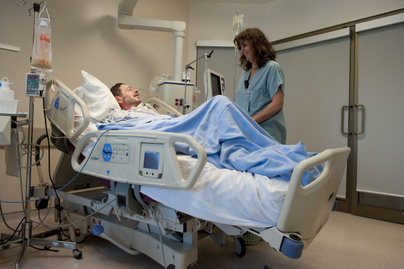
No Wrong Side To This Bad


US researchers have created a smart hospital bed that is aware of its surrounding. The intelligent bed will make important decisions regarding the patients' healthcare which will involve both improving and saving precious lives.
A prototype of the smart bed that is being tested at the University of New Hampshire could revolutionise health care In the future


John LaCourse, professor at the University of New Hampshire, is currently negotiating with hospital bed manufacturers

to adopt his prograq~.med algorithm, which could become the basis for computerised hospital beds.

These smart hospital beds would communicate with and respond to medical devices that monitor a patient's condition.

"Perhaps a sleeping patient moves, causing a drop in blood pressure. The blood pressure monitor would communicate

this change to the bed and the bed, in turn, would move up or down until the patients' blood pressure is stabilised," he says.

IMPROVED POST-SURGERY CARE

Post-surgical needs may also be met with this bed. "Procedures such as retinal surgery require exact blood pressure levels," says LaCourse. "A smart hospital bed would adjust itself to maintain these levels for patients."

Even quality-of-life conditions such as bed sores could be addressed. "Instead of requiring hospital staff to move the patient, monitors could send signals to the bed to roll the patient to his left or right to avoid bed sores," says LaCourse.

"Microprocessors installed into the bed can also sense respiration patterns to determine when breathing has ceased, the bed moves in such a way that the breathing resumes," said Jonathan Waters, who is working on modifying the bed for sleep apnoea.

PLUG AND PLAY

The ultimate success of LaCourse's project rests with the plug-and-play component. Plug-and-play means that medical devices – everything from blood pressure monitors to breathing machines- "can talk to each other and share patient information which greatly reduces care errors," explains LaCourse.

To realise plug-and-play capability, however, LaCourse's technology must become the industry standard for hospital bed manufacturers. 1n this way, medical devices could seamlessly connect to and exchange patient data.

LaCourse is hopeful that, within two to three years, his technology may be accepted by most if not all hospital bed companies.