BTemplates.com

Powered by Blogger.

Pageviews past week

Quantum mechanics

Auto News

artificial intelligence

About Me

Recommend us on Google!

Information Technology

Popular Posts

Saturday, October 29, 2011

Scientists Measure Dream Content for the First Time: Dreams Activate the Brain in a Similar Way to Real Actions


The ability to dream is a fascinating aspect of the human mind. However, how the images and emotions that we experience so intensively when we dream form in our heads remains a mystery. Up to now it has not been possible to measure dream content. Max Planck scientists working with colleagues from the Charité hospital in Berlin have now succeeded, for the first time, in analysing the activity of the brain during dreaming.

Top: Patient in a functional magnetic resonance imaging machine. Bottom: Activity in the motor cortex during the movement of the hands while awake (left) and during a dreamed movement (right). Blue areas indicate the activity during a movement of the right hand, which is clearly demonstrated in the left brain hemisphere, while red regions indicate the corresponding left-hand movements in the opposite brain hemisphere. (Credit: © MPI of Psychiatry)

They were able to do this with the help of lucid dreamers, i.e. people who become aware of their dreaming state and are able to alter the content of their dreams. The scientists measured that the brain activity during the dreamed motion matched the one observed during a real executed movement in a state of wakefulness.

The research is published in the journal Current Biology.

Methods like functional magnetic resonance imaging have enabled scientists to visualise and identify the precise spatial location of brain activity during sleep. However, up to now, researchers have not been able to analyse specific brain activity associated with dream content, as measured brain activity can only be traced back to a specific dream if the precise temporal coincidence of the dream content and measurement is known. Whether a person is dreaming is something that could only be reported by the individual himself.

Scientists from the Max Planck Institute of Psychiatry in Munich, the Charité hospital in Berlin and the Max Planck Institute for Human Cognitive and Brain Sciences in Leipzig availed of the ability of lucid dreamers to dream consciously for their research. Lucid dreamers were asked to become aware of their dream while sleeping in a magnetic resonance scanner and to report this "lucid" state to the researchers by means of eye movements. They were then asked to voluntarily "dream" that they were repeatedly clenching first their right fist and then their left one for ten seconds.


This enabled the scientists to measure the entry into REM sleep -- a phase in which dreams are perceived particularly intensively -- with the help of the subject's electroencephalogram (EEG) and to detect the beginning of a lucid phase. The brain activity measured from this time onwards corresponded with the arranged "dream" involving the fist clenching. A region in the sensorimotor cortex of the brain, which is responsible for the execution of movements, was actually activated during the dream. This is directly comparable with the brain activity that arises when the hand is moved while the person is awake. Even if the lucid dreamer just imagines the hand movement while awake, the sensorimotor cortex reacts in a similar way.

The coincidence of the brain activity measured during dreaming and the conscious action shows that dream content can be measured. "With this combination of sleep EEGs, imaging methods and lucid dreamers, we can measure not only simple movements during sleep but also the activity patterns in the brain during visual dream perceptions," says Martin Dresler, a researcher at the Max Planck Institute for Psychiatry.

The researchers were able to confirm the data obtained using MR imaging in another subject using a different technology. With the help of near-infrared spectroscopy, they also observed increased activity in a region of the brain that plays an important role in the planning of movements. "Our dreams are therefore not a 'sleep cinema' in which we merely observe an event passively, but involve activity in the regions of the brain that are relevant to the dream content," explains Michael Czisch, research group leader at the Max Planck Institute for Psychiatry.

Wednesday, October 26, 2011

Design Rules Will Enable Scientists to Use DNA to Build Nanomaterials With Desired Properties


Nature is a master builder. Using a bottom-up approach, nature takes tiny atoms and, through chemical bonding, makes crystalline materials, like diamonds, silicon and even table salt. In all of them, the properties of the crystals depend upon the type and arrangement of atoms within the crystalline lattice.
Abstract rendering of a DNA strand.
(Credit: iStockphoto/Johan Swanepoel)

Now, a team of Northwestern University scientists has learned how to top nature by building crystalline materials from nanoparticles and DNA, the same material that defines the genetic code for all living organisms.

Using nanoparticles as "atoms" and DNA as "bonds," the scientists have learned how to create crystals with the particles arranged in the same types of atomic lattice configurations as some found in nature, but they also have built completely new structures that have no naturally occurring mineral counterpart.

The basic design rules the Northwestern scientists have established for this approach to nanoparticle assembly promise the possibility of creating a variety of new materials that could be useful in catalysis, electronics, optics, biomedicine and energy generation, storage and conversion technologies.

The new method and design rules for making crystalline materials from nanostructures and DNA will be published Oct. 14 by the journal Science.

"We are building a new periodic table of sorts," said Professor Chad A. Mirkin, who led the research. "Using these new design rules and nanoparticles as 'artificial atoms,' we have developed modes of controlled crystallization that are, in many respects, more powerful than the way nature and chemists make crystalline materials from atoms. By controlling the size, shape, type and location of nanoparticles within a given lattice, we can make completely new materials and arrangements of particles, not just what nature dictates."

Mirkin is the George B. Rathmann Professor of Chemistry in the Weinberg College of Arts and Sciences and professor of medicine, chemical and biological engineering, biomedical engineering and materials science and engineering and director of Northwestern's International Institute for Nanotechnology (IIN).

"Once we have a certain type of lattice," Mirkin said, "the particles can be moved closer together or farther apart by changing the length of the interconnecting DNA, thereby providing near-infinite tunability."

"This work resulted from an interdisciplinary collaboration that coupled synthetic chemistry with theoretical model building," said coauthor George C. Schatz, a theoretician and the Charles E. and Emma H. Morrison Professor of Chemistry at Northwestern. "It was the back and forth between synthesis and theory that was crucial to the development of the design rules. Collaboration is a special aspect of research at Northwestern, and it worked very effectively for this project."


In the study, the researchers start with two solutions of nanoparticles coated with single-stranded DNA. They then add DNA strands that bind to these DNA-functionalized particles, which then present a large number of DNA "sticky ends" at a controlled distance from the particle surface; these sticky ends then bind to the sticky ends of adjacent particles, forming a macroscopic arrangement of nanoparticles.

Different crystal structures are achieved by using different combinations of nanoparticles (with varying sizes) and DNA linker strands (with controllable lengths). After a process of mixing and heating, the assembled particles transition from an initially disordered state to one where every particle is precisely located according to a crystal lattice structure. The process is analogous to how ordered atomic crystals are formed.

The researchers report six design rules that can be used to predict the relative stability of different structures for a given set of nanoparticle sizes and DNA lengths. In the paper, they use these rules to prepare 41 different crystal structures with nine distinct crystal symmetries. However, the design rules outline a strategy to independently adjust each of the relevant crystallographic parameters, including particle size (varied from 5 to 60 nanometers), crystal symmetry and lattice parameters (which can range from 20 to 150 nanometers). This means that these 41 crystals are just a small example of the near infinite number of lattices that could be created using different nanoparticles and DNA strands.

Mirkin and his team used gold nanoparticles in their work but note that their method also can be applied to nanoparticles of other chemical compositions. Both the type of nanoparticle assembled and the symmetry of the assembled structure contribute to the properties of a lattice, making this method an ideal means to create materials with predictable and controllable physical properties.

Mirkin believes that, one day soon, software will be created that allows scientists to pick the particle and DNA pairs required to make almost any structure on demand.

The Air Force Office of Scientific Research, the U.S. Department of Energy Office of Basic Energy Sciences and the National Science Foundation supported the research.

Sunday, October 23, 2011

Global Warming Is Real, New Analysis Confirms


Global warming is real, according to a major study released Oct. 20. Despite issues raised by climate change skeptics, the Berkeley Earth Surface Temperature study finds reliable evidence of a rise in the average world land temperature of approximately 1°C since the mid-1950s.

Comparison of data showing decadal land-surface average world temperature changes from 15 different sources, some going back as far as 1800. (Credit: Image courtesy of Berkeley Earth Surface Temperature)

Analyzing temperature data from 15 sources, in some cases going as far back as 1800, the Berkeley Earth study directly addressed scientific concerns raised by skeptics, including the urban heat island effect, poor station quality, and the risk of data selection bias.

On the basis of its analysis, according to Berkeley Earth's founder and scientific director, Professor Richard A. Muller, the group concluded that earlier studies based on more limited data by teams in the United States and Britain had accurately estimated the extent of land surface warming.

"Our biggest surprise was that the new results agreed so closely with the warming values published previously by other teams in the U.S. and the U.K.," Muller said. "This confirms that these studies were done carefully and that potential biases identified by climate change skeptics did not seriously affect their conclusions."

Previous studies, carried out by NOAA, NASA, and the Hadley Center, also found that land warming was approximately 1°C since the mid-1950s, and that the urban heat island effect and poor station quality did not bias the results. But their findings were criticized by skeptics who worried that they relied on ad-hoc techniques that meant that the findings could not be duplicated. Robert Rohde, lead scientist for Berkeley Earth, noted that "the Berkeley Earth analysis is the first study to address the issue of data selection bias, by using nearly all of the available data, which includes about 5 times as many station locations as were reviewed by prior groups."

Elizabeth Muller, co-founder and Executive Director of Berkeley Earth, said she hopes the Berkeley Earth findings will help "cool the debate over global warming by addressing many of the valid concerns of the skeptics in a clear and rigorous way." This will be especially important in the run-up to the COP 17 meeting in Durban, South Africa, later this year, where participants will discuss targets for reducing Greenhouse Gas (GHG) emissions for the next commitment period as well as issues such as financing, technology transfer and cooperative action.

The Berkeley Earth team includes physicists, climatologists, and statisticians from California, Oregon, and Georgia. Rohde led the development of a new statistical approach and what Richard Muller called "the Herculean labor" of merging the data sets. One member of the group, Saul Perlmutter, was recently announced as a winner of the 2011 Nobel Prize in Physics (for his work in cosmology).


The Berkeley Earth study did not assess temperature changes in the oceans, which according to the Intergovernmental Panel on Climate Change (IPCC) have not warmed as much as land. When averaged in, they reduce the global surface temperature rise over the past 50 years -- the period during which the human effect on temperatures is discernable -- to about two thirds of one degree Centigrade.

Specifically, the Berkeley Earth study concludes that:
  • The urban heat island effect is locally large and real, but does not contribute significantly to the average land temperature rise. That's because the urban regions of Earth amount to less than 1% of the land area.
  • About 1/3 of temperature sites around the world reported global cooling over the past 70 years (including much of the United States and northern Europe). But 2/3 of the sites show warming. Individual temperature histories reported from a single location are frequently noisy and/or unreliable, and it is always necessary to compare and combine many records to understand the true pattern of global warming.
  • The large number of sites reporting cooling might help explain some of the skepticism of global warming," Rohde commented. "Global warming is too slow for humans to feel directly, and if your local weather man tells you that temperatures are the same or cooler than they were a hundred years ago it is easy to believe him." In fact, it is very hard to measure weather consistently over decades and centuries, and the presence of sites reporting cooling is a symptom of the noise and local variations that can creep in. A good determination of the rise in global land temperatures can't be done with just a few stations: it takes hundreds -- or better, thousands -- of stations to detect and measure the average warming. Only when many nearby thermometers reproduce the same patterns can we know that the measurements were reliably made.
  • Stations ranked as "poor" in a survey by Anthony Watts and his team of the most important temperature recording stations in the U.S., (known as the USHCN -- the US Historical Climatology Network), showed the same pattern of global warming as stations ranked "OK." Absolute temperatures of poor stations may be higher and less accurate, but the overall global warming trend is the same, and the Berkeley Earth analysis concludes that there is not any undue bias from including poor stations in the survey.
Four scientific papers setting out these conclusions have been submitted for peer review and will form part of the literature for the next IPCC report on Climate Change. They can be accessed on: www.BerkeleyEarth.org. A video animation graphically shows global warming around the world since 1800.

Berkeley Earth is making its preliminary results public, together with its programs and dataset, in order to invite additional scrutiny. Elizabeth Muller said that "one of our goals is to make the science behind global warming readily accessible to the public." Most of the data were previously available on public websites, but in so many different locations and different formats that most people could access only a small subset of the data. The merged database, which combines 1.6 billion records, is now accessible from the Berkeley Earth website: www.BerkeleyEarth.org .

What Berkeley Earth has not done is make an independent assessment of how much of the observed warming is due to human actions, Richard Muller acknowledged. As a next step, Berkeley Earth plans to address the total warming of the oceans, with a view to obtaining a more accurate figure for the total amount of global warming observable.

More information about Berkeley Earth is available at www.BerkeleyEarth.org.

Saturday, October 22, 2011

Pi enthusiast calculates it to ten trillion digits


Shigeru Kondo is a seriously committed guy. Ever since discovering he had an interest in calculating pi (aka π) back in his college days, he’s been following the results achieved by others using massive supercomputers. Now, in his late 50's, with some help from Northwestern University grad school student Alexander Yee, he’s succeeded in calculating pi to ten trillion digits; on a home built PC yet.

Pi, the mathematical constant that describes the ratio of a circle’s circumference to its diameter, is generally rounded off to just two places, bringing it to 3.14. Believed to have been first described by Archimedes way back in the 3rd century BC, the ratio is used in all sorts of mathematical computations, not the least of which is in figuring out the area of a circle. But because pi is an irrational number, it’s value cannot be written as an fraction which means when written as a decimal approximation, it’s numbers go on infinitely, and perhaps more importantly, never repeat.

For hundreds of years, pi has held fascination for mathematicians, scientists, philosophers and even regular run of the mill people. Why this is so is hard to say, and so too is the seemingly endless progression of people that have set before themselves the task of calculating its digits. In spite of that, it’s possible that none has ever been so obsessed as Kondo. He’s spent the better part of a year with the singular task of finding the ten trillionth digit, and of course all those past the five trillionth and one digit leading up to the ten trillionth, since he found the five trillionth digit just last year.

Finding the value of pi to 10 trillion digits requires performing a lot of calculations (using software written by Yee), so many in fact, that Kondo had to add a lot more hard drive space than you’d find on your average PC. Forty eight terabytes to be exact. So intense was the computation that the computer alone caused the temperature in the room to hold steady at 104° F.

Also, it’s not as easy to keep a custom built super-sized PC going full steam ahead twenty four hours day for months on end, as it might seem. Hard drive failures and the threat of power disruption from the earthquake in Japan back in March threatened the project many times. And of course there was that power bill itself which ran to something close to $400 a month as the computer ground away.

But in the end, it was Kondo’s persistence that paid off. For his efforts he will be forever known (in the annals of science, and probably the Guinness Book of World Records) as the man who calculated the ten trillionth digit of pi. It’s 5.

More information: http://www.numberworld.org/misc_runs/pi-10t/details.html
http://ja0hxv.calico.jp/pai/estart.html


Monday, October 17, 2011

Mental time-travel in birds


Certain types of birds may track army ant swarms using sophisticated memory and the ability to plan for the future.
White Whiskered Puff bird Credit: Glenn M Duggan FZS

Some tropical birds collect their prey at army ant raids, where massive swarms of ants sweep through the forest and drive out insects. The behaviour of interest is called bivouac checking; it allows these birds to track the cyclical raid activity of army ant colonies.

Army ants have regular alternating periods of high and low raiding activity, and birds visit the ants’ temporary nest sites (bivouacs) to determine which colonies are raiding on a given day.

The new findings published today in the journal Behavioural Ecology, suggest that bivouac checking allows birds to keep track of multiple army ant colonies, keeping account of which colonies are in periods of high-raiding activity while avoiding colonies with low-raiding activity.

Recent research has discovered that birds check army ant bivouacs at the end of the day, after they have fed at the raid. They may use the information about the army ant nest location the next day to find the ants again, thus accessing a past memory (the nest location) to fulfil a future need (bird will be hungry tomorrow), also known as ‘mental time-travel’.

Two of the authors of the study Corina Logan of the University of Cambridge, and Sean O’Donnell of the University of Washington, observed bivouac checking behaviour in Monteverde, Costa Rica.

Mental time-travel consists of two elements: the ability to remember past events and the ability to anticipate and plan for future events. It has traditionally been considered a quality unique to humans. However, ever since Nicola Clayton of the University of Cambridge discovered that scrub jays (a species of large-brained crow) can remember the past and plan for the future, there have been a suite of studies showing evidence of this ability in other species as well. We now know that corvids (birds in the crow family), some primates, and possibly rats have all shown the ability to remember the past and plan for the future.

Corina Logan, said: “We suspect that future planning could be involved in bivouac-checking bird behaviour because the birds were checking bivouacs when they were not hungry, a behaviour that does not make sense until the next morning upon return to the bivouac when the bird finds the ants raiding again and encounters its next meal – a delayed benefit.”

Until recently, it has been difficult to find model systems for studying mental time travel in an ecologically relevant way. “The fact that we might have happened on a whole new system for exploring these capacities is quite exciting,” added Corina Logan.

Provided by University of Cambridge


Recommend this story on Facebook, Twitter, and Google +1

Saturday, October 15, 2011

From Blue Whales to Earthworms, a Common Mechanism Gives Shape to Living Beings


Why don't our arms grow from the middle of our bodies? The question isn't as trivial as it appears. Vertebrae, limbs, ribs, tailbone ... in only two days, all these elements take their place in the embryo, in the right spot and with the precision of a Swiss watch. Intrigued by the extraordinary reliability of this mechanism, biologists have long wondered how it works. Now, researchers at EPFL (Ecole Polytechnique Fédérale de Lausanne) and the University of Geneva (Unige) have solved the mystery.

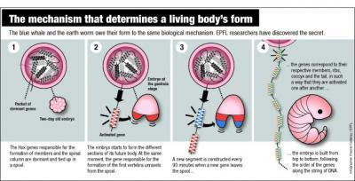
This is a diagram of the mechanism for form. The blue whale and the earth worm owe their form to the same biological mechanism. EPFL researchers have discovered the secret. (Credit: Infographic courtesy of Pascal Coderay, EPFL)

Their discovery will be published October 13, 2011 in the journal Science.

The embryo is built one layer at a time

During the development of an embryo, everything happens at a specific moment. In about 48 hours, it will grow from the top to the bottom, one slice at a time -- scientists call this the embryo's segmentation. "We're made up of thirty-odd horizontal slices," explains Denis Duboule, a professor at EPFL and Unige. "These slices correspond more or less to the number of vertebrae we have."

Every hour and a half, a new segment is built. The genes corresponding to the cervical vertebrae, the thoracic vertebrae, the lumbar vertebrae and the tailbone become activated at exactly the right moment one after another. "If the timing is not followed to the letter, you'll end up with ribs coming off your lumbar vertebrae," jokes Duboule. How do the genes know how to launch themselves into action in such a perfectly synchronized manner? "We assumed that the DNA played the role of a kind of clock. But we didn't understand how."

When DNA acts like a mechanical clock


Very specific genes, known as "Hox," are involved in this process. Responsible for the formation of limbs and the spinal column, they have a remarkable characteristic. "Hox genes are situated one exactly after the other on the DNA strand, in four groups. First the neck, then the thorax, then the lumbar, and so on," explains Duboule. "This unique arrangement inevitably had to play a role."

The process is astonishingly simple. In the embryo's first moments, the Hox genes are dormant, packaged like a spool of wound yarn on the DNA. When the time is right, the strand begins to unwind. When the embryo begins to form the upper levels, the genes encoding the formation of cervical vertebrae come off the spool and become activated. Then it is the thoracic vertebrae's turn, and so on down to the tailbone. The DNA strand acts a bit like an old-fashioned computer punchcard, delivering specific instructions as it progressively goes through the machine.

"A new gene comes out of the spool every ninety minutes, which corresponds to the time needed for a new layer of the embryo to be built," explains Duboule. "It takes two days for the strand to completely unwind; this is the same time that's needed for all the layers of the embryo to be completed."

This system is the first "mechanical" clock ever discovered in genetics. And it explains why the system is so remarkably precise.

This discovery is the result of many years of work. Under the direction of Duboule and Daniël Noordermeer, the team analyzed thousands of Hox gene spools. With assistance from the Swiss Institute for Bioinformatics, the scientists were able to compile huge quantities of data and model the structure of the spool and how it unwinds over time.

The snake: a veritable vertebral assembly line

The process discovered at EPFL is shared by numerous living beings, from humans to some kinds of worms, from blue whales to insects. The structure of all these animals -- the distribution of their vertebrae, limbs and other appendices along their bodies -- is programmed like a sheet of player-piano music by the sequence of Hox genes along the DNA strand.

The sinuous body of the snake is a perfect illustration. A few years ago, Duboule discovered in these animals a defect in the Hox gene that normally stops the vertebrae-making process.

"Now we know what's happening. The process doesn't stop, and the snake embryo just keeps on making vertebrae, all identical, until the process just runs out of steam."

The Hox clock is a demonstration of the extraordinary complexity of evolution. One notable property of the mechanism is its extreme stability, explains Duboule. "Circadian or menstrual clocks involve complex chemistry. They can thus adapt to changing contexts, but in a general sense are fairly imprecise. The mechanism that we have discovered must be infinitely more stable and precise. Even the smallest change would end up leading to the emergence of a new species."


Recommend this story on Facebook, Twitter, and Google +1

Friday, October 14, 2011

Dark Matter of the Genome Revealed


6MK9UNF4B2AX
An international team of researchers has discovered the vast majority of the so-called "dark matter" in the human genome, by means of a sweeping comparison of 29 mammalian genomes. The team, led by scientists from the Broad Institute, has pinpointed the parts of the human genome that control when and where genes are turned on. This map is a critical step in interpreting the thousands of genetic changes that have been linked to human disease.

Rendering of DNA. Researchers have discovered the vast majority of the so-called "dark matter" in the human genome, by means of a sweeping comparison of 29 mammalian genomes. (Credit: iStockphoto/Martin McCarthy)

Their findings appear online October 12 in the journal Nature.

Early comparison studies of the human and mouse genomes led to the surprising discovery that the regulatory information that controls genes dwarfs the information in the genes themselves. But, these studies were indirect: they could infer the existence of these regulatory sequences, but could find only a small fraction of them. These mysterious sequences have been referred to as the dark matter of the genome, analogous to the unseen matter and energy that make up most of the universe.

This new study enlisted a menagerie of mammals -- including rabbit, bat, elephant, and more -- to reveal these mysterious genomic elements.

Over the last five years, the Broad Institute, the Genome Institute at Washington University, and the Baylor College of Medicine Human Genome Sequencing Center have sequenced the genomes of 29 placental mammals. The research team compared all of these genomes, 20 of which are first reported in this paper, looking for regions that remained largely unchanged across species.

"With just a few species, we didn't have the power to pinpoint individual regions of regulatory control," said Manolis Kellis, last author of the study and associate professor of computer science at MIT. "This new map reveals almost 3 million previously undetectable elements in non-coding regions that have been carefully preserved across all mammals, and whose disruptions appear to be associated with human disease."

These findings could yield a deeper understanding of disease-focused studies, which look for genetic variants closely tied to disease.

"Most of the genetic variants associated with common diseases occur in non-protein coding regions of the genome. In these regions, it is often difficult to find the causal mutation," said first author Kerstin Lindblad-Toh, scientific director of vertebrate genome biology at the Broad and a professor in comparative genomics at Uppsala University, Sweden. "This catalog will make it easier to decipher the function of disease-related variation in the human genome."

This new map helps pinpoint those mutations that are likely responsible for disease, as they have been preserved across millions of years of evolution, but are commonly disrupted in individuals that suffer from a given disease. Knowing the causal mutations and their likely functions can then help uncover the underlying disease mechanisms and reveal potential drug targets.

The scientists were able to suggest possible functions for more than half of the 360 million DNA letters contained in the conserved elements, revealing the hidden meaning behind the As, Cs, Ts, and Gs. These revealed:
  • Almost 4,000 previously undetected exons, or segments of DNA that code for protein
  • 10,000 highly conserved elements that may be involved in how proteins are made
  • More than 1,000 new families of RNA secondary structures with diverse roles in gene regulation
  • 2.7 million predicted targets of transcription factors, proteins that control gene expression

"We can use this treasure trove of new elements to revisit disease association studies, focusing on those that disrupt conserved elements and trying to discern their likely functions," said Kellis. "Using a single genome, the language of DNA seems cryptic. When studied through the lens of evolution, words light up and gain meaning."

The researchers were also able to harness this collection of genomes to look back in time, across more than 100 million years of evolution, to uncover the fundamental changes that shaped mammalian adaptation to different environments and lifestyles. The researchers revealed specific proteins under rapid evolution, including some related to the immune system, taste perception, and cell division. They also uncovered hundreds of protein domains within genes that are evolving rapidly, some of which are related to bone remodeling and retinal functions.

"The comparison of mammalian genomes reveals the regulatory controls that are common across all mammals," said Eric Lander, director of the Broad Institute and the third corresponding author of the paper. "These evolutionary innovations were devised more than 100 million years ago and are still at work in the human population today."

In addition to finding the DNA controls that are common across all mammals, the comparison highlighted areas that have been changing rapidly only in the human and primate genomes. Researchers had previously uncovered two hundred of these regions, some of which are linked to brain and limb development. The expanded list -- which now includes more than 1,000 regions -- will give scientists new starting points for understanding human evolution.

The comparison of many complete genomes is beginning to offer a clear view of once indiscernible genomic regions, and with additional genomes, that resolution will only increase. "The power of this resource is that it continues to improve with the inclusion of more species," said Lindblad-Toh. "It's a very systematic and unbiased approach that will only become more powerful with the inclusion of additional genomes."

Other Broad researchers who contributed to this work include Manuel Garber, Or Zuk, Michael F. Lin, Pouya Kheradpour, Jason Ernst, Evan Mauceli, Lucas D. Ward, Michele Clamp, Sante Gnerre, Jessica Alföldi, Jean Chang, Federica Di Palma, Mitchell Guttman, David B. Jaffe, Irwin Jungreis, Marcia Lara, Jim Robinson, Xiaohui Xie, Michael C. Zody, and members of the Broad Institute Sequencing Platform and Whole Genome Assembly Team.

This project was supported by the National Human Genome Research Institute, National Institute for General Medicine, the European Science Foundation, National Science Foundation, the Sloan Foundation, an Erwin Schrödinger Fellowship, the Gates Cambridge Trust, Novo Nordisk Foundation, University of Copenhagen, the David and Lucile Packard Foundation, the Danish Council for Independent Research Medical Sciences, and The Lundbeck Foundation.

Recommend this story on Facebook, Twitter, and Google +1


Tuesday, October 11, 2011

LHSee - Large Hadron Collider app - Big bang science in your pocket


Want to find out how to Hunt the Higgs Boson using your phone? Ever wondered how the Large Hadron Collider experiments work, and what the collisions look like?


Scientists at the world's biggest scientific experiment - the Large Hadron Collider (LHC)at CERN, Geneva - are trying to answer fundamental questions about the nature of the Universe, the origin of mass, the structure of space and time, and the conditions of the early universe. For those of us not lucky enough to have the world's highest energy particle smasher in our own back gardens, we can still get close to the action using an exciting new smartphone App.

The new App, called 'LHSee', makes the LHC accessible to anybody with a smartphone or tablet PC running the Google Android operating system. Written by Oxford University scientists in collaboration with the ATLAS, one of the four LHC experiments at CERN, it has been designed for experts and non-experts alike.

For the first time you can now grab live collision events from the underground detectors in Geneva, and beam them direct to your own device. As well as a variety of educational resources, the application allows you to interact with the collision events in full 3D graphics. You can also find out how the different parts of the detector work, learn how to identify different types of collision, and even put your new skills to the test by playing the 'Hunt the Higgs' game.

Dr Alan Barr of the University of Oxford says: "I love the detail in the live displays - it's amazing to see that you can pick out the different individual proton collisions."

With help from their international friends within the ATLAS collaboration, the developers offer the App with language support not just in English, but also in French, German, Italian, Spanish and Swedish.

The App is free to download from the Google Android Marketplace.

Paralyzed man uses mind-powered robot arm to touch


Giving a high-five. Rubbing his girlfriend's hand. Such ordinary acts - but a milestone for a paralyzed man.

True, a robotic arm parked next to his wheelchair did the touching, painstakingly, palm to palm. But Tim Hemmes made that arm move just by thinking about it.

Emotions surged. For the first time in the seven years since a motorcycle accident left him a quadriplegic, Hemmes was reaching out to someone - even if it was only temporary, part of a monthlong science experiment at the University of Pittsburgh.

"It wasn't my arm but it was my brain, my thoughts. I was moving something," Hemmes says. "I don't have one single word to give you what I felt at that moment. That word doesn't exist."

The Pennsylvania man is among the pioneers in an ambitious quest for thought-controlled prosthetics to give the paralyzed more independence - the ability to feed themselves, turn a doorknob, hug a loved one.

The goal is a Star Trek-like melding of mind and machine, combining what's considered the most humanlike bionic arm to date - even the fingers bend like real ones - with tiny chips implanted in the brain. Those electrodes tap into electrical signals from brain cells that command movement. Bypassing a broken spinal cord, they relay those signals to the robotic third arm.
This research is years away from commercial use, but numerous teams are investigating different methods.

At Pittsburgh, monkeys learned to feed themselves marshmallows by thinking a robot arm into motion. At Duke University, monkeys used their thoughts to move virtual arms on a computer and got feedback that let them distinguish the texture of what they "touched."

Through a project known as BrainGate and other research, a few paralyzed people outfitted with brain electrodes have used their minds to work computers, even make simple movements with prosthetic arms.

But can these neuroprosthetics ever offer the complex, rapid movements that people would need for more practical, everyday use?

"We really are at a tipping point now with this technology," says Michael McLoughlin of the Johns Hopkins University Applied Physics Laboratory, which developed the humanlike arm in a $100 million project for DARPA, the Pentagon's research agency.

Pittsburgh is helping to lead a closely watched series of government-funded studies over the next two years to try to find out. A handful of quadriplegic volunteers will train their brains to operate the DARPA arm in increasingly sophisticated ways, even using sensors implanted in its fingertips to try to feel what they touch, while scientists explore which electrodes work best.

"Imagine all the joints that are in your hand. There's 20 motions around all those joints," says Pittsburgh neurobiologist Andrew Schwartz. "It's not just reaching out and crudely grasping something. We want them to be able to use the fingers we've worked so hard on."

The 30-year-old Hemmes' task was a much simpler first step. He was testing whether a new type of chip, which for safety reasons the Food and Drug Administration let stay on this initial volunteer's brain for just a month, could allow for three-dimensional arm movement.

He surprised researchers the day before the electrodes were removed. The robotic arm whirred as Hemmes' mind pushed it forward to hesitantly tap palms with a scientist. Then his girlfriend beckoned. The room abruptly hushed. Hemmes painstakingly raised the black metal hand again and slowly rubbed its palm against hers a few times.

These emotional robotic touches have inspired researchers now recruiting volunteers for soon-to-start yearlong experiments.

"It was awesome," is the decidedly unscientific description from the normally reserved Dr. Michael Boninger, rehabilitation chief at the University of Pittsburgh Medical Center. "To interact with a human that way. ... This is the beginning."

---


Hemmes' journey began in 2004. He owned an auto-detailing shop and rode his motorcycle in his spare time. Then one summer evening he swerved to miss a deer. His bike struck a guardrail. His neck snapped.

His determination didn't. Paralyzed below the shoulders, he's tried other experimental procedures in hopes, so far unrealized, of regaining some arm function.

"I always tell people your legs are great ... but they just get you from here to there," Hemmes says as his caregiver waits to feed him a bite of a cheeseburger near his home in Butler, north of Pittsburgh. "Your arms and fingers and hands do everything else. I have to get those back, I absolutely have to."

His ultimate goal is to hug his 8-year-old daughter. "I'm going to do whatever it takes, as long as it takes, to do that again."

Hemmes entered an operating room at UPMC with a mix of nerves and excitement.

"It's good anxiety," he says. "There is so much riding on this."

---

Think "I want that apple," and your arm reaches out and grasps it. You're not aware that neurons are instantaneously firing in patterns that send commands down the spinal cord - make the shoulder raise the arm, extend the elbow, flex the wrist and all five fingers.

A very similar firing occurs when you imagine movement or watch the movement you'd like to perform, explains Boninger, who with Schwartz is leading the Pittsburgh research together with a team of bioengineers, neuroscientists and physicians.

The DARPA arm was developed primarily for amputees. Separate research is under way to help them move it by using transplanted nerves to sense those brain commands. The paralyzed pose a more difficult challenge: getting those signals around a broken spinal cord.

For quadriplegic patients, scientists use implanted electrodes, called a "brain-computer interface" or BCI, to record that electrical activity. The signals move down through wires that tunnel under the skin and out by the collarbone, and are plugged into a computer or a robotic arm.

Until now, researchers mostly have tested miniature electrodes that poke inside the brain's motor cortex and record from individual cells, presumably allowing for precise movements. Pittsburgh's next test-patient will have two penetrating grids implanted in different parts of the cortex for a year to record from 200 cells altogether.

In contrast, Hemmes' chip sat on the surface of his motor cortex, a less invasive method that records from groups of cells. The size of two postage stamps, it's based on a kind of electrical signal mapping used to track seizures in epilepsy patients.

Both approaches need study, says Daofen Chen of the National Institutes of Health, who oversees neurorehabilitation research. He compares the options to eavesdropping on a party by sending in individual microphones or setting up a recorder at the window.

Boninger adds that scar tissue can blunt the penetrating electrodes over time, and the surface chips may be easier to convert to a wireless system, which is important for commercial use.

---

Hemmes' operation took two hours. He had practiced imagining arm movements inside brain scanners, to see where the electrical signals concentrated. That's where neurosurgeon Elizabeth Tyler-Kabara cut, attaching the chip through an inch-wide opening on the left side of Hemmes' skull.

Two days later, Hemmes was hooked to a computer, beginning simple cursor movements. The next week, it was time to test if he could trigger real-life movement using the DARPA arm.

Hemmes reclined in his wheelchair, the robot arm bolted to a steel rod nearby. The task: make the arm reach out to grasp a ball mounted on a board.

The arm whirs forward, then stops, then goes again, then suddenly pulls back.

"It's doing the opposite of what I ask it do," Hemmes says in frustration. "When I think about reaching back, it goes forward."

Dr. Wei Wang, a member of the research team, watches Hemmes' brain patterns on a nearby computer screen, trying to match them to the robotic movements. Focus on your elbow, Wang advises.

Hemmes takes a deep breath and tries. The arm whirs forward this time, reaching the ball. The fingers clench around it.

"There's no owner's manual," Hemmes says, thrilled that the back-and-forth pays off. "I'm training my brain to figure how to do all this."

Letting go is harder, the motor growling as the arm tugs backward before the fingers fully release. Hemmes starts imagining his hand relaxing before pulling backward, and the robot hand follows.

---

Sure, a robotic hand that one day mounts to a wheelchair could be useful. But no matter how well today's prosthetics move, they've got a problem: They don't sense what they touch. Normally, instant messages flash from the skin up to the brain to say "squeeze tighter" so we don't drop that coffee cup, or "tight enough" so we don't hug too hard.

Besides, Hemmes shares the dream of many quadriplegics. He doesn't want a bionic third hand. He wants to move his own hands again.

"These are all scientific goals that are very real," Boninger says.

Recreating sensation means crafting a two-way highway with those brain chips. That's what Duke University, in a study published last week in the journal Nature, did with its two monkeys. When the animals "touched" objects on a computer screen with their video game-like arms, electrical signals flashed back up to implanted electrodes - different signals for different textures, to tell the objects apart.

Sensors in the DARPA arm's fingertips allow for that same kind of feedback. McLoughlin says the plan is for one of the Pittsburgh study patients to begin testing touch capability next year, with a similar attempt at the California Institute of Technology to follow.

What about moving paralyzed limbs? Duke's plan is to turn its research into a robotic exoskeleton that would help the paralyzed move their bodies.

Hemmes is more intrigued by what's called functional electrical stimulation, zapping muscles with electrical currents to make them move. At Hemmes' request, Boninger's team attempted to fit his hand with a stimulator glove that might be linked to his electrode, but it was unsuccessful. The NIH's Chen says still other researchers are working on that kind of approach.

---

Hemmes likened moving the DARPA arm to learning to drive a car with a manual transmission. It took practice, but by week four he was moving the arm sideways as well as back and forth.

The fingers still clenched pretty tight, though. So when his girlfriend Katie Schaffer spoke up - "I want to hold your hand," she said on his last day of testing - Hemmes didn't dare bend them.

The two met after his accident, so he'd never before reached out to her.

"I was just trying to be gentle. I didn't want to hurt her, and I finally got there," Hemmes says. "Definitely the tears were flowing."

He says he was ready for a break after almost daily scientific testing, so removing the electrode and wires the next day wasn't a disappointment. He's confident the researchers will call him back once the technology advances.

Monday, October 10, 2011

Graphene's 'Big Mac' creates next generation of chips


The world's thinnest, strongest and most conductive material, discovered in 2004 at the University of Manchester by Professor Andre Geim and Professor Kostya Novoselov, has the potential to revolutionize material science.
Artistic impression of graphene molecules.
Image: University of Manchester

Demonstrating the remarkable properties of graphene won the two scientists the Nobel Prize for Physics last year and UK's Chancellor of the Exchequer George Osborne has just announced plans for a £50m graphene research hub to be set up.

Now, writing in the journal Nature Physics, the University of Manchester team have for the first time demonstrated how graphene inside electronic circuits will probably look like in the future.

By sandwiching two sheets of graphene with another two-dimensional material, boron nitrate, the team created the graphene 'Big Mac' – a four-layered structure which could be the key to replacing the silicon chip in computers.

Because there are two layers of graphene completed surrounded by the boron nitrate, this has allowed the researchers for the first time to observe how graphene behaves when unaffected by the environment.

Dr Leonid Ponomarenko, the leading author on the paper, said: "Creating the multilayer structure has allowed us to isolate graphene from negative influence of the environment and control graphene's electronic properties in a way it was impossible before.

"So far people have never seen graphene as an insulator unless it has been purposefully damaged, but here high-quality graphene becomes an insulator for the first time."

The two layers of boron nitrate are used not only to separate two graphene layers but also to see how graphene reacts when it is completely encapsulated by another material.

Professor Geim said: "We are constantly looking at new ways of demonstrating and improving the remarkable properties of graphene."

"Leaving the new physics we report aside, technologically important is our demonstration that graphene encapsulated within boron nitride offers the best and most advanced platform for future graphene electronics. It solves several nasty issues about graphene's stability and quality that were hanging for long time as dark clouds over the future road for graphene electronics.

We did this on a small scale but the experience shows that everything with graphene can be scaled up."

"It could be only a matter of several months before we have encapsulated graphene transistors with characteristics better than previously demonstrated."

Graphene is a novel two-dimensional material which can be seen as a monolayer of carbon atoms arranged in a hexagonal lattice.

Its remarkable properties could lead to bendy, touch screen phones and computers, lighter aircraft, wallpaper-thin HD TV sets and superfast internet connections, to name but a few.

Provided by University of Manchester

Thursday, October 6, 2011

World's highest webcam brings Everest to Internet


The world's highest webcam has been installed in the Nepalese Himalayas, beaming live images of Mount Everest back to scientists studying the effects of climate change on the planet's tallest peak.

This undated image released by German surveillance firm Mobotix shows two scientists from the Ev-K2 National Research Council installing a solar-powered webcam to film Mt. Everest from the summit of the nearby Mt. Kala Patthar. The webcam will beam images of Everest across the web which could provide vital clues on the effects of climate change on the Himalayas.

The solar-powered camera, set at 5,675 metres (18,618 feet) on Kala Patthar, a smaller mountain facing Everest, will withstand temperatures as low as minus 30 degrees Celsius (minus 22 Fahrenheit) and operates during daylight hours.

The device, developed by German surveillance firm Mobotix, is more than a kilometre higher than the previous record for a high-altitude webcam set by a 4,389-metre-altitude camera at the base camp of Mount Aconcagua in Argentina.

"We spent months developing the perfect set-up for the installation and invested a lot of time testing and verifying the system," said Giampietro Kohl of Ev-K2-CNR, the mountain research group which installed the camera.

"It inspired us on to set a record: operating the highest webcam in the world."

The webcam operates from 6:00 am to 6:00 pm Nepalese time (0015 to 1215 GMT) from the Kala Patthar summit, recording stunning images of 8,848-metre Mount Everest as well as the South Col.

The image is updated every five minutes, allowing climatologists to track the movement of the clouds around the mountain's summit.

"Researchers selected Kala Patthar as the camera location because it offers an excellent view of the western side of Mount Everest, including the north and southwest faces of the mountain and the West Ridge," a spokesman for Mobotix said in a statement.

This undated image released by German surveillance firm Mobotix shows
scientists from the Ev-K2 National Research Council installing a solar
powered webcam at the summit of Mt. Kala Patthar. The webcam operates
from 6:00 am to 6:00 pm Nepalese time from the Kala Patthar summit,
recording stunning images of 8,848-metre Mt. Everest as well as the
South Col.

The camera, which went live in September, uses a wireless connection to transmit images to the Ev-K2-CNR Pyramid Laboratory, located at an altitude of 5,050 metres.

The footage is then analysed by scientists in Italy who hope to learn more about climate change and global warming using the images in conjunction with meteorological data gathered from Everest.

The exact height of the world's tallest peak is also being re-measured in a separate Nepali project attempting to end confusion on the issue.

The mountain, which straddles Nepal and China, is generally thought to stand at 8,848 metres after an Indian survey in 1954, but other more recent measurements have varied by several metres.

Last year, officials from Nepal and China reached a compromise under which Nepal measured the height of Everest's snowcap at 8,848 metres and China measured the rock peak at 8,844 metres.

The final result will be known in two years' time after reference points are set up on Everest and then global-positioning system satellites are used to calculate the precise measurement.

The first measurement of Everest was made in 1856. It was conquered in 1953 by Sherpa Tenzing Norgay and Edmund Hillary, and has since been climbed by more than 3,000 people. 

Images from the world's highest webcam can be seen at:http://www.evk2cnr.org/WebCams/PyramidOne/everest-webcam.html

Recommend this story on Facebook, Twitter, and Google +1

Wednesday, October 5, 2011

India to launch $45 tablet computer


India is set Wednesday to launch its long-awaited low-cost computer, a $45 tablet device designed to bring the information technology revolution to tens of millions of students.
Indian customers visit the computer section at the Croma electronics mega-store in Mumbai in September 2011. India is set Wednesday to launch its long-awaited low-cost computer, a $45 tablet device designed to bring the information technology revolution to tens of millions of students.

The touchscreen computer has a seven-inch (18-centimetre) screen, Wi-Fi Internet access, a media player and 180 minutes of battery power, according to official specifications.

Called the "Akash" ("Sky"), the locally-made device will be launched in New Delhi by Human Resources Development Minister Kapil Sibal after years of delays.

"It will cost 2,200 rupees ($45) and the first batch of 500 tablets will be handed over to students after the release," ministry spokeswoman Mamata Varma told AFP.

"Initially, 700 Akash tablets will be made every day and we expect the production to pick up when more companies join in to manufacture the device," she said.

The commercial marketing strategy for the Akash remains unclear, but most of the computers are likely to be sold through universities and colleges rather than shops.

Canada-based Datawind, the current manufacturer, said the tablet used an Android 2.2 operating system, had video-conferencing capability, two USB ports and a 32GB expandable memory.

But experts warned its 256-megabyte random access memory (RAM) would limit performance.

Commercial manufacturers are hoping Indian customers will leapfrog personal computers to buy tablets, as millions did by buying mobile telephones instead of waiting for a landline.

Apple's internationally-popular iPad computers costs a minimum of $600 in India, with competitor Reliance Communications selling a rival tablet device at about $290.

The Akash is part of a push to increase the number of students in higher education and to give them the technological skills needed to further boost the country's recent rapid economic growth.

India, where the 61 percent literacy rate lags far behind many other developing nations such as China with 92 percent, is making major efforts to improve its education system.

The government had promised to release the first 100,000 Akash computers by January 2011, but uncertainty over the level of government subsidy is thought to have delayed mass production.

The much-hyped "computer for the masses" was said to be on the brink of release in both 2005 and 2009 -- only for it never to materialise. Industry observers say rising labour charges, cheap imports, and more sophisticated tablets could undermine the Akash among India's tech-savvy youngsters.

Electricity from the nose: Engineers make power from human respiration


The same piezoelectric effect that ignites your gas grill with the push of a button could one day power sensors in your body via the respiration in your nose.
Graduate Student Jian Shi and Materials Science and
Engineering Assistant Professor Xudong Wang
demonstrate a material that could be used to
capture energy from respiration.

Writing in the September issue of the journal Energy and Environmental Science, Materials Science and Engineering Assistant Professor Xudong Wang, postdoctoral Researcher Chengliang Sun and graduate student Jian Shi report creating a plastic microbelt that vibrates when passed by low-speed airflow such as human respiration.

In certain materials, such as the polyvinylidene fluoride (PVDF) used by Wang’s team, an electric charge accumulates in response to applied mechanical stress. This is known as the piezoelectric effect. The researchers engineered PVDF to generate sufficient electrical energy from respiration to operate small electronic devices.


“Basically, we are harvesting mechanical energy from biological systems. The airflow of normal human respiration is typically below about two meters per second,” says Wang. “We calculated that if we could make this material thin enough, small vibrations could produce a microwatt of electrical energy that could be useful for sensors or other devices implanted in the face.”

Researchers are taking advantage of advances in nanotechnology and miniaturized electronics to develop a host of biomedical devices that could monitor blood glucose for diabetics or keep a pacemaker battery charged so that it would not need replacing. What’s needed to run these tiny devices is a miniscule power supply. Waste energy in the form or blood flow, motion, heat, or in this case respiration, offers a consistent source of power.

Wang’s team used an ion-etching process to carefully thin material while preserving its piezoelectric properties. With improvements, he believes the thickness can be controlled down to the submicron level. Because PVDF is biocompatible, he says the development represents a significant advance toward creating a practical micro-scale device for harvesting energy from respiration.

Provided by University of Wisconsin-Madison