BTemplates.com

Powered by Blogger.

Pageviews past week

Quantum mechanics

Auto News

artificial intelligence

About Me

Recommend us on Google!

Information Technology

Popular Posts

Showing posts with label DNA. Show all posts

Wednesday, October 26, 2011

Design Rules Will Enable Scientists to Use DNA to Build Nanomaterials With Desired Properties


Nature is a master builder. Using a bottom-up approach, nature takes tiny atoms and, through chemical bonding, makes crystalline materials, like diamonds, silicon and even table salt. In all of them, the properties of the crystals depend upon the type and arrangement of atoms within the crystalline lattice.
Abstract rendering of a DNA strand.
(Credit: iStockphoto/Johan Swanepoel)

Now, a team of Northwestern University scientists has learned how to top nature by building crystalline materials from nanoparticles and DNA, the same material that defines the genetic code for all living organisms.

Using nanoparticles as "atoms" and DNA as "bonds," the scientists have learned how to create crystals with the particles arranged in the same types of atomic lattice configurations as some found in nature, but they also have built completely new structures that have no naturally occurring mineral counterpart.

The basic design rules the Northwestern scientists have established for this approach to nanoparticle assembly promise the possibility of creating a variety of new materials that could be useful in catalysis, electronics, optics, biomedicine and energy generation, storage and conversion technologies.

The new method and design rules for making crystalline materials from nanostructures and DNA will be published Oct. 14 by the journal Science.

"We are building a new periodic table of sorts," said Professor Chad A. Mirkin, who led the research. "Using these new design rules and nanoparticles as 'artificial atoms,' we have developed modes of controlled crystallization that are, in many respects, more powerful than the way nature and chemists make crystalline materials from atoms. By controlling the size, shape, type and location of nanoparticles within a given lattice, we can make completely new materials and arrangements of particles, not just what nature dictates."

Mirkin is the George B. Rathmann Professor of Chemistry in the Weinberg College of Arts and Sciences and professor of medicine, chemical and biological engineering, biomedical engineering and materials science and engineering and director of Northwestern's International Institute for Nanotechnology (IIN).

"Once we have a certain type of lattice," Mirkin said, "the particles can be moved closer together or farther apart by changing the length of the interconnecting DNA, thereby providing near-infinite tunability."

"This work resulted from an interdisciplinary collaboration that coupled synthetic chemistry with theoretical model building," said coauthor George C. Schatz, a theoretician and the Charles E. and Emma H. Morrison Professor of Chemistry at Northwestern. "It was the back and forth between synthesis and theory that was crucial to the development of the design rules. Collaboration is a special aspect of research at Northwestern, and it worked very effectively for this project."


In the study, the researchers start with two solutions of nanoparticles coated with single-stranded DNA. They then add DNA strands that bind to these DNA-functionalized particles, which then present a large number of DNA "sticky ends" at a controlled distance from the particle surface; these sticky ends then bind to the sticky ends of adjacent particles, forming a macroscopic arrangement of nanoparticles.

Different crystal structures are achieved by using different combinations of nanoparticles (with varying sizes) and DNA linker strands (with controllable lengths). After a process of mixing and heating, the assembled particles transition from an initially disordered state to one where every particle is precisely located according to a crystal lattice structure. The process is analogous to how ordered atomic crystals are formed.

The researchers report six design rules that can be used to predict the relative stability of different structures for a given set of nanoparticle sizes and DNA lengths. In the paper, they use these rules to prepare 41 different crystal structures with nine distinct crystal symmetries. However, the design rules outline a strategy to independently adjust each of the relevant crystallographic parameters, including particle size (varied from 5 to 60 nanometers), crystal symmetry and lattice parameters (which can range from 20 to 150 nanometers). This means that these 41 crystals are just a small example of the near infinite number of lattices that could be created using different nanoparticles and DNA strands.

Mirkin and his team used gold nanoparticles in their work but note that their method also can be applied to nanoparticles of other chemical compositions. Both the type of nanoparticle assembled and the symmetry of the assembled structure contribute to the properties of a lattice, making this method an ideal means to create materials with predictable and controllable physical properties.

Mirkin believes that, one day soon, software will be created that allows scientists to pick the particle and DNA pairs required to make almost any structure on demand.

The Air Force Office of Scientific Research, the U.S. Department of Energy Office of Basic Energy Sciences and the National Science Foundation supported the research.

Saturday, October 15, 2011

From Blue Whales to Earthworms, a Common Mechanism Gives Shape to Living Beings


Why don't our arms grow from the middle of our bodies? The question isn't as trivial as it appears. Vertebrae, limbs, ribs, tailbone ... in only two days, all these elements take their place in the embryo, in the right spot and with the precision of a Swiss watch. Intrigued by the extraordinary reliability of this mechanism, biologists have long wondered how it works. Now, researchers at EPFL (Ecole Polytechnique Fédérale de Lausanne) and the University of Geneva (Unige) have solved the mystery.

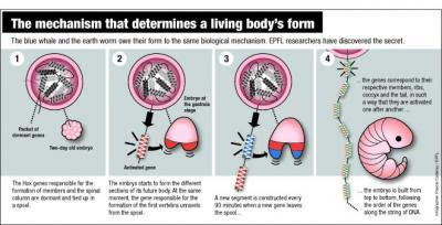
This is a diagram of the mechanism for form. The blue whale and the earth worm owe their form to the same biological mechanism. EPFL researchers have discovered the secret. (Credit: Infographic courtesy of Pascal Coderay, EPFL)

Their discovery will be published October 13, 2011 in the journal Science.

The embryo is built one layer at a time

During the development of an embryo, everything happens at a specific moment. In about 48 hours, it will grow from the top to the bottom, one slice at a time -- scientists call this the embryo's segmentation. "We're made up of thirty-odd horizontal slices," explains Denis Duboule, a professor at EPFL and Unige. "These slices correspond more or less to the number of vertebrae we have."

Every hour and a half, a new segment is built. The genes corresponding to the cervical vertebrae, the thoracic vertebrae, the lumbar vertebrae and the tailbone become activated at exactly the right moment one after another. "If the timing is not followed to the letter, you'll end up with ribs coming off your lumbar vertebrae," jokes Duboule. How do the genes know how to launch themselves into action in such a perfectly synchronized manner? "We assumed that the DNA played the role of a kind of clock. But we didn't understand how."

When DNA acts like a mechanical clock


Very specific genes, known as "Hox," are involved in this process. Responsible for the formation of limbs and the spinal column, they have a remarkable characteristic. "Hox genes are situated one exactly after the other on the DNA strand, in four groups. First the neck, then the thorax, then the lumbar, and so on," explains Duboule. "This unique arrangement inevitably had to play a role."

The process is astonishingly simple. In the embryo's first moments, the Hox genes are dormant, packaged like a spool of wound yarn on the DNA. When the time is right, the strand begins to unwind. When the embryo begins to form the upper levels, the genes encoding the formation of cervical vertebrae come off the spool and become activated. Then it is the thoracic vertebrae's turn, and so on down to the tailbone. The DNA strand acts a bit like an old-fashioned computer punchcard, delivering specific instructions as it progressively goes through the machine.

"A new gene comes out of the spool every ninety minutes, which corresponds to the time needed for a new layer of the embryo to be built," explains Duboule. "It takes two days for the strand to completely unwind; this is the same time that's needed for all the layers of the embryo to be completed."

This system is the first "mechanical" clock ever discovered in genetics. And it explains why the system is so remarkably precise.

This discovery is the result of many years of work. Under the direction of Duboule and Daniël Noordermeer, the team analyzed thousands of Hox gene spools. With assistance from the Swiss Institute for Bioinformatics, the scientists were able to compile huge quantities of data and model the structure of the spool and how it unwinds over time.

The snake: a veritable vertebral assembly line

The process discovered at EPFL is shared by numerous living beings, from humans to some kinds of worms, from blue whales to insects. The structure of all these animals -- the distribution of their vertebrae, limbs and other appendices along their bodies -- is programmed like a sheet of player-piano music by the sequence of Hox genes along the DNA strand.

The sinuous body of the snake is a perfect illustration. A few years ago, Duboule discovered in these animals a defect in the Hox gene that normally stops the vertebrae-making process.

"Now we know what's happening. The process doesn't stop, and the snake embryo just keeps on making vertebrae, all identical, until the process just runs out of steam."

The Hox clock is a demonstration of the extraordinary complexity of evolution. One notable property of the mechanism is its extreme stability, explains Duboule. "Circadian or menstrual clocks involve complex chemistry. They can thus adapt to changing contexts, but in a general sense are fairly imprecise. The mechanism that we have discovered must be infinitely more stable and precise. Even the smallest change would end up leading to the emergence of a new species."


Recommend this story on Facebook, Twitter, and Google +1

Friday, October 14, 2011

Dark Matter of the Genome Revealed


6MK9UNF4B2AX
An international team of researchers has discovered the vast majority of the so-called "dark matter" in the human genome, by means of a sweeping comparison of 29 mammalian genomes. The team, led by scientists from the Broad Institute, has pinpointed the parts of the human genome that control when and where genes are turned on. This map is a critical step in interpreting the thousands of genetic changes that have been linked to human disease.

Rendering of DNA. Researchers have discovered the vast majority of the so-called "dark matter" in the human genome, by means of a sweeping comparison of 29 mammalian genomes. (Credit: iStockphoto/Martin McCarthy)

Their findings appear online October 12 in the journal Nature.

Early comparison studies of the human and mouse genomes led to the surprising discovery that the regulatory information that controls genes dwarfs the information in the genes themselves. But, these studies were indirect: they could infer the existence of these regulatory sequences, but could find only a small fraction of them. These mysterious sequences have been referred to as the dark matter of the genome, analogous to the unseen matter and energy that make up most of the universe.

This new study enlisted a menagerie of mammals -- including rabbit, bat, elephant, and more -- to reveal these mysterious genomic elements.

Over the last five years, the Broad Institute, the Genome Institute at Washington University, and the Baylor College of Medicine Human Genome Sequencing Center have sequenced the genomes of 29 placental mammals. The research team compared all of these genomes, 20 of which are first reported in this paper, looking for regions that remained largely unchanged across species.

"With just a few species, we didn't have the power to pinpoint individual regions of regulatory control," said Manolis Kellis, last author of the study and associate professor of computer science at MIT. "This new map reveals almost 3 million previously undetectable elements in non-coding regions that have been carefully preserved across all mammals, and whose disruptions appear to be associated with human disease."

These findings could yield a deeper understanding of disease-focused studies, which look for genetic variants closely tied to disease.

"Most of the genetic variants associated with common diseases occur in non-protein coding regions of the genome. In these regions, it is often difficult to find the causal mutation," said first author Kerstin Lindblad-Toh, scientific director of vertebrate genome biology at the Broad and a professor in comparative genomics at Uppsala University, Sweden. "This catalog will make it easier to decipher the function of disease-related variation in the human genome."

This new map helps pinpoint those mutations that are likely responsible for disease, as they have been preserved across millions of years of evolution, but are commonly disrupted in individuals that suffer from a given disease. Knowing the causal mutations and their likely functions can then help uncover the underlying disease mechanisms and reveal potential drug targets.

The scientists were able to suggest possible functions for more than half of the 360 million DNA letters contained in the conserved elements, revealing the hidden meaning behind the As, Cs, Ts, and Gs. These revealed:
  • Almost 4,000 previously undetected exons, or segments of DNA that code for protein
  • 10,000 highly conserved elements that may be involved in how proteins are made
  • More than 1,000 new families of RNA secondary structures with diverse roles in gene regulation
  • 2.7 million predicted targets of transcription factors, proteins that control gene expression

"We can use this treasure trove of new elements to revisit disease association studies, focusing on those that disrupt conserved elements and trying to discern their likely functions," said Kellis. "Using a single genome, the language of DNA seems cryptic. When studied through the lens of evolution, words light up and gain meaning."

The researchers were also able to harness this collection of genomes to look back in time, across more than 100 million years of evolution, to uncover the fundamental changes that shaped mammalian adaptation to different environments and lifestyles. The researchers revealed specific proteins under rapid evolution, including some related to the immune system, taste perception, and cell division. They also uncovered hundreds of protein domains within genes that are evolving rapidly, some of which are related to bone remodeling and retinal functions.

"The comparison of mammalian genomes reveals the regulatory controls that are common across all mammals," said Eric Lander, director of the Broad Institute and the third corresponding author of the paper. "These evolutionary innovations were devised more than 100 million years ago and are still at work in the human population today."

In addition to finding the DNA controls that are common across all mammals, the comparison highlighted areas that have been changing rapidly only in the human and primate genomes. Researchers had previously uncovered two hundred of these regions, some of which are linked to brain and limb development. The expanded list -- which now includes more than 1,000 regions -- will give scientists new starting points for understanding human evolution.

The comparison of many complete genomes is beginning to offer a clear view of once indiscernible genomic regions, and with additional genomes, that resolution will only increase. "The power of this resource is that it continues to improve with the inclusion of more species," said Lindblad-Toh. "It's a very systematic and unbiased approach that will only become more powerful with the inclusion of additional genomes."

Other Broad researchers who contributed to this work include Manuel Garber, Or Zuk, Michael F. Lin, Pouya Kheradpour, Jason Ernst, Evan Mauceli, Lucas D. Ward, Michele Clamp, Sante Gnerre, Jessica Alföldi, Jean Chang, Federica Di Palma, Mitchell Guttman, David B. Jaffe, Irwin Jungreis, Marcia Lara, Jim Robinson, Xiaohui Xie, Michael C. Zody, and members of the Broad Institute Sequencing Platform and Whole Genome Assembly Team.

This project was supported by the National Human Genome Research Institute, National Institute for General Medicine, the European Science Foundation, National Science Foundation, the Sloan Foundation, an Erwin Schrödinger Fellowship, the Gates Cambridge Trust, Novo Nordisk Foundation, University of Copenhagen, the David and Lucile Packard Foundation, the Danish Council for Independent Research Medical Sciences, and The Lundbeck Foundation.

Recommend this story on Facebook, Twitter, and Google +1


Friday, September 23, 2011

All-access genome: New study explores packaging of DNA


While efforts to unlock the subtleties of DNA have produced remarkable insights into the code of life, researchers still grapple with fundamental questions. For example, the underlying mechanisms by which human genes are turned on and off -- generating essential proteins, determining our physical traits, and sometimes causing disease -- remain poorly understood.

Fluorescence resonance energy transfer (FRET): experimental design. Nucleosomes are constructed having a fluorescent donor (cyan) attached to one end of the DNA, and a fluorescent acceptor (magenta) attached nearby on the histone protein core. In the middle diagram, spontaneous partial unwrapping of the DNA thread exposes a hidden DNA target site (hatched area), which is site-specific for the DNA binding protein LexA. When LexA is added in sufficient concentration, nucleosomes are temporarily trapped in their unwrapped state. The distance between the two fluorescent molecules changes as the DNA unwraps and rewraps, allowing the process to be precisely measured. Credit: Reprinted from: Journal of Molecular Biology, volume 411(2), Tims HS, Gurunathan K, Levitus M, Widom J, Dynamics of nucleosome invasion by DNA binding proteins, pgs 430-48, with permission from Elsevier.

Biophysicists Marcia Levitus and Kaushik Gurunathan at the Biodesign Institute at Arizona State University along with their colleagues Hannah S. Tims, and Jonathan Widom of Northwestern University in Evanston, Illinois have been preoccupied with tiny, spool-like entities known as nucleosomes. Their latest insights into how these structures wrap and unwrap, permitting regulatory proteins to access, bind with and act on regions of DNA, recently appeared in the Journal of Molecular Biology.

Nucleosomes, Levitus explains, are essential components of the genome, acting to regulate access to DNA and protect it from harm. Nucleosome structure permits the entire strand of human DNA, roughly 6 feet in length, to be densely packed into the nucleus of every cell—an area just 10 microns in diameter. This occurs after nucleosomes assemble and fold into higher order structures, culminating in the formation of chromosomes.

Each nucleosome (there are roughly 30 million per cell) consists of a 147 base pair segment of DNA. This length of DNA thread is wound 1.67 times around the spool-like protein units, known as histones. The histone complex, together with its windings of DNA, forms the nucleosome core particle.

A multitude of proteins must act on regions of the DNA strand, by binding with appropriate target sites. Essential functions rely on these operations, including gene expression, replication and repair of damaged regions of the DNA molecule. But in eukaryotic cells like those of humans, some 75-80 percent of the DNA strand is curled up and hidden in the nucleosomes—inaccessible to protein binding interactions.

In earlier work, the group was able to show that nucleosomes are dynamic structures, quite different from the static pictures produced by X-ray crystallography. Lengths of DNA make themselves available for protein interaction by unwrapping and rewrapping around the histone core. When nucleosomes unwrap, proteins present in sufficient concentration can find their DNA targets and bind with them.

In order to observe and characterize the dynamic behavior of nucleosomes, the team relied on a versatile imaging method known as Fluorescence Resonance Energy Transfer or FRET. The technique allows researchers to look at a pair of fluorescent molecules or fluorophores, one of which is attached to the end of the exposed DNA strand, the other, to one of the histones around which the DNA is coiled, (see figure 1).

As Levitus explains, spontaneous unwrapping and rewrapping of DNA changes the distance between fluorophores, signaling that the process has occurred and allowing the group to quantify the frequency and rate of DNA exposure and concealment. "Although FRET has been used for decades to measure molecular distances in biological systems, dynamic biomolecules such as nucleosomes present particular challenges," notes Levitus. Traditionally, FRET experiments are performed with protein solutions containing many billions of particles. In the case of nucleosomes however, the dynamic behavior of each particle is crucial and bulk measurements using FRET are not effective.
This graphic shows the time elapsed during DNA unwrapping (a) and re-wrapping (b) as measured by FRET analysis. FRET works by measuring the distance between a pair of fluorescent molecules of fluorophores -- one attached to the end of the DNA and the other attached to the histone protein spool around which the DNA "thread" winds and unwinds. Credit: Reprinted from: Journal of Molecular Biology, volume 411(2), Tims HS, Gurunathan K, Levitus M, Widom J, Dynamics of nucleosome invasion by DNA binding proteins, pgs 430-48, with permission from Elsevier.


"In simple terms, if one wanted to understand how humans clap, it would be useless to listen to the whole planet clapping at once. Instead, one would listen to a few individuals, and that is exactly what we did with nucleosomes," Levitus says.

The results of initial studies were revealing. For base pair sequences along the nucleosomes' outer rind, spontaneous DNA unwrapping occurs at a rapid rate— about 4 times per second. This corresponds to a period of only 250 milliseconds during which this region of DNA remains fully wrapped and occluded by the histone complex. Once unwrapped, the DNA remains exposed for 10-50 milliseconds.

These findings present a plausible mechanism to allow protein binding with unwrapped DNA in vivo, so long as the binding sites occur near the ends of wrapped nucleosomal DNA.

The new study also examines, for the first time, the condition of DNA sequences occurring further along the wound length of nucleosomal DNA, that is, closer to the nucleosome's center. Here, rates of DNA unwrapping decreased by orders of magnitude, (see figure 2).

To examine this phenomenon, the group used a site-specific binding protein of Escherichia coli (known as LexA) to identify binding site exposure caused by nucleosome unwrapping. The nucleosomes were labeled with a FRET dye, which allowed the binding process of LexA and its target to be visualized. In successive experiments, the team shifted the binding sites in 10 base pair increments from the end of the nucleosome toward the middle.

The changes in unwrapping rate observed as the binding site was successively moved further inside the nucleosome were dramatic. In one case, a change in position of just 10 base pairs could produce a 250-fold decrease in unwrapping rate of the binding region.

These results prompt the question of how DNA binding sites more deeply wound within the nucleosome are able to successfully interact with their respective protein binders in vivo. The team proposes several possible mechanisms that would permit rapid access to hidden DNA binding regions, even where intrinsic rates of nucleosome unwrapping are low.

One hypothesis is that two or more proteins with target sites on the same nucleosome can act cooperatively, with one protein holding the momentarily unwrapped DNA open as the other enters the nucleosome and invades more inward regions of the DNA sequence, in what the authors describe as a ratcheting process.

Jonathan Widom, Dr. Levitus' collaborator and a co-author of the new study was responsible for much of the pathbreaking research into nucleosome complexity. Dr. Widom died unexpectedly this past month. He was honored for his generosity, prolific research and outstanding contributions to biology in the August 25th issue of the journal Nature.

"I consider myself tremendously fortunate to have had the chance to collaborate with Jon Widom," Levitus says. "Jon has been, and will continue to be, an incredible role model. His generosity, humility, and scientific genius has touched my life in many ways, and his death will leave a void that will be felt for many years to come."

Ongoing research into the subtleties of nucleosome behavior promises to yield rich dividends for genomic science in general and provide a deeper appreciation for foundational issues of health and disease.

Provided by Arizona State University


Saturday, August 20, 2011

Biologists Discovery May Force Revision of Biology Textbooks: Novel Chromatin Particle Halfway Between DNA and a Nucleosome


Basic biology textbooks may need a bit of revising now that biologists at UC San Diego have discovered a never-before-noticed component of our basic genetic material.
Biologists have discovered a novel chromatin 
particle halfway between DNA and a nucleosome. 
While it looks like a nucleosome, it is in fact a 
distinct particle of its own, researchers say. 
(Credit: James Kadonaga, UC San Diego)

According to the textbooks, chromatin, the natural state of DNA in the cell, is made up of nucleosomes. And nucleosomes are the basic repeating unit of chromatin.

When viewed by a high powered microscope, nucleosomes look like beads on a string. But in the Aug. 19 issue of the journal Molecular Cell, UC San Diego biologists report their discovery of a novel chromatin particle halfway between DNA and a nucleosome. While it looks like a nucleosome, they say, it is in fact a distinct particle of its own.

"This novel particle was found as a precursor to a nucleosome," said James Kadonaga, a professor of biology at UC San Diego who headed the research team and calls the particle a "pre-nucleosome." "These findings suggest that it is necessary to reconsider what chromatin is. The pre-nucleosome is likely to be an important player in how our genetic material is duplicated and used."


The biologists say that while the pre-nucleosome may look something like a nucleosome under the microscope, biochemical tests have shown that it is in reality halfway between DNA and a nucleosome.

These pre-nucleosomes, the researchers say, are converted into nucleosomes by a motor protein that uses the energy molecule ATP.

"The discovery of pre-nucleosomes suggests that much of chromatin, which has been generally presumed to consist only of nucleosomes, may be a mixture of nucleosomes and pre-nucleosomes," said Kadonaga. "So, this discovery may be the beginning of a revolution in our understanding of what chromatin is."

"The packaging of DNA with histone proteins to form chromatin helps stabilize chromosomes and plays an important role in regulating gene activities and DNA replication," said Anthony Carter, who oversees chromatin grants at the National Institute of General Medical Sciences of the National Institutes of Health, which funded the research. "The discovery of a novel intermediate DNA-histone complex offers intriguing insights into the nature of chromatin and may help us better understand how it impacts these key cellular processes."

Saturday, August 13, 2011

Supergene is key to copycat butterflies


Since Charles Darwin, biologists have pondered the mystery of "mimicry butterflies", which survive by copying the wing patterns of other butterflies that taste horrible to their predators, birds.
This undated handout photo released by the CNRS shows butterflies, Melinaea mneme (top) and Heliconius numata. The mystery of how a butterfly has changed its wing patterns to mimic neighbouring species and avoid being eaten by birds has been solved by a team of European scientists.

The answer, according to a study released on Friday, lies in an astonishing cluster of about 30 genes in a single chromosome.

"We were blown away by what we found," said Mathieu Joron of France's National Museum of Natural History, who led the probe into what is being called a "supergene".

"These butterflies are the 'transformers' of the insect world," said Joron.

"But instead of being able to turn from a car into a robot with the flick of a switch, a single genetic switch allows these insects to morph into several different mimetic forms.

"It is amazing, and the stuff of science fiction. Now we are starting to understand how this switch can have such a pervasive effect."


The trick, known as Muellerian mimicry, was investigated by French and British scientists, who focussed on a species of Amazonian rainforest butterfly, Heliconius numata.

It is able to copy the colour patterns of several species of the Melinaea butterfly which are unpalatable to birds.

The "supergene" comprises a tightly packed region of genes on a single chromosome which control different elements of the wing pattern.

"By changing just one gene, the butterfly is able to fool its predators," explained Richard ffrench-Constant of the University of Exeter, southwestern England.

Even more astonishing is that three versions of the chromosome exist within this species, with each version controlling distinct wing-pattern forms.

Even though the butterflies look quite different from each other, they have the same DNA.

The supergene apparently transmits in a block from generation to generation, rather than go through recombination -- the mingling of genes from both parents.

The "supergene" also appears important in other species, say the authors.

One such species, the peppered moth, developped black wings in 19th-century Britain as a means of gaining camouflage in the sooty industrial environment.

"It's a gene that really packs an evolutionary punch," said ffrench-Constant. The paper is published online by the British science journal Nature.

Thursday, July 7, 2011

The turn of the corkscrew : Structural analysis uncovers mechanisms of gene expression


The diverse functions of living cells are all based on the information encoded in the structure of the hereditary material DNA. Gene expression must therefore be tightly controlled, and this task is accomplished by the binding of regulatory proteins to, and their removal from, specific DNA sequences. One class of large molecular machines known as Swi2/Snf2 remodelers plays a central role in modulating these processes. However, until now, it was not clear how Swi2/Snf2 remodelers actually work. A team led by Professor Karl-Peter Hopfner at the Gene Center at Ludwig-Maximilians-Universität (LMU) in Munich has clarified the structure and function of the remodeler Mot1 (Modifier of Transcription 1), which binds directly to DNA. It turns out that Mot1 acts like a molecular corkscrew that migrates along the DNA, following its helical contour. During its progress, Mot1 displaces a crucial transcription factor called TBP (for “TATA Box Binding Protein”) from the DNA. Removal of TBP from a TATA box represses transcription of the adjacent gene, and the protein encoded by that gene is no longer synthesized. At the same time, TBP is stabilized and its binding specificity is changed, which facilitates the expression of genes that lack TATA boxes and code for other proteins. (Nature 6 July 2011)

The DNA in the cells of higher organisms is tightly wrapped around protein complexes called nucleosomes. This type of structural organization not only makes it possible to package the long DNA molecules in a highly compact form, it also provides the basis for the controlled expression of genetic information. Densely packed sections of the molecule are effectively in a repressed state, and genes located in these DNA segments cannot be transcribed. Activation of repressed genes depends on the intervention of complex molecular machines, so-called Swi2/Snf2 remodelers, which reorganize condensed stretches of DNA so as to make them accessible for transcription. The precise mode of action of remodelers has so far been unclear, mainly because most of them are made up of several components and the active complexes are difficult to study. This is why Hopfner chose to study Mot1, which is a comparatively simple representative of the family that functions as an Swi2/Snf2 remodeler on its own, and can serve as a guide to understanding the more complicated members of the class. Mot1 is known to participate in the control of gene expression, but how exactly it does so is not well understood.

The first stage of the process that leads to the synthesis of a given protein is the transcription of the specific segment of DNA that codes for it into molecules of messenger RNA. This initial step requires the action of so-called transcription factors. One of the most important of these is TBP, which binds preferentially to DNA sequences called TATA boxes that are located near the beginnings of many genes. Binding of TBP introduces a kink into the DNA, and this landmark serves as a platform for the binding of further proteins, ultimately leading to the assembly of the complex necessary for the initiation of transcription. Mot1 regulates transcription by actively removing TBP from the DNA, using ATP as a source of energy. “How Mot1 dissociates the TBP-DNA complex was completely unclear up to now,” says Hopfner. With the aid of so-called hybrid methods - in which data obtained from high-resolution X-ray diffraction analysis of the crystallized protein complex with images of the same molecular complex taken with the electron microscope were combined - Hopfner’s team was able to define the three-dimensional structure of the Mot1-TBP complex for the first time. This revealed how Mot1 recognizes the surface of the DNA-bound TBP. “Once Mot1 has recognized TBP, it binds to the adjacent DNA and begins to migrate along the DNA strand, using the energy released by the hydrolysis of ATP to power its movement. This helical movement, which is reminiscent of the insertion of a corkscrew, causes TBP to detach from the DNA,” explains Dr Petra Wollmann, who is first author on the new study. The researchers were surprised to find that Mot1 contains a strikingly extended loop. After TBP has dissociated from the DNA, this loop masks TBP’s DNA binding site and prevents the protein from reoccupying it.


Previous studies had reported what appeared to be paradoxical observations, which indicated that Mot1 inhibits transcription of TATA box-containing genes while facilitating the expression of genes that lack canonical TATA boxes. “Our results suggest that Mot1 also stabilizes the DNA-free conformation of TBP, increasing the probability that it can reach, bind to and activate genes that lack TATA boxes,” explains Hopfner. In other words, Mot1 is also a redistribution factor, which enables TBP to bind to different sequences and thus controls its association with other cellular components. This combination of detachment and redistribution functions may be a common feature of remodeling complexes, and would help to explain how they mediate the large-scale redistribution of DNA-binding regulatory proteins. (göd/PH)

The project was carried out under the auspices of two Clusters of Excellence - the Center for Integrated Protein Science Munich (CiPSM) and the Munich Centre for Advanced Photonics (MAP). The work was also supported by the German Research Foundation (DFG) as part of the Collaborate Research Centers (SFB) 646 and TR5, and by the LMUexcellent Investment Fund.

Wednesday, July 6, 2011

Environs Prompt Advantageous Gene Mutations as Plants Grow; Changes Passed to Progeny


If a person were to climb a towering redwood and take a sample from the top and a sample from the bottom of the tree, a comparison would show that the two DNA samples are different.
Research shows that if a person were to climb a towering redwood and take a sample from the top and a sample from the bottom of the tree, a comparison would show that the two DNA samples are different. (Credit: © Galyna Andrushko / Fotolia)

Christopher A. Cullis, chair of biology at Case Western Reserve University, explains that this is the basis of his controversial research findings.

Cullis, who has spent over 40 years studying mutations within plants, most recently flax (Linum usitatissimum), has found that the environment not only weeds out harmful and useless mutations through natural selection, but actually influences helpful mutations.

Cullis published his findings in the International Journal of Genetics and Molecular Biology and repeated them in the Journal of Visualized Experiments, where he challenged other scientists to repeat his experiment themselves.

Specifically, Cullis focuses on mutations involving the appearance of a small sequence of DNA known as LIS-1 and how the environment affects these changes.

The controversy stems from the idea that the environment changes organisms as they grow and these changes are passed on.

While originally accepted, the theory was eventually thrown out because science revealed that animals pass along DNA through their gamete or sex cells, which are not affected by the environment. This concept was assumed to be the same for plants, but Cullis's research says otherwise.

In his second study, three separate strands (the plastic strand, short strand, and tall strand) of the Stormont Cirrus variety of flax were grown under three separate conditions.

Each of the strands had been bred over multiple generations under different conditions: The plastic strand's ancestors were grown under control conditions, the short strand's ancestors were grown under low-nutrient conditions, and the tall strand's ancestors were grown under high-nutrient conditions.


The experiment showed each strand responded to each condition in a different way, corresponding to the environment its ancestors were grown in. The plastic strand outgrew the other strands under control conditions, the short strand outgrew the other strands when few nutrients were available, and the tall strand grew best when nutrients were readily available.

All this information does not completely explain Cullis's assertion that the environment can in a single generation help sift out the useful mutations.

This is where polymerase chain reaction (PCR) amplification of DNA comes in. Through this process, the researchers could see when a specific DNA sequence (in this case LIS-1) appears or disappears.

When the plastic strand is grown under low nutrient conditions, the LIS-1 sequence, which had been absent, appears and continues for future generations. Since the LIS-1 sequence helps plants survive when there is a shortage of nutrients, its presence helps confirm Cullis's belief that the environment can act on how a plant mutates and keep helpful mutations, even within one generation.

These findings help explain why the top of a redwood is genetically different from the bottom. Young redwoods grow by the tips of the existing branches budding into meristems. Each new meristem is different from the tree because the environment has affected its genetic makeup. And as the redwood grows, the top becomes more and more genetically different from the bottom.

Due to the controversy surrounding Cullis's findings, many scientists are hesitant to accept them as true. Cullis himself recalls at first being skeptical and thinking, "If this really works… [we can] get a plant that's better adapted to its environment in one generation."

These adapted plants have practical uses. Cullis hopes to identify the specific gene sequence responsible for flax's ability to withstand harsh environments and insert it into the DNA sequence of other plants so that they too can withstand trying environments.

This would bypass the current method of genetically engineering plants, which involves isolating specific DNA sequences that control heat-resistance, cold-resistance, pest-resistance, etc., and instead narrows the effort down to one DNA sequence.

By inserting this sequence into the plant and growing it in a specific trying environment, scientists could make the plant resistant to what they want. All of that plant's offspring would be adapted to the environment and ready to grow.

By making the plant do all the work, the price of producing better crops would be greatly reduced. This would greatly benefit developing nations that need a large supply of food in an otherwise harsh environment. The DNA sequence may no longer just help the plant survive, but can now help entire countries thrive.

Monday, July 4, 2011

DNA cages 'can survive inside living cells'


Scientists at Oxford University have shown for the first time that molecular cages made from DNA can enter and survive inside living cells.
Human embryonic kidney cells were used to test the DNA cages

The work, a collaboration between physicists and molecular neuroscientists at Oxford, shows that artificial DNA cages that could be used to carry cargoes of drugs can enter living cells, potentially leading to new methods of drug delivery.

A report of the research is published online in the journal ACS Nano.

The cages developed by the researchers are made from four short strands of synthetic DNA. These strands are designed so that they naturally assemble themselves into a tetrahedron (a pyramid with four triangular faces) around 7 nanometres tall.

The Oxford researchers have previously shown that it is possible to assemble these cages around protein molecules, so that the protein is trapped inside, and that DNA cages can be programmed to open when they encounter specific ‘trigger’ molecules that are found inside cells.


In the new experiment they introduced fluorescently-labelled DNA tetrahedrons into human kidney cells grown in the laboratory. They then examined the cells under the microscope and found that the cages remained substantially intact, surviving attack by cellular enzymes, for at least 48 hours. This is a crucial advance: to be useful as a drug delivery vehicle, a DNA cage must enter cells efficiently and survive until it can release its cargo where and when it is needed.

‘At the moment we are only testing our ability to create and control cages made of DNA,’ said Professor Andrew Turberfield of Oxford University’s Department of Physics, who led the work. ‘However, these results are an important first step towards proving that DNA cages could be used to deliver cargoes, such as drugs, inside living cells.’

Professor Turberfield said: ‘Previous studies have shown that the size of particles is an important factor in whether or not they can easily enter cells, with particles with a radius less than 50 nanometres proving much more successful at gaining entry than larger particles. At 7 nanometres across our DNA tetrahedrons are compact enough to easily enter cells but still large enough to carry a useful cargo. More work is now needed to understand exactly how these DNA cages manage to find their way inside living cells.’
Provided by Oxford University

Sunday, July 3, 2011

Researchers decipher protein structure of key molecule in DNA transcription system


The research adds an important link to discoveries that have enabled scientists to gain a deeper understanding of how cells translate genetic information into the proteins and processes of life. The findings, published in the July 3 advance online issue of the journal Nature, were reported by a research team led by Yuichiro Takagi, Ph.D., assistant professor of biochemistry and molecular biology at Indiana University School of Medicine.
Scientists have deciphered the structure of an essential 
part of Mediator, a complex molecular machine that plays 
a vital role in regulating the transcription of DNA.

The fundamental operations of all cells are controlled by the genetic information – the genes –stored in each cell's DNA, a long double-stranded chain. Information copied from sections of the DNA – through a process called transcription – leads to synthesis of messenger RNA, eventually enabling the production of proteins necessary for cellular function. Transcription is undertaken by the enzyme called RNA polymerase II.

As cellular operations proceed, signals are sent to the DNA asking that some genes be activated and others be shut down. The Mediator transcription regulator accepts and interprets those instructions, telling RNA polymerase II where and when to begin the transcription process.

Mediator is a gigantic molecular machine composed of 25 proteins organized into three modules known as the head, the middle, and the tail. Using X-ray crystallography, the Takagi team was able to describe in detail the structure of the Mediator Head module, the most important for interactions with RNA polymerase II.

"It's turned out to be extremely novel, revealing how a molecular machine is built from multiple proteins," said Takagi.

"As a molecular machine, the Mediator head module needs to have elements of both stability and flexibility in order to accommodate numerous interactions. A portion of the head we named the neck domain provides the stability by arranging the five proteins in a polymer-like structure," he said.


"We call it the alpha helical bundle," said Dr. Takagi. "People have seen structures of alpha helical bundles before but not coming from five different proteins."
A research team led by Yuichiro Takagi, Ph.D.,
Indiana University School of Medicine, has
deciphered the structure of an essential part
of Mediator, a complex molecular machine
that plays a vital role in regulating the
transcription of DNA.

"This is a completely noble structure," he said.

One immediate benefit of the research will be to provide detailed mapping of previously known mutations that affect the regulation of the transcription process, he said.

The ability to solve such complex structures will be important because multi-protein complexes such as Mediator will most likely become a new generation of drug targets for treatment of disease, he said.

Previously, the structure of RNA polymerase II was determined by Roger Kornberg of Stanford University, with whom Dr. Takagi worked prior to coming to IU School of Medicine. Kornberg received the Nobel Prize in 2006 for his discoveries. The researchers who described the structure of the ribosome, the protein production machine, were awarded the Nobel Prize in 2009. The structure of the entire Mediator has yet to be described, and thus remains the one of grand challenges in structure biology. Dr. Takagi's work on the Mediator head module structure represents a major step towards a structure determination of the entire Mediator. In addition to Dr. Takagi as a senior author, the lead author for the Nature paper was Tsuyoshi Imasaki, Ph.D., of the IU School of Medicine. Other collaborators included researchers at The Scripps Research Institute, Stanford University, Memorial Sloan-Kettering Cancer Center and the European Molecular Biology Laboratory.

Wednesday, June 22, 2011

Non-coding RNA has role in inherited neurological disorder -- and maybe other brain diseases too


A team of scientists, led by researchers at the University of California, San Diego School of Medicine, have uncovered a novel mechanism regulating gene expression and transcription linked to Spinocerebellar ataxia 7, an inherited neurological disorder. The discovery promises to have broad ramifications, suggesting that abundant non-coding transcripts of ribonucleic acid (RNA) may be key players in neurological development and function, and could be powerful targets for future clinical therapies.
Researchers have discovered that expression
of the ataxin‑7 gene - the cause of the
neurological disorder spinocerebellar
ataxia type 7 - has two regulators: a highly
conserved, multi‑tasking protein called
CTCF and, surprisingly, an adjacent promoter
containing non‑coding RNA.
Credit: Illustration courtesy of Christina
Takamatsu‑Butler, UC San Diego.

The research, headed by Albert La Spada, MD, PhD, chief of the division of genetics in the UCSD department of pediatrics, and professor of cellular and molecular medicine, neurosciences and biological sciences, is published in the June 22 issue of the journal Neuron.

"Our paper highlights a number of important emerging themes in our understanding of gene regulation in the brain," said La Spada, who is also associate director of the UCSD Institute for Genomic Medicine.

"With the advent of new technologies, science has learned that the vast majority of our transcripts are non-coding," said La Spada. "The challenge going forward is to determine what they do do, and if they have specific functions. It now seems increasingly likely that a multitude of these non-coding RNAs help finely tune transcription regulation in the brain, and perturbation of their work is linked to disease. If we can figure out exactly how, we should be able to gain new insights into how the brain is so precisely regulated – knowledge that may help us better understand how the brain works."


Spinocerebellar ataxia 7 is one of several types of spinocerebellar ataxia (SCA), genetic degenerative disorders characterized by atrophy in the cerebellum of the brain, progressive loss of physical coordination – and in the case of type 7 – retinal degeneration that can result in blindness. There is currently no known cure.

Many SCAs are classified as polyglutamine diseases, caused when a protein associated with the disease contains too many repeats of the amino acid glutamine. Polyglutamine diseases are also known as "CAG Triplet Repeat Disorders" because CAG is the sequence of nucleic acids that codes for glutamine.

La Spada and colleagues have long studied SCA. In 2001, they were the first to demonstrate that SCA7 retinal degeneration was the result of transcription dysregulation of ataxin-7, the protein associated with SCA7. Following up, they decided to learn how the gene that expresses ataxin-7 is itself regulated.

The researchers found not one, but two, regulators. The first is called CTCF, a highly conserved protein that regulates a variety of transcriptional processes, most notable establishing insulator domains and controlling genomic imprinting. But they also discovered an adjacent, alternative promoter dubbed intron 2 promoter (P2A) and a transcribed antisense, non-coding RNA, which they labeled SpinoCerebellarAtaxia-AntisenseNoncodingTranscript1 or SCAANT1.

Antisense RNA is single-stranded ribonucleic acid whose primary function appears to be as an inhibitor or suppressor of a gene, though sometimes it can promote gene expression instead. Most antisense RNAs are non-coding, meaning that their sequences do not provide information for making proteins. Even though non-coding RNAs do not provide instructions for the production of vital proteins, they comprise the bulk of the human genome. A major challenge for biomedical research in the 21st century is to figure what they do, and how they do it.

In their Neuron paper, La Spada and colleagues highlight one function, at least for SCAANT1. When they investigated how CTCF regulated ataxin-7 gene expression in transgenic mice, they discovered that CTCF promotes the production of SCAANT1 which in turn represses the newly discovered ataxin-7 sense promoter P2A. In mice lacking SCAANT1, sense promoter P2A is de-repressed, allowing a mutant ataxin-7 gene to be expressed, resulting in mice with a version of SCA7. The scientists found a similar lack of antisense SCAANT1 in the fibroblasts and white blood cells taken from human patients with SCA7, implicating deregulation of this pathway in the disease process.

As many inherited neurological disorders are now known to exhibit such overlapping "bidirectional" transcription, the findings in SCA7 could shed light on similar abnormalities with non-coding RNA function in a number of brain diseases.

Provided by University of California - San Diego

Wednesday, June 15, 2011

New Clues About Aging: Genetic Splicing Mechanism Triggers Both Premature Aging Syndrome and Normal Cellular Aging


National Institutes of Health researchers have identified a new pathway that sets the clock for programmed aging in normal cells. The study provides insights about the interaction between a toxic protein called progerin and telomeres, which cap the ends of chromosomes like aglets, the plastic tips that bind the ends of shoelaces.
The 46 human chromosomes are shown in blue, with 
the telomeres appearing as white pinpoints. The DNA 
has already been copied, so each chromosome is 
actually made up of two identical lengths of DNA, 
each with its own two telomeres. 
(Credit: Hesed Padilla-Nash and Thomas Ried)

The study by researchers from the National Human Genome Research Institute (NHGRI) appears online in the Journal of Clinical Investigation.

Telomeres wear away during cell division. When they degrade sufficiently, the cell stops dividing and dies. The researchers have found that short or dysfunctional telomeres activate production of progerin, which is associated with age-related cell damage. As the telomeres shorten, the cell produces more progerin.

Progerin is a mutated version of a normal cellular protein called lamin A, which is encoded by the normal LMNA gene. Lamin A helps to maintain the normal structure of a cell's nucleus, the cellular repository of genetic information.

In 2003, NHGRI researchers discovered that a mutation in LMNA causes the rare premature aging condition, progeria, formally known as known as Hutchinson-Gilford progeria syndrome. Progeria is an extremely rare disease in which children experience symptoms normally associated with advanced age, including hair loss, diminished subcutaneous fat, premature atherosclerosis and skeletal abnormalities. These children typically die from cardiovascular complications in their teens.

"Connecting this rare disease phenomenon and normal aging is bearing fruit in an important way," said NIH Director Francis S. Collins, M.D., Ph.D., a senior author of the current paper. "This study highlights that valuable biological insights are gained by studying rare genetic disorders such as progeria. Our sense from the start was that progeria had a lot to teach us about the normal aging process and clues about more general biochemical and molecular mechanisms."


Collins led the earlier discovery of the gene mutation responsible for progeria and subsequent advances at NIH in understanding the biochemical and molecular underpinnings of the disease.

In a 2007 study, NIH researchers showed that normal cells of healthy people can produce a small amount of progerin, the toxic protein, even when they do not carry the mutation. The more cell divisions the cell underwent, the shorter the telomeres and the greater the production of progerin. But a mystery remained: What was triggering the production of the toxic progerin protein?

The current study shows that the mutation that causes progeria strongly activates the splicing of lamin A to produce the toxic progerin protein, leading to all of the features of premature aging suffered by children with this disease. But modifications in the splicing of LMNA are also at play in the presence of the normal gene.

The research suggests that the shortening of telomeres during normal cell division in individuals with normal LMNA genes somehow alters the way a normal cell processes genetic information when turning it into a protein, a process called RNA splicing. To build proteins, RNA is transcribed from genetic instructions embedded in DNA. RNA does not carry all of the linear information embedded in the ribbon of DNA; rather, the cell splices together segments of genetic information called exons that contain the code for building proteins, and removes the intervening letters of unused genetic information called introns. This mechanism appears to be altered by telomere shortening, and affects protein production for multiple proteins that are important for cytoskeleton integrity. Most importantly, this alteration in RNA splicing affects the processing of the LMNA messenger RNA, leading to an accumulation of the toxic progerin protein.

Cells age as part of the normal cell cycle process called senescence, which progressively advances through a limited number of divisions in the cell lifetime. "Telomere shortening during cellular senescence plays a causative role in activating progerin production and leads to extensive change in alternative splicing in multiple other genes," said lead author Kan Cao, Ph.D., an assistant professor of cell biology and molecular genetics at the University of Maryland, College Park.

Telomerase is an enzyme that can extend the structure of telomeres so that cells continue to maintain the ability to divide. The study supplied support for the telomere-progerin link, showing that cells that have a perpetual supply of telomerase, known as immortalized cells, produce very little progerin RNA. Most cells of this kind are cancer cells, which do not reach a normal cell cycle end point, and instead replicate out of control.

The researchers also conducted laboratory tests on normal cells from healthy individuals using biochemical markers to indicate the occurrence of progerin-generating RNA splicing in cells. The cell donors ranged in age from 10 to 92 years. Regardless of age, cells that passed through many cell cycles had progressively higher progerin production. Normal cells that produce higher concentrations of progerin also displayed shortened and dysfunctional telomeres, the tell-tale indication of many cell divisions.

In addition to their focus on progerin, the researchers conducted the first systematic analysis across the genome of alternative splicing during cellular aging, considering which other protein products are affected by jumbled instructions as RNA molecules assemble proteins through splicing. Using laboratory techniques that analyze the order of chemical units of RNA, called nucleotides, the researchers found that splicing is altered by short telomeres, affecting lamin A and a number of other genes, including those that encode proteins that play a role in the structure of the cell.

The researchers suggest that the combination of telomere fraying and loss with progerin production together induces cell aging. This finding lends insights into how progerin may participate in the normal aging process.

For more about Hutchinson-Gilford progeria syndrome, see: http://www.genome.gov/11007255.

Sunday, January 2, 2011

Your Genome in Minutes: New Technology Could Slash Sequencing Time


Scientists from Imperial College London are developing technology that could ultimately sequence a person's genome in mere minutes, at a fraction of the cost of current commercial techniques.
Dr Joshua Edel shows the prototype chip,
and an array of the chips prior to use.
(Credit: Image courtesy of Imperial College London)

The researchers have patented an early prototype technology that they believe could lead to an ultrafast commercial DNA sequencing tool within ten years. Their work is described in a study published this month in the journal Nano Letters.

The research suggests that scientists could eventually sequence an entire genome in a single lab procedure, whereas at present it can only be sequenced after being broken into pieces in a highly complex and time-consuming process. Fast and inexpensive genome sequencing could allow ordinary people to unlock the secrets of their own DNA, revealing their personal susceptibility to diseases such as Alzheimer's, diabetes and cancer. Medical professionals are already using genome sequencing to understand population-wide health issues and research ways to tailor individualised treatments or preventions.

Dr Joshua Edel, one of the authors on the study from the Department of Chemistry at Imperial College London, said: "Compared with current technology, this device could lead to much cheaper sequencing: just a few dollars, compared with $1m to sequence an entire genome in 2007. We haven't tried it on a whole genome yet but our initial experiments suggest that you could theoretically do a complete scan of the 3,165 million bases in the human genome within minutes, providing huge benefits for medical tests, or DNA profiles for police and security work. It should be significantly faster and more reliable, and would be easy to scale up to create a device with the capacity to read up to 10 million bases per second, versus the typical 10 bases per second you get with the present day single molecule real-time techniques."

In the new study, the researchers demonstrated that it is possible to propel a DNA strand at high speed through a tiny 50 nanometre (nm) hole -- or nanopore -- cut in a silicon chip, using an electrical charge. As the strand emerges from the back of the chip, its coding sequence (bases A, C, T or G) is read by a 'tunnelling electrode junction'. This 2 nm gap between two wires supports an electrical current that interacts with the distinct electrical signal from each base code. A powerful computer can then interpret the base code's signal to construct the genome sequence, making it possible to combine all these well-documented techniques for the first time.

Sequencing using nanopores has long been considered the next big development for DNA technology, thanks to its potential for high speed and high-capacity sequencing. However, designs for an accurate and fast reader have not been demonstrated until now.

Co-author Dr Emanuele Instuli, from the Department of Chemistry at Imperial College London, explained the challenges they faced in this research: "Getting the DNA strand through the nanopore is a bit like sucking up spaghetti. Until now it has been difficult to precisely align the junction and the nanopore. Furthermore, engineering the electrode wires with such dimensions approaches the atomic scale and is effectively at the limit of existing instrumentation. However in this experiment we were able to make two tiny platinum wires into an electrode junction with a gap sufficiently small to allow the electron current to flow between them."

This technology would have several distinct advantages over current techniques, according to co-author, Aleksandar Ivanov from the Department of Chemistry at Imperial College London: "Nanopore sequencing would be a fast, simple procedure, unlike available commercial methods, which require time-consuming and destructive chemical processes to break down and replicate small sections of the DNA molecules to determine their sequence. Additionally, these silicon chips are incredibly durable compared with some of the more delicate materials currently used. They can be handled, washed and reused many times over without degrading their performance."

Dr Tim Albrecht, another author on the study, from the Department of Chemistry at Imperial College London, says: "The next step will be to differentiate between different DNA samples and, ultimately, between individual bases within the DNA strand (ie true sequencing). I think we know the way forward, but it is a challenging project and we have to make many more incremental steps before our vision can be realised."

This work was supported by the Wellcome Trust Translational Award and the Corrigan Foundation.