BTemplates.com

Powered by Blogger.

Pageviews past week

Quantum mechanics

Auto News

artificial intelligence

About Me

Recommend us on Google!

Information Technology

Popular Posts

Friday, June 18, 2010

Nanoparticles: Glimpse Into Never-Before-Seen


Scientists can now peer into the inner workings of catalyst nanoparticles 3,000 times smaller than a human hair within nanoseconds.
Image
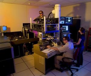
Making adjustments to the dynamic transmission electron 
microscope. From left: Curtis Brown, Thomas LaGrange and 
Judy Kim. (Credit: Image courtesy of DOE/Lawrence 
Livermore National Laboratory)

The findings point the way toward future work that could greatly improve catalyst efficiency in a variety of processes that are crucial to the world's energy security, such as petroleum catalysis and catalyst-based nanomaterial growth for next-generation rechargeable batteries. The work was performed in a collaborative effort by Lawrence Livermore National Laboratory and the University of California at Davis.

Using a new imaging technique on Lawrence Livermore's Dynamic Transmission Electron Microscope (DTEM), researchers have achieved unprecedented spatial and temporal resolution in single-shot images of nanoparticulate catalysts.

The DTEM uses a laser-driven photocathode to produce short pulses of electrons capable of recording electron micrographs with 15-nanosecond (one billionth of a second) exposure time. The recent addition of an annular dark field (ADF) aperture to the instrument has greatly improved its ability to time-resolve images of nanoparticles as small as 30 nanometers in diameter.

"Nanoparticles in this size range are of crucial importance to a wide variety of catalytic process of keen interest to energy and nanotechnology researchers," said UC Davis' Dan Masiel, formerly of LLNL and lead author of a paper appearing in the journal, ChemPhysChem. "Time-resolved imaging of such materials will allow for unprecedented insight into the dynamics of their behavior."

Previously, particles smaller than 50 nanometers could not be resolved in the 15-nanosecond exposure because of the limited signal and low contrast without ADF aperature. But by using DTEM's ADF, almost every 50-nanometer particle and many 30-nanometer ones became clearly visible because of the fast time resolution and high contrast.

"The stark difference between these two images clearly demonstrates the efficacy of annual dark field imaging when imaging samples with feature sizes near the resolution limit of DTEM," Masiel said.

The new technique makes it easier to discern significant features when compared to bright field pulsed imaging. It allows for vastly improved contrast for smaller particles, widening the range of catalyst systems that can be studied using DTEM.

DTEM can record images with six orders of magnitude higher temporal resolution than conventional TEM and can provide important insights into processes such as phase transformations, chemical reactions and nanowire and nanotube growth.

Co-authors include LLNL's Bryan Reed, Thomas LaGrange, Geoffrey Campbell, Ting Guo and Nigel Browning. The work was funded by the Department of Energy's Office of Science, Office of Basic Energy Sciences, Materials Sciences and Engineering Division.

The article appears in the May 27 online edition of ChemPhys Chem.
Enhanced by Zemanta

Thursday, June 17, 2010

Inbred Sperm Fertilize Fewer Eggs, Beetle Study Finds


Inbred male sperm have been found to fertilize fewer eggs when in competition with non-inbred males, according to a new study by the University of East Anglia.
Image
Red flour beetle. (Credit: Image courtesy of 
University of East Anglia)

Research into the breeding habits of the red flour beetle, published in Proceedings of the Royal Society B, shows that the reduced fitness of inbred beetles, known as 'inbreeding depression', reveals itself in competitive scenarios.

Inbreeding is a potentially important problem in declining species across the world, and conserving genetic variation is now recognized as a priority by the International Union for Conservation of Nature. The new research is potentially vital for the successful implementation of recovery programs of inbred species.

When populations deplete or fragment, relatives can be forced into reproduction, often leading to inbreeding depression.

Led by Dr. Matt Gage, the new research into the promiscuous red flour beetle (Tribolium castaneum) measured how male reproduction responded to forced inbreeding.

After mating brothers with sisters for eight generations, the research found no changes in male fertility or mating behavior.

However, inbred male sperm fertilized fewer eggs when in competition with another non-inbred male, and sperm became more variable in size.

Dr. Gage said: "The experiment was designed to make comparisons with non-inbred control lines. Using multiple inbred lines, we measured the effects of inbreeding on pre- and post-mating success, in the absence and presence of male-male competition."

The results showed no differences between inbred and non-inbred males in terms of mating success, latency, duration, the number of mounts or persistency in a non-competitive setting.

However inbred males suffered significantly reduced sperm competitiveness, fathering an average of 15 per cent fewer offspring than non-inbred males across 330 sperm competition comparisons.

Dr. Gage said: "It seems that inbreeding depression in sperm competitiveness was caused by a decrease in either sperm quantity or quality that is critical for relative competitiveness, but still allows full male fertilization success to be achieved under benign, competition-free conditions.

"We have shown that male fertility and mating competence are not affected by inbreeding and that any decline in sperm quality under inbreeding is only detectable when sperm competition is invoked.

"One limitation to this study is that the ancestral laboratory stock we have used is likely to carry relatively reduced genetic diversity. Also insect sperm do not generally manifest cellular abnormalities akin to those commonly found in more complex mammalian sperm," he added.

The next stage of the research will explore ways that female beetles use multiple mating to generate sperm competition and thereby avoid inbreeding depression of their own fertility.

The research is part of a three-year £400,000 project funded by the Natural Environmental Research Council (NERC). The overall results will help managers of conservation and captive breeding projects recognize when inbreeding is a problem, how it progresses and how best to manage or reverse it.
Enhanced by Zemanta

Hand Study Reveals Brain's Distorted Body Model


Our brains contain a highly distorted model of our own bodies, according to scientists at UCL (University College London).
Image
Locations of knuckles and tips of each finger for 
the actual hand (black dots) and localisation 
judgments (white dots) superposed to highlight 
differences in shape. Average hand shape is 
displayed as solid lines for the actual hand and 
as dotted lines for the localisation judgments. 
The hand representation revealed by the localisation 
judgments is distorted, being represented as too wide 
and the fingers too short. (Credit: Image courtesy 
of University College London)

A new study on the brain's representation of the hand found that our model of our bodies is out of sync with reality -- with a strong tendency to think that the hands are shorter and fatter than their true shape.

The results of the study, published in the journal Proceedings of the National Academy of Sciences, show that the brain maintains a model of the hand in which our fingers are perceived to be shorter and our hands fatter than they are. Neuroscientists suspect the reason for these distortions may lie in the way the brain receives information from different regions of the skin.

Dr Matthew Longo, lead author from the UCL Institute of Cognitive Neuroscience, said: "The phrase "I know the town like the back of my hand" suggests that we have near-perfect knowledge of the size and position of our own body parts but these results show that this is far from being the case."

"Our results show dramatic distortions of hand shape, which were highly consistent across participants. The hand appears to be represented as wider than it actually is and the fingers as shorter than they actually are -- a finding that might also apply to other parts of the body," added Dr Longo.

Participants in the study were asked to put their left hands palm down under a board and judge the location of the covered hand's knuckles and fingertips by pointing to where they perceived each of these landmarks to be. A camera situated above the experiment recorded where the participant pointed. By putting together the locations of all the landmarks, the researchers reconstructed the brain's model of the hand, and revealed its striking distortions.

The research, which was funded by the Biotechnology and Biological Science Research Council (BBSRC), aims to find out how the brain knows where all parts of the body are in space even when your eyes are closed -- an ability known as 'position sense'. Neuroscientists think that position sense requires two distinct kinds of information. Signals that the brain receives from muscles and joints play an important role in position sense, but the brain also needs a model of the shape and size of each body part. For example, to know where the fingertip is in space, the brain needs to know the angles of joints in the arm and hand, but also the length of the arm, hand, and finger.

It is this model of our body's size and shape that is investigated in the study. "Of course, we know what our hand really looks like" said Dr Longo, "and our participants were very accurate in picking out a photo of their own hand from a set of photos with various distortions of hand shape. So there is clearly a conscious visual image of the body as well. But that visual image seems not to be used for position sense."

The results showed that in this task people estimated that their hands were about two-thirds wider and about one-third shorter than actual measurements.

Neuroscientists suspect that the brain's distorted model of body shape result is due to the way the brain represents different parts of the skin. For example, the size of the brain representation of the five fingers gets progressively smaller for each finger between the thumb and the little finger, mirroring the relative size of fingers in the body model reported in this study.

"These findings may well be relevant to psychiatric conditions involving body image such as anorexia nervosa, as there may be a general bias towards perceiving the body to be wider than it is. Our healthy participants had a basically accurate visual image of their own body, but the brain's model of the hand underlying position sense was highly distorted. This distorted perception could come to dominate in some people, leading to distortions of body image as well, such as in eating disorders," said Dr Longo.
Enhanced by Zemanta

Wednesday, June 16, 2010

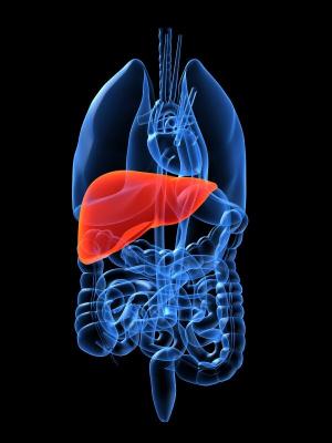
Discarded Livers: Replacement Organs?


A team led by researchers from the Center for Engineering in Medicine at Massachusetts General Hospital (MGH) has developed a technique that someday may allow growth of transplantable replacement livers. In a study appearing in Nature Medicine, the investigators describe using the structural tissue of rat livers as scaffolding for the growth of tissue regenerated from liver cells introduced through a novel reseeding process.
Image
Researchers have developed a technique that someday 
may allow growth of transplantable replacement livers. 
(Credit: iStockphoto/Sebastian Kaulitzki)

"Having the detailed microvasculature of the liver within a biocompatible, natural scaffold is a major advantage to growing liver tissue in a synthetic environment," says Basak Uygun, PhD, research associate at the MGH Center for Engineering in Medicine (MGH-CEM) and the paper's lead author. "Our technique of 'decellularizing' organs leaves the vascular system intact, which facilitates repopulation of the structural matrix and the subsequent survival and function of the introduced liver cells."

Liver transplantation is the only effective treatment for liver failure but is greatly limited by the shortage of donor organs. Each year 4,000 individuals who might have survived with a liver transplant die in the U.S. The shortage of donor livers and other organs is a major force behind the emerging field of tissue engineering and regenerative medicine. Efforts to build tissues from the ground up have not yet approached the goal of transplantable replacement organs, and replacing the liver -- in which each cell is a metabolic factory requiring constant, direct contact with the vascular system -- has been particularly challenging.

The current report describes a refinement of an approach to re-engineering replacement rat hearts that was reported in 2008 by University of Minnesota researchers. Since liver tissue is much more delicate than the muscular structure of the heart, the MGH-CEM team developed a gentler way of flushing living cells out of the liver's structural matrix, which is primarily made of connective tissue like collagen. After the cells were removed, the lobular structure of the liver and its extracellular matrix remained. Containing specific biochemical signals and cues that would direct liver cells to travel to the correct location and resume function -- something quite difficult to replicate using synthetic methods -- the matrix also maintained the organ's intricate network of blood vessels.

Another novel technique was used to reintroduce hepatocytes, the cells that carry out most of the liver's primary functions, into the decellularized matrix. The MGH-CEM approach actually caused cells to penetrate the vascular network and become embedded in the matrix, leaving major vessels clear to carry the essential blood supply. The repopulated matrix displayed normal liver function for up to 10 days in culture, and recellularized grafts were successfully connected to the circulation of live rats with minimal cellular damage and normal hepatocyte function.

"As far as we know, a transplantable liver graft has never been constructed in a laboratory setting before," explains Korkut Uygun, PhD, of the MGH-CEM, the paper's senior author. "Even though this is very exciting and promising, it is a proof-of-concept study only. Much more work will be required to make long-term functional liver grafts that can actually be transplanted into humans. We haven't been able to go beyond several hours in the rats, but it's a great start."

Martin Yarmush, MD, PhD, director of the MGH-CEM and a co-author of the Nature Medicine study, explains that the quarter of a million donor livers discarded each year because they are not suitable for transplantation would be an obvious source of supply for the creation of whole-organ scaffolds. "There is great potential for constructing full-fledged liver lobes containing animal or human cells, but several thorny issues must first be tackled, including formation of a layer of endothelial cells to line graft blood vessels," he says. "Given enough careful work, this approach could ultimately revolutionize tissue engineering and provide real working grafts for the liver and other complex tissues." Yarmush and Korkut Uygun both have faculty appointments at Harvard Medical School.

Additional co-authors of the Nature Medicine report are Alejandro Soto-Gutierrez, MD, PhD, Hiroshi Yagi, MD, Maria-Louisa Izamis, Maria Guzzardi, Carley Shulman, Jack Milwid, Arno Tilles, MD, Francois Berthiaume, PhD, and Yaahov Nahmias, PhD, MGH Center for Engineering in Medicine; Martin Hertl, MD, MGH Surgery; and Naoya Kobyashi, MD, PhD, Okayama University School of Medicine and Dentistry, Japan. The study was partially supported by grants from the National Institutes of Health, National Science Foundation and Shriners Hospitals for Children.
Enhanced by Zemanta

High-Yield Agriculture Slows Global Warming


Advances in high-yield agriculture over the latter part of the 20th century have prevented massive amounts of greenhouse gases from entering the atmosphere -- the equivalent of 590 billion metric tons of carbon dioxide -- according to a new study led by two Stanford Earth scientists.

Image
Increased yields of crops -- such as this maize in Kenya -- 
have not only helped feed the world, but have reduced 
greenhouse gas emissions. (Credit: Marshall Burke)

The yield improvements reduced the need to convert forests to farmland, a process that typically involves burning of trees and other plants, which generates carbon dioxide and other greenhouse gases.

The researchers estimate that if not for increased yields, additional greenhouse gas emissions from clearing land for farming would have been equal to as much as a third of the world's total output of greenhouse gases since the dawn of the Industrial Revolution in 1850.

The researchers also calculated that for every dollar spent on agricultural research and development since 1961, emissions of the three principal greenhouse gases -- methane, nitrous oxide and carbon dioxide -- were reduced by the equivalent of about a quarter of a ton of carbon dioxide -- a high rate of financial return compared to other approaches to reducing the gases.

"Our results dispel the notion that modern intensive agriculture is inherently worse for the environment than a more 'old-fashioned' way of doing things," said Jennifer Burney, lead author of a paper describing the study that will be published online by the Proceedings of the National Academy of Sciences.

Adding up the impact

The researchers calculated emissions of carbon dioxide, methane and nitrous oxide, converting the amounts of the latter two gases into the quantities of carbon dioxide that would have an equivalent impact on the atmosphere, to facilitate comparison of total greenhouse gas outputs.

Burney, a postdoctoral researcher with the Program on Food Security and the Environment at Stanford, said agriculture currently accounts for about 12 percent of human-caused greenhouse gas emissions. Although greenhouse gas emissions from the production and use of fertilizer have increased with agricultural intensification, those emissions are far outstripped by the emissions that would have been generated in converting additional forest and grassland to farmland.

"Every time forest or shrub land is cleared for farming, the carbon that was tied up in the biomass is released and rapidly makes its way into the atmosphere -- usually by being burned," she said. "Yield intensification has lessened the pressure to clear land and reduced emissions by up to 13 billion tons of carbon dioxide a year."

"When we look at the costs of the research and development that went into these improvements, we find that funding agricultural research ranks among the cheapest ways to prevent greenhouse gas emissions," said Steven Davis, a co-author of the paper and a postdoctoral researcher at the Carnegie Institution at Stanford.

To evaluate the impact of yield intensification on climate change, the researchers compared actual agricultural production between 1961 and 2005 with hypothetical scenarios in which the world's increasing food needs were met by expanding the amount of farmland rather than by the boost in yields produced by the Green Revolution.

"Even without higher yields, population and food demand would likely have climbed to levels close to what they are today," said David Lobell, also a coauthor and assistant professor of environmental Earth system science at Stanford.

"Lower yields per acre would likely have meant more starvation and death, but the population would still have increased because of much higher birth rates," he said. "People tend to have more children when survival of those children is less certain."

Avoiding the need for more farmland

The researchers found that without the advances in high-yield agriculture, several billion additional acres of cropland would have been needed.

Comparing emissions in the theoretical scenarios with real-world emissions from 1961 to 2005, the researchers estimated that the actual improvements in crop yields probably kept greenhouse gas emissions equivalent to at least 317 billion tons of carbon dioxide out of the atmosphere, and perhaps as much as 590 billion tons.

Without the emission reductions from yield improvements, the total amount of greenhouse gas pumped into the atmosphere over the preceding 155 years would have been between 18 and 34 percent greater than it has been, they said.

To calculate how much money was spent on research for each ton of avoided emissions, the researchers calculated the total amount of agricultural research funding related to yield improvements since 1961 through 2005. That produced a price between approximately $4 and $7.50 for each ton of carbon dioxide that was not emitted.

"The size and cost-effectiveness of this carbon reduction is striking when compared with proposed mitigation options in other sectors," said Lobell. "For example, strategies proposed to reduce emissions related to construction would cut emissions by a little less than half the amount that we estimate has been achieved by yield improvements and would cost close to $20 per ton."

The authors also note that raising yields alone won't guarantee lower emissions from land use change.

"It has been shown in several contexts that yield gains alone do not necessarily stop expansion of cropland," Lobell said. "That suggests that intensification must be coupled with conservation and development efforts.

"In certain cases, when yields go up in an area, it increases the profitability of farming there and gives people more incentive to expand their farm. But in general, high yields keep prices low, which reduces the incentive to expand."

The researchers concluded that improvement of crop yields should be prominent among a portfolio of strategies to reduce global greenhouse gases emissions.

"The striking thing is that all of these climate benefits were not the explicit intention of historical investments in agriculture. This was simply a side benefit of efforts to feed the world," Burney noted. "If climate policy intentionally rewarded these kinds of efforts, that could make an even bigger difference. The question going forward is how climate policy might be designed to achieve that."
Enhanced by Zemanta

Monday, June 14, 2010

Replace Silicon With Graphene on Nanocircuitry


Scientists have made a breakthrough toward creating nanocircuitry on graphene, widely regarded as the most promising candidate to replace silicon as the building block of transistors. They have devised a simple and quick one-step process based on thermochemical nanolithography (TCNL) for creating nanowires, tuning the electronic properties of reduced graphene oxide on the nanoscale and thereby allowing it to switch from being an insulating material to a conducting material.
Image
In a technique known as thermochemical 
nanolithography, the tip of an atomic force microscope 
uses heat to turn graphene oxide into reduced 
graphene oxide, a substance that can be used to produce 
nanocircuits and nanowires with controllable conductivity. 
(Credit: University of Illinois at Urbana-Champaign)

The technique works with multiple forms of graphene and is poised to become an important finding for the development of graphene electronics. The research appears in the June 11, 2010, issue of the journal Science.

Scientists who work with nanocircuits are enthusiastic about graphene because electrons meet with less resistance when they travel along graphene compared to silicon and because today's silicon transistors are nearly as small as allowed by the laws of physics. Graphene also has the edge due to its thickness -- it's a carbon sheet that is a single atom thick. While graphene nanoelectronics could be faster and consume less power than silicon, no one knew how to produce graphene nanostructures on such a reproducible or scalable method. That is until now.

"We've shown that by locally heating insulating graphene oxide, both the flakes and epitaxial varieties, with an atomic force microscope tip, we can write nanowires with dimensions down to 12 nanometers. And we can tune their electronic properties to be up to four orders of magnitude more conductive. We've seen no sign of tip wear or sample tearing," said Elisa Riedo, associate professor in the School of Physics at the Georgia Institute of Technology.

On the macroscale, the conductivity of graphene oxide can be changed from an insulating material to a more conductive graphene-like material using large furnaces.

Now, the research team used TCNL to increase the temperature of reduced graphene oxide at the nanoscale, so they can draw graphene-like nanocircuits. They found that when it reached 130 degrees Celsius, the reduced graphene oxide began to become more conductive.

"So the beauty of this is that we've devised a simple, robust and reproducible technique that enables us to change an insulating sample into a conducting nanowire. These properties are the hallmark of a productive technology," said Paul Sheehan, head of the Surface Nanoscience and Sensor Technology Section at the Naval Research Laboratory in Washington, D.C.

The research team tested two types of graphene oxide -- one made from silicon carbide, the other with graphite powder.

"I think there are three things about this study that make it stand out," said William P. King, associate professor in the Mechanical Science and Engineering department at the University of Illinois at Urbana-Champaign. "First, is that the entire process happens in one step. You go from insulating graphene oxide to a functional electronic material by simply applying a nano-heater. Second, we think that any type of graphene will behave this way. Third, the writing is an extremely fast technique. These nanostructures can be synthesized at such a high rate that the approach could be very useful for engineers who want to make nanocircuits."

"This project is an excellent example of the new technologies that epitaxial graphene electronics enables," said Walt de Heer, Regent's Professor in Georgia Tech's School of Physics and the original proponent of epitaxial graphene in electronics. His study led to the establishment of the Materials Research Science and Engineering Center two years ago. "The simple conversion from graphene oxide to graphene is an important and fast method to produce conducting wires. This method can be used not only for flexible electronics, but it is possible, sometime in the future, that the bio-compatible graphene wires can be used to measure electrical signals from single biological cells."

Sunday, June 13, 2010

Powerful New Science Tools


With controlled stretching of molecules, Cornell researchers have demonstrated that single-molecule devices can serve as powerful new tools for fundamental science experiments. Their work has resulted in detailed tests of long-existing theories on how electrons interact at the nanoscale.

Image
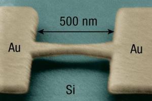
A scanning electron micrograph of a gold bridge 
suspended 40 nanometers above a silicon substrate.
In the experiment, the bridge is severed in the middle, 
a single molecule is suspended across the gap, and 
the substrate is bent to stretch the molecule while 
simultaneously measuring the electron current 
through the molecule. (Credit: J.J. Parks)

The work, led by professor of physics Dan Ralph, is published in the June 10 online edition of the journal Science. First author is J.J. Parks, a former graduate student in Ralph's lab.

The scientists studied particular cobalt-based molecules with so-called intrinsic spin -- a quantized amount of angular momentum.

Theories first postulated in the 1980s predicted that molecular spin would alter the interaction between electrons in the molecule and conduction electrons surrounding it, and that this interaction would determine how easily electrons flow through the molecule. Before now, these theories had not been tested in detail because of the difficulties involved in making devices with controlled spins.

Understanding single-molecule electronics requires expertise in both chemistry and physics, and Cornell's team has specialists in both.

"People know about high-spin molecules, but no one has been able to bring together the chemistry and physics to make controlled contact with these high-spin molecules," Ralph said.

The researchers made their observations by stretching individual spin-containing molecules between two electrodes and analyzing their electrical properties. They watched electrons flow through the cobalt complex, cooled to extremely low temperatures, while slowly pulling on the ends to stretch it. At a particular point, it became more difficult to pass current through the molecule. The researchers had subtly changed the magnetic properties of the molecule by making it less symmetric.

After releasing the tension, the molecule returned to its original shape and began passing current more easily -- thus showing the molecule had not been harmed. Measurements as a function of temperature, magnetic field and the extent of stretching gave the team new insights into exactly what is the influence of molecular spin on the electron interactions and electron flow.

The effects of high spin on the electrical properties of nanoscale devices were entirely theoretical issues before the Cornell work, Ralph said. By making devices containing individual high-spin molecules and using stretching to control the spin, the Cornell team proved that such devices can serve as a powerful laboratory for addressing these fundamental scientific questions.

The study was funded primarily by the National Science Foundation.
Enhanced by Zemanta

Thursday, June 10, 2010

Genetic 'Parts' List Now Available for Hypothalamus


A Johns Hopkins and Japanese research team has generated the first comprehensive genetic "parts" list of a mouse hypothalamus, an enigmatic region of the brain -- roughly cherry-sized, in humans -- that controls hunger, thirst, fatigue, body temperature, wake-sleep cycles and links the central nervous system to control of hormone levels.
Image
A. A diagram of the developing mouse brain at 12 
days gestation. The hypothalamus is outlined in 
red, while cells that express the Shh gene, which 
was used as a landmark in this study, are shown in 
brown. B. Various different genes that are turned on 
in different parts of the hypothalamus are shown in 
purple, while Shh is shown in brown. (Credit: Image 
courtesy of Johns Hopkins Medical Institutions)

Flaws in hypothalamus development may underlie both inborn and acquired metabolic balance problems that can lead to obesity, diabetes, mood disorders and high blood pressure, according to a report on the study published May 2 in the advance online publication of Nature Neuroscience.

"Knowing how cells develop in this part of the brain will help us understand how they regulate behavior, mood and metabolism," says Seth Blackshaw, Ph.D., an assistant professor in the Solomon H. Snyder Department of Neuroscience, Johns Hopkins University School of Medicine.

The hypothalamus is one of the most diverse and complex parts of the brain, and having an index of the genes involved in producing its many cell types is a toolbox that researchers can use to manipulate the activity of brain cells by turning them on and off, or tracing their connections. This may ultimately lead to better diagnostic and treatment options for a variety of disorders.

"The study of hypothalamic development, particularly of cell specification, will help us to understand how hypothalamic neurons function to regulate behavior and physiology," says Blackshaw. "Because of when and where we saw certain genes turn on, we now have identified a set of candidate players that guide the assembly of the different parts of the hypothalamus and that specify the many individual cell types within it."

The hypothalamus is composed of at least dozens of types of neurons -- and more likely hundreds -- each of which corresponds to a gene that has remained unidentified until now. Its cellular arrangement is more akin to a bowl of spaghetti than a neatly organized club sandwich, according to Blackshaw. The catalog of molecular markers identified here helps unravel this complexity.

The team's first challenge was to dissect away, at the very start of neural development, the part of the mouse brain which develops into the hypothalamus, and then cut tiny slices of this region for use in microarray analysis, a technology that reveals multiple gene activity. By analyzing all the roughly 20,000 genes in the mouse genome, the team identified 1200 as strongly activated in developing hypothalamus and characterized the cells within the hypothalamus in which they were activated. The team then characterized the expression of the most interesting 350 genes in detail using another gene called Shh, for sonic hedgehog, as a landmark to identify the precise region of the hypothalamus in which these genes were turned on. This involved processing close to 20,000 tissue sections -- painstakingly sliced at one-fiftieth of a millimeter thickness and then individually examined.

"We were able to use this data to find genes whose expression matched every individual hypothalamic nucleus and essentially assemble a jigsaw puzzle of gene expression patterns that completely covered the developing hypothalamus," Blackshaw says. "Now that we have a complete set of molecular landmarks, along with an extensive molecular parts list, we can begin to learn how all these parts fit together to create this essential and highly complex brain region."

Authors of the paper, in addition to Blackshaw, are Daniel A. Lee, Ana Miranda-Angulo, Yangqin Yang, Aya C. Yoshida, Hong Wang, Hiromi Mashiko, Lizhi Jiang, Marina Avetisyan, Lixin Qi, and Jiang Qian, all of Johns Hopkins; Ayane Kataoka and Tomomi Shimogori of RIKEN-BSI, 2-1 Hirosawa, Wako-shi, Saitama, Japan.

This research was supported by March of Dimes, the Klingenstein Fund, the W.M. Keck Foundation, and the Japan Society for Promotion of Science.

Wednesday, June 9, 2010

How the Brain Recognizes Objects


Researchers at MIT's McGovern Institute for Brain Research have developed a new mathematical model to describe how the human brain visually identifies objects. The model accurately predicts human performance on certain visual-perception tasks, which suggests that it's a good indication of what actually happens in the brain, and it could also help improve computer object-recognition systems.
Image
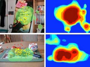
A new computational model of how the primate brain 
recognizes objects creates a map of "interesting" features 
(right) for a given image. The model's predictions of which 
parts of the image will attract a viewer's attention 
(green clouds, left) accord well with experimental data 
(yellow and red dots). 
(Credit: Images courtesy of Sharat Chikkerur)

The model was designed to reflect neurological evidence that in the primate brain, object identification -- deciding what an object is -- and object location -- deciding where it is -- are handled separately. "Although what and where are processed in two separate parts of the brain, they are integrated during perception to analyze the image," says Sharat Chikkerur, lead author on a paper appearing this week in the journal Vision Research, which describes the work. "The model that we have tries to explain how this information is integrated."

The mechanism of integration, the researchers argue, is attention. According to their model, when the brain is confronted by a scene containing a number of different objects, it can't keep track of all of them at once. So instead it creates a rough map of the scene that simply identifies some regions as being more visually interesting than others. If it's then called upon to determine whether the scene contains an object of a particular type, it begins by searching -- turning its attention toward -- the regions of greatest interest.

Chikkerur and Tomaso Poggio, the Eugene McDermott Professor in the Department of Brain and Cognitive Sciences and at the Computer Science and Artificial Intelligence Laboratory, together with graduate student Cheston Tan and former postdoc Thomas Serre, implemented the model in software, then tested its predictions against data from experiments with human subjects. The subjects were asked first to simply regard a street scene depicted on a computer screen, then to count the cars in the scene, and then to count the pedestrians, while an eye-tracking system recorded their eye movements. The software predicted with great accuracy which regions of the image the subjects would attend to during each task.

The software's analysis of an image begins with the identification of interesting features -- rudimentary shapes common to a wide variety of images. It then creates a map that depicts which features are found in which parts of the image. But thereafter, shape information and location information are processed separately, as they are in the brain.

The software creates a list of all the interesting features in the feature map, and from that, it creates another list, of all the objects that contain those features. But it doesn't record any information about where or how frequently the features occur.

At the same time, it creates a spatial map of the image that indicates where interesting features are to be found, but not what sorts of features they are.

It does, however, interpret the "interestingness" of the features probabilistically. If a feature occurs more than once, its interestingness is spread out across all the locations at which it occurs. If another feature occurs at only one location, its interestingness is concentrated at that one location.

Mathematically, this is a natural consequence of separating information about objects' identity and location and interpreting the results probabilistically. But it ends up predicting another aspect of human perception, a phenomenon called "pop out." A human subject presented with an image of, say, one square and one star will attend to both objects about equally. But a human subject presented an image of one square and a dozen stars will tend to focus on the square.

Like a human asked to perform a visual-perception task, the software can adjust its object and location models on the fly. If the software is asked to identify only the objects at a particular location in the image, it will cross off its list of possible objects any that don't contain the features found at that location.

By the same token, if it's asked to search the image for a particular kind of object, the interestingness of features not found in that object will go to zero, and the interestingness of features found in the object will increase proportionally. This is what allows the system to predict the eye movements of humans viewing a digital image, but it's also the aspect of the system that could aid the design of computer object-recognition systems. A typical object-recognition system, when asked to search an image for multiple types of objects, will search through the entire image looking for features characteristic of the first object, then search through the entire image looking for features characteristic of the second object, and so on. A system like Poggio and Chikkerur's, however, could limit successive searches to just those regions of the image that are likely to have features of interest.

Funding: DARPA, the Honda Research Institute USA, NEC, Sony and the Eugene McDermott Foundation

Secondhand Smoke Associated With Psychiatric Distress, Illness


Exposure to secondhand smoke appears to be associated with psychological distress and the risk of future psychiatric hospitalization among healthy adults, according to a report posted online that will appear in the August print issue of Archives of General Psychiatry.

Me
Second hand smoke exposure is associated with psychological distress and risk of future psychiatric illness (Credit: iStockphoto/Michael Bodmann)

"A growing body of literature has demonstrated the harmful physical health effects of secondhand smoke exposure," the authors write as background information in the article. "Given the highly prevalent exposure to secondhand smoke -- in the United States, an estimated 60 percent of American non-smokers had biological evidence of exposure to secondhand smoke -- even a low level of risk may have a major public health impact."

Mark Hamer, Ph.D., of University College London, and colleagues studied 5,560 non-smoking adults (average age 49.8) and 2,595 smokers (average age 44.8) who did not have a history of mental illness and participated in the Scottish Health Survey in 1998 or 2003. Participants were assessed with a questionnaire about psychological distress, and admissions to psychiatric hospitals were tracked over six years of follow-up. Exposure to secondhand smoke among non-smokers was assessed using saliva levels of cotinine -- the main product formed when nicotine is broken down by the body -- "a reliable and valid circulating biochemical marker of nicotine exposure," the authors write.

A total of 14.5 percent of the participants reported psychological distress. Non-smokers with a high exposure to secondhand smoke (cotinine levels between 0.70 and 15 micrograms per liter) had higher odds of psychological distress when compared with those who had no detectable cotinine.

Over the six-year follow-up, 41 individuals were newly admitted to psychiatric hospitals. Smokers and non-smokers with high exposure to secondhand smoke were both more likely than non-smokers with low levels of secondhand smoke exposure to be hospitalized for depression, schizophrenia, delirium or other psychiatric conditions.

Animal data have suggested that tobacco may induce a negative mood, and some human studies have also identified a potential association between smoking and depression. "Taken together, therefore, our data are consistent with other emerging evidence to suggest a causal role of nicotine exposure in mental health," the authors write.

"To our knowledge, this is the first study to demonstrate a prospective association between objectively assessed secondhand smoke exposure and mental health in a representative sample of a general population," they conclude.
Reblog this post [with Zemanta]