BTemplates.com

Powered by Blogger.

Pageviews past week

Quantum mechanics

Auto News

artificial intelligence

About Me

Recommend us on Google!

Information Technology

Popular Posts

Showing posts with label Stem cells. Show all posts

Tuesday, April 16, 2013

Brain Development Is Guided by Junk DNA That Isn't Really Junk


Specific DNA once dismissed as junk plays an important role in brain development and might be involved in several devastating neurological diseases, UC San Francisco scientists have found.
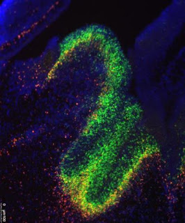
UCSF researchers have uncovered a role in brain development and in neurological disease for little appreciated molecules called long noncoding RNA. In this image, fluorescent dyes track the presence of the RNA molecules and the genes they affect in the developing mouse brain.
UCSF researchers have uncovered a role in brain development and in neurological disease for little appreciated molecules called long noncoding RNA. In this image, fluorescent dyes track the presence of the RNA molecules and the genes they affect in the developing mouse brain. (Credit: Image courtesy of Alexander Ramos)
Their discovery in mice is likely to further fuel a recent scramble by researchers to identify roles for long-neglected bits of DNA within the genomes of mice and humans alike.

While researchers have been busy exploring the roles of proteins encoded by the genes identified in various genome projects, most DNA is not in genes. This so-called junk DNA has largely been pushed aside and neglected in the wake of genomic gene discoveries, the UCSF scientists said.

In their own research, the UCSF team studies molecules called long noncoding RNA (lncRNA, often pronounced as "link" RNA), which are made from DNA templates in the same way as RNA from genes.

"The function of these mysterious RNA molecules in the brain is only beginning to be discovered," said Daniel Lim, assistant professor of neurological surgery, a member of the Eli and Edythe Broad Center of Regeneration Medicine and Stem Cell Research at UCSF, and the senior author of the study, published online April 11 in the journal Cell Stem Cell.

Alexander Ramos, a student enrolled in the MD/PhD program at UCSF and first author of the study, conducted extensive computational analysis to establish guilt by association, linking lncRNAs within cells to the activation of genes.

Ramos looked specifically at patterns associated with particular developmental pathways or with the progression of certain diseases. He found an association between a set of 88 long noncoding RNAs and Huntington's disease, a deadly neurodegenerative disorder. He also found weaker associations between specific groups of long noncoding RNAs and Alzheimer's disease, convulsive seizures, major depressive disorder and various cancers.

"Alex was the team member who developed this new research direction, did most of the experiments, and connected results to the lab's ongoing work," Lim said. The study was mostly funded through Lim's grant - a National Institutes of Health (NIH) Director's New Innovator Award, a competitive award for innovative projects that have the potential for unusually high impact.

Unlike messenger RNA, which is transcribed from the DNA in genes and guides the production of proteins, lncRNA molecules do not carry the blueprints for proteins. Because of this fact, they were long thought to not influence a cell's fate or actions.

Nonetheless, lncRNAs also are transcribed from DNA in the same way as messenger RNA, and they, too, consist of unique sequences of nucleic acid building blocks.

Evidence indicates that lncRNAs can tether structural proteins to the DNA-containing chromosomes, and in so doing indirectly affect gene activation and cellular physiology without altering the genetic code. In other words, within the cell, lncRNA molecules act "epigenetically" -- beyond genes -- not through changes in DNA.

The brain cells that the scientists focused on the most give rise to various cell types of the central nervous system. They are found in a region of the brain called the subventricular zone, which directly overlies the striatum. This is the part of the brain where neurons are destroyed in Huntington's disease, a condition triggered by a single genetic defect.

Ramos combined several advanced techniques for sequencing and analyzing DNA and RNA to identify where certain chemical changes happen to the chromosomes, and to identify lncRNAs on specific cell types found within the central nervous system. The research revealed roughly 2,000 such molecules that had not previously been described, out of about 9,000 thought to exist in mammals ranging from mice to humans.

In fact, the researchers generated far too much data to explore on their own. The UCSF scientists created a website through which their data can be used by others who want to study the role of lncRNAs in development and disease.

"There's enough here for several labs to work on," said Ramos, who has training grants from the California Institute for Regenerative Medicine (CIRM) and the NIH.

"It should be of interest to scientists who study long noncoding RNA, the generation of new nerve cells in the adult brain, neural stem cells and brain development, and embryonic stem cells," he said.

Other co-authors who worked on the study include UCSF postdoctoral fellows Aaron Diaz, PhD, Abhinav Nellore, PhD, Michael Oldham, PhD, Jun Song, PhD, Ki-Youb Park, PhD, and Gabriel Gonzales-Roybal, PhD; and MD/PhD student Ryan Delgado. Additional funders of the study included the Sontag Foundation and the Sandler Foundation.

UCSF is a leading university dedicated to promoting health worldwide through advanced biomedical research, graduate-level education in the life sciences and health professions, and excellence in patient care. 

================
Follow UCSF UCSF.edu | Facebook.com/ucsf | Twitter.com/ucsf | YouTube.com/ucsf

Friday, April 5, 2013

3-D Printer Can Build Synthetic Tissues


A custom-built programmable 3D printer can create materials with several of the properties of living tissues, Oxford University scientists have demonstrated.
A custom-built programmable 3D printer can create materials with several of the properties of living tissues, Oxford University scientists have demonstrated: Droplet network c.500 microns across with electrically conductive pathway between electrodes mimicking nerve.
A custom-built programmable 3D printer can create materials with several of the properties of living tissues, Oxford University scientists have demonstrated: Droplet network c.500 microns across with electrically conductive pathway between electrodes mimicking nerve. (Credit: Oxford University/G Villar)

The new type of material consists of thousands of connected water droplets, encapsulated within lipid films, which can perform some of the functions of the cells inside our bodies.

These printed 'droplet networks' could be the building blocks of a new kind of technology for delivering drugs to places where they are needed and potentially one day replacing or interfacing with damaged human tissues. Because droplet networks are entirely synthetic, have no genome and do not replicate, they avoid some of the problems associated with other approaches to creating artificial tissues -- such as those that use stem cells.

The team report their findings in this week's Science.

'We aren't trying to make materials that faithfully resemble tissues but rather structures that can carry out the functions of tissues,' said Professor Hagan Bayley of Oxford University's Department of Chemistry, who led the research. 'We've shown that it is possible to create networks of tens of thousands connected droplets. The droplets can be printed with protein pores to form pathways through the network that mimic nerves and are able to transmit electrical signals from one side of a network to the other.'

Each droplet is an aqueous compartment about 50 microns in diameter. Although this is around five times larger than living cells the researchers believe there is no reason why they could not be made smaller. The networks remain stable for weeks.

'Conventional 3D printers aren't up to the job of creating these droplet networks, so we custom built one in our Oxford lab to do it,' said Professor Bayley. 'At the moment we've created networks of up to 35,000 droplets but the size of network we can make is really only limited by time and money. For our experiments we used two different types of droplet, but there's no reason why you couldn't use 50 or more different kinds.'

The unique 3D printer was built by Gabriel Villar, a DPhil student in Professor Bayley's group and the lead author of the paper.

The droplet networks can be designed to fold themselves into different shapes after printing -- so, for example, a flat shape that resembles the petals of a flower is 'programmed' to fold itself into a hollow ball, which cannot be obtained by direct printing. The folding, which resembles muscle movement, is powered by osmolarity differences that generate water transfer between droplets.

Gabriel Villar of Oxford University's Department of Chemistry said: 'We have created a scalable way of producing a new type of soft material. The printed structures could in principle employ much of the biological machinery that enables the sophisticated behaviour of living cells and tissues.'

Saturday, July 21, 2012

Entire Genetic Sequence of Individual Human Sperm Determined


The entire genomes of 91 human sperm from one man have been sequenced by Stanford University researchers. The results provide a fascinating glimpse into naturally occurring genetic variation in one individual, and are the first to report the whole-genome sequence of a human gamete -- the only cells that become a child and through which parents pass on physical traits.

Entire Genetic Sequence of Individual Human Sperm Determined
Every sperm cell looks essentially the same, with that characteristic tadpole appearance. But inside, sperm cells carry differences within their genes -- even cells from the same man. Now, researchers provide a detailed picture of how the cell's DNA varies in a new study published in the July 20, 2012 issue of the Cell Press journal Cell. The techniques used could be helpful for understanding male reproductive disorders or, when applied to other areas of research, for characterizing normal and diseased cells in the body. (Credit: iStockphoto/Alexandr Mitiuc)

"This represents the culmination of nearly a decade of work in my lab," said Stephen Quake, PhD, the Lee Otterson Professor in the School of Engineering and professor of bioengineering and of applied physics. "We now have devices that will allow us to routinely amplify and sequence to a high degree of accuracy the entire genomes of single cells, which has far-ranging implications for the study of cancer, infertility and many other disorders."

Quake is the senior author of the research, published July 20 in Cell. Graduate student Jianbin Wang and former graduate student H. Christina Fan, PhD, now a senior scientist at ImmuMetrix, share first authorship of the paper.

Sequencing sperm cells is particularly interesting because of a natural process called recombination that ensures that a baby is a blend of DNA from all four of his or her grandparents. Until now, scientists had to rely on genetic studies of populations to estimate how frequently recombination had occurred in individual sperm and egg cells, and how much genetic mixing that entailed.

"Single-sperm sequencing will allow us to chart and understand how recombination differs between individuals at the finest scales. This is an important proof of principle that will allow us to study both fundamental dynamics of recombination in humans and whether it is involved in issues relating to male infertility," said Gilean McVean, PhD, professor of statistical genetics at the Wellcome Trust Centre for Human Genetics. McVean was not involved in the research.

The Stanford study showed that the previous, population-based estimates were, for the most part, surprisingly accurate: on average, the sperm in the sample had each undergone about 23 recombinations, or mixing events. However, individual sperm varied greatly in the degree of genetic mixing and in the number and severity of spontaneously arising genetic mutations. Two sperm were missing entire chromosomes. The study has long-ranging implication for infertility doctors and researchers.

"For the first time, we were able to generate an individual recombination map and mutation rate for each of several sperm from one person," said study co-author Barry Behr, PhD, HCLD, professor of obstetrics and gynecology and director of Stanford's in vitro fertilization laboratory. "Now we can look at a particular individual, make some calls about what they would likely contribute genetically to an embryo and perhaps even diagnose or detect potential problems."

Most cells in the human body have two copies of each of 23 chromosomes, and are known as "diploid" cells. Recombination occurs during a process called meiosis, which partitions a single copy of each chromosome into a sperm (in a man) or egg (in a woman) cell. When a sperm and an egg join, the resulting fertilized egg again has a full complement of DNA.

To ensure an orderly distribution during recombination, pairs of chromosomes are lined up in tight formation along the midsection of the cell. During this snug embrace, portions of matching chromosomes are sometimes randomly swapped. The process generates much more genetic variation in a potential offspring than would be possible if only intact chromosomes were segregated into the reproductive cells.

"The exact sites, frequency and degree of this genetic mixing process is unique for each sperm and egg cell," said Quake, "and we've never before been able to see it with this level of detail. It's very interesting that what happens in one person's body mirrors the population average."

Major problems with the recombination process can generate sperm missing portions or even whole chromosomes, making them incapable of or unlikely to fertilize an egg. But it can be difficult for fertility researchers to identify potential problems.

"Most of the techniques we currently use to assess sperm viability are fairly crude," said Quake.

To conduct the research, Wang, Quake and Behr first isolated and sequenced nearly 100 sperm cells from the study subject, a 40-year-old man. The man has healthy offspring, and the semen sample appeared normal. His whole-genome sequence (obtained from diploid cells) has been previously sequenced to a high level of accuracy.

They then compared the sequence of the sperm with that of the study subject's diploid genome. They could see, by comparing the sequences of the chromosomes in the diploid cells with those in the haploid sperm cells, where each recombination event took place. The researchers also identified 25 to 36 new single nucleotide mutations in each sperm cell that were not present in the subject's diploid genome. Such random mutations are another way to generate genetic variation, but if they occur at particular points in the genome they can have deleterious effects.

It's important to note that individual sperm cells are destroyed by the sequencing process, meaning that they couldn't go on to be used for fertilization. However, the single-cell sequencing described in the paper could potentially be used to diagnose male reproductive disorders and help infertile couples assess their options. It could also be used to learn more about how male fertility and sperm quality change with increasing age.

"This could serve as a new kind of early detection system for men who may have reproductive problems," said Behr, who also co-directs Stanford's reproductive endocrinology and infertility program. "It's also possible that we could one day use other, correlating features to harmlessly identify healthy sperm for use in IVF. In the end, the DNA is the raw material that ultimately defines a sperm's potential. If we can learn more about this process, we can better understand human fertility."

The research was supported by the National Institute of Health, the Chinese Scholarship Council and the Siebel Foundation.

Saturday, July 7, 2012

Diabetes Drug Makes Brain Cells Grow


The widely used diabetes drug metformin comes with a rather unexpected and alluring side effect: it encourages the growth of new neurons in the brain. The study reported in the July 6th issue of Cell Stem Cell, a Cell Press publication, also finds that those neural effects of the drug also make mice smarter.

New research finds that the widely used diabetes drug metformin comes with a rather unexpected and alluring side effect: it encourages the growth of new neurons in the brain.
New research finds that the widely used diabetes drug 
metformin comes with a rather unexpected and alluring 
side effect: it encourages the growth of new neurons in 
the brain. (Credit: iStockphoto/Guido Vrola)
The discovery is an important step toward therapies that aim to repair the brain not by introducing new stem cells but rather by spurring those that are already present into action, says the study's lead author Freda Miller of the University of Toronto-affiliated Hospital for Sick Children. The fact that it's a drug that is so widely used and so safe makes the news all that much better.

Earlier work by Miller's team highlighted a pathway known as aPKC-CBP for its essential role in telling neural stem cells where and when to differentiate into mature neurons. As it happened, others had found before them that the same pathway is important for the metabolic effects of the drug metformin, but in liver cells.

"We put two and two together," Miller says. If metformin activates the CBP pathway in the liver, they thought, maybe it could also do that in neural stem cells of the brain to encourage brain repair.

The new evidence lends support to that promising idea in both mouse brains and human cells. Mice taking metformin not only showed an increase in the birth of new neurons, but they were also better able to learn the location of a hidden platform in a standard maze test of spatial learning.

While it remains to be seen whether the very popular diabetes drug might already be serving as a brain booster for those who are now taking it, there are already some early hints that it may have cognitive benefits for people with Alzheimer's disease. It had been thought those improvements were the result of better diabetes control, Miller says, but it now appears that metformin may improve Alzheimer's symptoms by enhancing brain repair.

Miller says they now hope to test whether metformin might help repair the brains of those who have suffered brain injury due to trauma or radiation therapies for cancer.

Tuesday, July 19, 2011

Stem Cells: Nearing Goal of Using Patient's Own Cells to Make Stem Cells to Replace Lost or Diseased Tissue


Scientists at the Salk Institute for Biological Studies have developed an improved technique for generating large numbers of blood cells from a patient's own cells. The new technique will be immediately useful in further stem cell studies, and when perfected, could be used in stem cell therapies for a wide variety of conditions including cancers and immune ailments.
Round hematopoietic (blood) cells emerge from 
differentiating human pluripotent stem cells. 
(Credit: Courtesy of Aaron Parker, Salk Institute 
for Biological Studies)

"There are further improvements that we need to make, but this takes us a significant step closer to the ultimate goal, which is to be able to take ordinary cells from a patient, induce them to become stem cells, and then use those stem cells to rebuild lost or diseased tissues, for example the patient's bone marrow," says Inder M. Verma, PhD, Irwin and Joan Jacobs Chair in Exemplary Life Science and American Cancer Society Professor of Molecular Biology at the Salk Institute Laboratory of Genetics. Verma is senior author of the report, which is published in the July edition of the journal Stem Cells.

Stem cell researchers have been racing towards this goal since 2006, when techniques for turning ordinary skin cells into induced pluripotential stem cells (iPSCs) were first reported. In principle, iPSCs mimic the embryonic stem cells (ESCs) from which organisms develop. Researchers now want to find the precise mixes and sequences of chemical compounds needed to coax iPSCs to mature into the tissue-specific stem cells of their choice. The latter are self-renewing, and can be transplanted into the body to produce the 'progenitor' cells that multiply locally and produce mature tissue cells.

However, researchers don't know yet how to induce iPSCs to become tissue-specific stem cells or mature tissue cells with high efficiency. "We've been producing these cells in quantities that are too low to enable them to be studied easily, much less used for therapies," says Aaron Parker, PhD, a former graduate student and now a postdoctoral researcher in Verma's lab. Parker is a co-lead-author of the paper, with Niels-Bjarne Woods, PhD, who was a postdoctoral researcher in the Verma lab at the outset of the project, and is now an assistant professor at Lund University in Sweden.

Like many other stem cell research laboratories, the Verma lab has been trying to find more efficient ways to turn iPSCs into blood-forming 'hematopoietic' stem cells (HSCs). These may be more valuable medically than any other tissue-specific stem cell, because they can supply not only oxygen-carrying red blood cells but also all the white blood cells of the immune system. "There would be an almost unlimited number of usages for true HSCs," says Verma.

For the present study, the research team sought to do a better job of mimicking the changing conditions that naturally direct ESCs to become HSCs in the womb. "We took seven lines of human ESCs and iPSCs, and experimented with different combinations and sequences of growth factors and other chemical compounds that are known to be present as ESCs move to the HSC state in a developing human," says Parker.


Applying cocktails of these factors, Parker and Woods and their colleagues induced the iPSCs and ESCs to form colonies of cells that bore the distinctive molecular markers of blood cells. With their best such cocktail they were able to detect blood-specific markers on 84% of their cells after three weeks. "That's a big jump in efficiency from what we saw in the field just a few years ago," says Parker.

The technique still has room for improvement. The researchers detected progenitor cells and mature cells from only one category or lineage: myeloid cells, which include red blood cells and primitive immune cells such as macrophages. "We didn't see any cells from the lymphoid lineage, meaning T-cells and B-cells," Parker says.

Another drawback was that the blood cell population they produced from ESCs and iPSCs contained short-lived progenitors and mature blood cells but no indefinitely renewing, transplantable HSCs. Their cocktail, they believed, either pushed the cells past the HSC state to the progenitor state too quickly, or made the maturing cells skip the HSC state entirely.

From this and other labs' results, the team hypothesized the existence of an intermediate, pre-hematopoietic type of stem cell, produced by ESCs and iPSCs and in turn producing HSCs. "We know that HSCs appear in a particular region of mammals during embryonic development, and our idea is that these pre-hematopoietic stem cells are there and are somehow made to mature into HSCs," says Parker. "So our lab is now going to focus on finding the precise maturation signals provided by that embryonic region to produce these true, transplantable HSCs."

Once that is done, researchers will need to make a number of further refinements to improve the safety of HSCs intended for human patients. "But we're now tantalizingly close to our ultimate goal," says Verma.

The other authors who contributed to the work were Roksana Moraghebi, of Lund University's Stem Cell Center; Margaret K. Lutz, Amy L. Firth, Kristen J. Brennand, W. Travis Berggren and Fred H. Gage of the Salk Institute Laboratory for Genetics; Juan Carlos Izpisúa Belmonte of the Salk Institute Gene Expression Laboratory; and Angel Raya of the Center of Regenerative Medicine in Barcelona, Spain.

Funding for this research was provided by the National Institutes for Health, the California Institute for Regenerative Medicine, the Leducq Foundation, the Merieux Foundation, the Ellison Medical Foundation, Ipsen/Biomeasure, Sanofi Aventis, the Prostate Cancer Foundation, the H.N. and Frances C. Berger Foundation, The Royal Physiographic Society of Sweden, the Lund University Medical Faculty, and the Lars Hierta Memorial Foundation, and the H.A. and Mary K. Chapman Charitable Trust.

Sunday, July 10, 2011

A Change of Heart: Researchers Reprogram Brain Cells to Become Heart Cells


For the past decade, researchers have tried to reprogram the identity of all kinds of cell types. Heart cells are one of the most sought-after cells in regenerative medicine because researchers anticipate that they may help to repair injured hearts by replacing lost tissue. Now, researchers at the Perelman School of Medicine at the University of Pennsylvania are the first to demonstrate the direct conversion of a non-heart cell type into a heart cell by RNA transfer.
Cardiomyocyte (center), showing protein distribution (green and red colors) indicative of a young cardiomyocyte. (Credit: Tae Kyung Kim, PhD, Perelman School of Medicine, University of Pennsylvania)

Working on the idea that the signature of a cell is defined by molecules called messenger RNAs (mRNAs), which contain the chemical blueprint for how to make a protein, the investigators changed two different cell types, an astrocyte (a star-shaped brain cell) and a fibroblast (a skin cell), into a heart cell, using mRNAs.

James Eberwine, PhD, the Elmer Holmes Bobst Professor of Pharmacology, Tae Kyung Kim, PhD, post-doctoral fellow, and colleagues report their findings online in the Proceedings of the National Academy of Sciences. This approach offers the possibility for cell-based therapy for cardiovascular diseases.

"What's new about this approach for heart-cell generation is that we directly converted one cell type to another using RNA, without an intermediate step," explains Eberwine. The scientists put an excess of heart cell mRNAs into either astrocytes or fibroblasts using lipid-mediated transfection, and the host cell does the rest. These RNA populations (through translation or by modulation of the expression of other RNAs) direct DNA in the host nucleus to change the cell's RNA populations to that of the destination cell type (heart cell, or tCardiomyocyte), which in turn changes the phenotype of the host cell into the destination cell.


The method the group used, called Transcriptome Induced Phenotype Remodeling, or TIPeR, is distinct from the induced pluripotent stem cell (iPS) approach used by many labs in that host cells do not have to be dedifferentiated to a pluripotent state and then redifferentiated with growth factors to the destination cell type. TIPeR is more similar to prior nuclear transfer work in which the nucleus of one cell is transferred into another cell where upon the transferred nucleus then directs the cell to change its phenotype based upon the RNAs that are made. The tCardiomyocyte work follows directly from earlier work from the Eberwine lab, where neurons were converted into tAstrocytes using the TIPeR process.

The team first extracted mRNA from a heart cell, then put it into host cells. Because there are now so many more heart-cell mRNAs versus astrocyte or fibroblast mRNAs, they take over the indigenous RNA population. The heart-cell mRNAs are translated into heart-cell proteins in the cell cytoplasm. These heart-cell proteins then influence gene expression in the host nucleus so that heart-cell genes are turned on and heart-cell-enriched proteins are made.

To track the change from an astrocyte to heart cell, the team looked at the new cells' RNA profile using single cell microarray analysis; cell shape; and immunological and electrical properties. While TIPeR-generated tCardiomyocytes are of significant use in fundamental science it is easy to envision their potential use to screen for heart cell therapeutics, say the study authors. What's more, creation of tCardiomyoctes from patients would permit personalized screening for efficacy of drug treatments; screening of new drugs; and potentially as a cellular therapeutic.

These studies were enabled through the collaboration of a number of investigators spanning multiple disciplines including Vickas Patel, MD and Nataliya Peternko from the Division of Cardiovascular Medicine, Miler Lee, PhD and Junhyong Kim, PhD from the Department of Biology and Jai-Yoon Sul, PhD and Jae Hee Lee, PhD also from the Department of Pharmacology, all from Penn. This work was funded by grants from the W. M. Keck Foundation, the National Institutes of Health Director's Office, and the Commonwealth of Pennsylvania.

Sunday, August 10, 2008

Stem cells for 10 disorders created


US stem cell experts have produced a library of the powerful cells using ordinary skin and bone marrow cells from patients.

They used a new method to re-program ordinary cells so they look and act like embryonic stem cells - the master cells of the body with the ability to produce any type of tissue or blood cell. The new cells come from patients with 10 incurable genetic diseases and conditions, including Parkinson's, the paralyzing disease amyotrophic lateral sclerosis, or ALS, juvenile diabetes and Down's Syndrome.

The team at Harvard Medical School and Children's Hospital in Boston said the point is not yet to treat anyone, but to get as many researchers as possible experimenting with these cells in lab dishes to better understand the diseases.