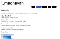
BTemplates.com

Powered by Blogger.

Pageviews past week

Quantum mechanics

Auto News

artificial intelligence

About Me

Recommend us on Google!

Information Technology

Popular Posts

Wednesday, June 24, 2009

Fotografix


Ultra-small, ultra-lightweight photo editor with an incredible number of options…


Adobe Photoshop and Gimp are great tools to have for anyone who likes to experiment with and edit images. However, these bulky software packages take up a lot of hard disk space and memory on your PC. So if you perform simpler tasks, such as cropping, small touches, etc, you can always opt to install something like Paint.NET .

Still, when it comes to portability, there is no software, which is worthwhile in this regard. There is a portable version of Gimp that you can carry around on your pen drive, but it’s very bulky and thus generally slow in actual usage.

Developer L. Madhavan decided to fill the void with his amazing new tool, Fotografix – a portable photo-editing application that requires no installation and takes up just 347KB of disk space!

Firing up the program, you are in for a surprise: At this light weight, the package still comes fully equipped with all of your regular image-manipulation tools, such as selection tools, brushes, clone tools, layer tools, etc.

Users of advanced software like Photoshop will have absolutely no issue in starting off, and will feel quite at home with Fotografix – a pane on the left with the necessary tools, the centre pane reserved for the photo and the right-most part has small windows to note your history and scripts.

Of course, it has its drawbacks. Those who like to freely crop their pictures will find the lasso tool sorely missing in this package, while the lack of keyboard and mouse shortcuts will irk power users.

But overall, this application misses out on very little from what is offered by the big guys; and considering its light -weight and zippy interface, it’s a must-have for your pen drive.

Rating: 4.5/5
Download: lmadhavan.com/software/fotografix/
Size: 347KB

If you like this post, buy me a beer at $3!
Reblog this post [with Zemanta]

Model Livers to Replace Lab Rats


German researchers have developed an external model of the liver, which could provide a safer alternative to animal experiments

If you have fever, headaches or a cold, it’s only a short way to the nearest chemist. The drugs, on the other hand, can take up to eight to ten years to develop.

Animal testing has been an essential step in testing drugs, but it continues to raise ethical issues. Furhermore,
humans and animals have different metabolisms, and around 30 per cent of side effects come to light in trials following animal experiments.

Researchers at the the Fraunhofer Institute have developed a model of the liver, which is viable outside the body and is suitable for testing drugs.

“Our artificial organ systems offer an alternative to animal experiments,” said Heike Mertsching, Professor at the Interfacial Engineering and Biotechnology Institute at Fraunhofer.

The test system, which Professor Mertsching has developed jointly with Dr Johanna Schanz, should in future give pharmaceutical companies more security and shorten the path to developing and releasing new drugs.

“Our liver model is a functioning system of blood vessels,” said Dr. Schanz. “We don’t build artificial blood vessels, but use existing ones – from a piece of pig’s intestine.”

All of the pig cells are removed, but the blood vessels are preserved. Human cells are then seeded onto this structure – hepatocytes, which are responsible for breaking down drugs, and endothelial cells, which act as a barrier between blood and tissue cells.

In order to simulate circulation, the researchers put the model into a computer-controlled bioreactor with a flexible tube pump. This enables the nutrient solution to be fed in and carried away in the same way as in veins and arteries in humans.

“The cells were active for up to three weeks,” said Dr. Schanz. “This time was sufficient to analyse and evaluate the functions. A longer period of activity is possible, however.”

The researchers established that the cells work in a similar way to those in the body. They detoxify, break down drugs and build up proteins. These are important pre-conditions for drug tests or transplants, as the effect of a substance can change when transformed or broken down. Many drugs are only metabolised into their therapeutic active form in the liver, while others can develop poisonous substances.

At the moment, the test system is being examined. The researchers say it could provide an alternative to animal testing within two years.


If you like this post, buy me a beer at $3!
Reblog this post [with Zemanta]

Thursday, June 18, 2009

New Nanoparticles Could Lead To End Of Chemotherapy


Nanoparticles specially engineered by University of Central Florida Assistant Professor J. Manuel Perez and his colleagues could someday target and destroy tumors, sparing patients from toxic, whole-body chemotherapies.

Dr. Manuel Perez and his team have been investigating
the use of nanoparticles for medicine for years.
(Credit: Jacque Brund)


Perez and his team used a drug called Taxol for their cell culture studies, recently published in the journal Small, because it is one of the most widely used chemotherapeutic drugs. Taxol normally causes many negative side effects because it travels throughout the body and damages healthy tissue as well as cancer cells.


The Taxol-carrying nanoparticles engineered in Perez's laboratory are modified so they carry the drug only to the cancer cells, allowing targeted cancer treatment without harming healthy cells. This is achieved by attaching a vitamin (folic acid) derivative that cancer cells like to consume in high amounts.


Because the nanoparticles also carry a fluorescent dye and an iron oxide magnetic core, their locations within the cells and the body can be seen by optical imaging and magnetic resonance imaging (MRI). That allows a physician to see how the tumor is responding to the treatment.


The nanoparticles also can be engineered without the drug and used as imaging (contrast) agents for cancer. If there is no cancer, the biodegradable nanoparticles will not bind to the tissue and will be eliminated by the liver. The iron oxide core will be utilized as regular iron in the body.


"What's unique about our work is that the nanoparticle has a dual role, as a diagnostic and therapeutic agent in a biodegradable and biocompatible vehicle," Perez said.


Perez has spent the past five years looking at ways nanotechnology can be used to help diagnose, image and treat cancer and infectious diseases. It's part of the quickly evolving world of nanomedicine.


The process works like this. Cancer cells in the tumor connect with the engineered nanoparticles via cell receptors that can be regarded as "doors" or "docking stations." The nanoparticles enter the cell and release their cargo of iron oxide, fluorescent dye and drugs, allowing dual imaging and treatment.


"Although the results from the cell cultures are preliminary, they are very encouraging," Perez said.


A new chemistry called "click chemistry" was utilized to attach the targeting molecule (folic acid) to the nanoparticles. This chemistry allows for the easy and specific attachment of molecules to nanoparticles without unwanted side products. It also allows for the easy attachment of other molecules to nanoparticles to specifically seek out particular tumors and other malignancies.


Perez's study builds on his prior research published in the prestigious journal Angewandte Chemie Int. Ed. His work has been partially funded by a National Institutes of Health grant and a Nanoscience Technology Center start-up fund.


"Our work is an important beginning, because it demonstrates an avenue for using nanotechnology not only to diagnose but also to treat cancer, potentially at an early stage," Perez said.


Perez, a Puerto Rico native, joined UCF in 2005. He works at UCF's NanoScience Technology Center and Chemistry Department and in the Burnett School of Biomedical Sciences in the College of Medicine. He has a Ph.D. from Boston University in Biochemistry and completed postdoctoral training at Massachusetts General Hospital, Harvard Medical School's teaching and research hospital.


If you like this post, buy me a beer at $3!
Reblog this post [with Zemanta]

Monday, June 15, 2009

WinCDEmu : Mounting a disc image was never so easy – just double-click and you’re done!


If you play video games on your PC on a regular basis, you are bound to need a disc-mounting tool.

Honestly, playing a game off its ISO image loaded on your hard disk is so much faster than playing the same through its DVD. Disc images also go a long way in preserving your DVD – and when you are buying a game for around Rs 1,000, you want to make sure the disc lasts for as long as it can!

So after you create a disc image, how do you load it up on to your computer? The normal course is to use a ‘mounting’ tool such as Daemon Tools or Alcohol.

These programs create a virtual optical disc drive on your computer and load, or ‘mount’, the disc image onto it. The final product is as good as having a DVD in a drive! But the process to use Daemon Tools and Alcohol is long and tedious.

First, you have to run the program; then create a virtual drive through a series of three clicks; and then go on to load the image through a further four clicks. Things have got to be simpler than this, right? Well, that’s where WinCDEmu comes in.

Cutting out all the various steps in mounting a disc image, this software works automatically to give you the most streamlined interface possible. All you have to do is install the program and double-click your ISO image for the program to automatically create a virtual drive and load up the image.

It’s really that simple! And the tedious process of unmounting a disc and getting rid of the virtual drive has also been cut down: just right click the virtual drive, hit ‘eject’ and you’re done.

Supporting ISO, CUE, BIN, RAW and IMG file formats, the application can create an unlimited number of virtual drives at a time. A must-have for any gamer!

Rating: 5/5
Download: wincdemu.sysprogs.org
Direct Download: biznetnetworks.dl.sourceforge.net/sourceforge/wincdemu/WinCDEmu-2.2.exe
Size: 782.10 KB


If you like this post, buy me a beer at $3!
Reblog this post [with Zemanta]

Friday, June 12, 2009

Scientists compute odds of interplanetary merger


Clash of the titans: Artist illustration depicts
collision between Earth and Venus

According to a study released Wednesday, there is a small, one-in-2500 chance that Earth will collide with Venus or Mars in 3.5 billion years.

Using powerful computers, Jacques Laskar, a researcher at the Observatoire de Paris, France, generated numerical simulations of orbital instability over the next five billion years.

Unlike previous models, they took into account Albert Einstein’s theory of general relativity. It resulted in dramatically different orbital paths.

The researchers looked at 2,501 possible scenarios, 25 of which ended with a severely disrupted Solar System.

“There is one scenario in which Mars passes 794 kilometres from Earth,” said Laskar. “When you come that close, it is the same as a collision as the planets gets torn apart.”

Laskar and colleagues ran additional two hundred computer models, slightly changing the path of Mars each time.

All but five of them ended in a two-way collision involving the Sun, Earth, Mercury, Venus or Mars. A quarter of them saw Earth smashed to pieces.

The cause of orbital chaos was the planet Mercury, found the study, published in the British journal Nature.

“Mercury is the trigger, and would be the first planet to be destabilised because it has the smallest mass,” explained Laskar. At some point Mercury’s orbit would get into resonance with that of Jupiter, throwing the smaller orb even more out of kilter, he said.


If you like this post, buy me a beer at $3!
Reblog this post [with Zemanta]

Monday, June 8, 2009

A cheaper, lighter and longer-lasting alternative to modern batteries


MOBILE phones looked like bricks in the 1980s. Indeed, they were so cumbersome that most were installed in cars. That was because the batteries required to power them were so hefty. When lithium-ion batteries were invented, mobile phones became small enough to be carried in a pocket or slipped into a handbag. Now a new design of battery that uses oxygen from ambient air to power devices could provide even smaller and lighter sources of power. Not only that, such batteries would be cheaper and last for longer between charges.


Lithium-ion batteries have two electrodes immersed in an electrically conductive solution, called an electrolyte. One of the electrodes, the cathode, is made of lithium cobalt oxide; the other, the anode, is composed of carbon. When the battery is being charged, positively charged lithium ions break away from the cathode and travel in the electrolyte to the anode, where they meet electrons brought there by a charging device. When electricity is needed, the anode releases the lithium ions, which rapidly move back to the cathode. As they do so, the electrons that were paired with them in the anode during the charging process are released. These electrons travel around an external circuit to which the battery is attached and power it.


Peter Bruce and his colleagues at the University of St Andrews in Scotland came up with the idea of replacing the lithium cobalt oxide electrode with a cheaper and lighter alternative. They designed an electrode made from porous carbon and lithium oxide. They knew that lithium oxide forms naturally from lithium ions, electrons and oxygen, but, to their surprise, they found that it could also be made to separate easily when an electric current passed through it. They exposed one side of their porous carbon electrode to an electrolyte rich in lithium ions and put a mesh window on the other side of the electrode through which air could be drawn. Oxygen from the air took the place of the cobalt oxide.


When they charged their battery, the lithium ions migrated to the anode where they combined with electrons from the charging device. When they discharged it, lithium ions and electrons were released from the anode. The ions crossed the electrolyte and the electrons travelled round the external circuit. The ions and electrons then met at the cathode, where they combined with the oxygen to form lithium oxide that filled the pores in the carbon.


Because the oxygen being used by the battery comes from the surrounding air rather than having to be carried around inside the battery, the device that Dr Bruce’s team has designed can be a mere one-eighth to one-tenth the size and weight of modern batteries, while still carrying the same charge. Alternatively, batteries using the new technology would last eight to ten times longer than modern batteries of the same size.


Performance and size are not the only expected improvements. Although they are not yet commercially available, making such a battery is expected to be cheaper. Lithium cobalt oxide accounts for 30% of the cost of a lithium-ion battery. Air, however, is free.


If you like this post, buy me a beer at $3!
Reblog this post [with Zemanta]

Saturday, June 6, 2009

Stem Cells Cultured On Contact Lens Restore Sight In Patients With Blinding Corneal Disease


In a world-first breakthrough, University of New South Wales (UNSW) medical researchers have used stem cells cultured on a simple contact lens to restore sight to sufferers of blinding corneal disease.

Dr. Di Girolamo and Dr. Watson.
(Credit: Image courtesy of University of New South Wales)


Sight was significantly improved within weeks of the procedure, which is simple, inexpensive and requires a minimal hospital stay.


The research team from UNSW’s School of Medical Sciences harvested stem cells from patients’ own eyes to rehabilitate the damaged cornea. The stem cells were cultured on a common therapeutic contact lens which was then placed onto the damaged cornea for 10 days, during which the cells were able to re-colonise the damaged eye surface.


While the novel procedure was used to rehabilitate damaged corneas, the researchers say it offers hope to people with a range of blinding eye conditions and could have applications in other organs.


A paper detailing the breakthrough appears in the journal Transplantation this week.


The trial was conducted on three patients; two with extensive corneal damage resulting from multiple surgeries to remove ocular melanomas, and one with the genetic eye condition aniridia. Other causes of cornea damage can include chemical or thermal burns, bacterial infection and chemotherapy.


“The procedure is totally simple and cheap,” said lead author of the study, UNSW’s Dr Nick Di Girolamo. “Unlike other techniques, it requires no foreign human or animal products, only the patient’s own serum, and is completely non-invasive.


The surgeon who carried out the procedure and managed the patients was UNSW senior lecturer, Dr Stephanie Watson.


"The operation is relatively non-invasive. The patient merely comes into the hospital for a couple of hours to have their eye prepared and the lens put in place, and then they're able to go home," she said.


“There’s no suturing, there is no major operation: all that’s involved is harvesting a minute amount – less than a millimeter – of tissue from the ocular surface,” said Dr Di Girolamo.


“If you’re going to be treating these sorts of diseases in third world countries all you need is the surgeon and a lab for cell culture. You don’t need any fancy equipment.”


Because the procedure uses the patient’s own stem cells harvested from their eye, it is ideal for sufferers of unilateral eye disease. However, it also works in patients who have had both eyes damaged, Dr Di Girolamo said.


“One of our patients had aniridia, a congenital condition affecting both eyes. In that case, instead of taking the stem cells from the other cornea, we took them from another part of the eye altogether – the conjunctiva – which also harbours stem cells.


“The stem cells were able to change from the conjunctival phenotype to a corneal phenotype after we put them onto the cornea. That’s the beauty of stem cells,” Dr Di Girolamo said.


The therapeutic contact lens used in the trial was of a type commonly used worldwide after ocular surface surgery. However, of the several brands on the market, only one was suitable for growing the stem cells.


“We don’t know why. It’s probably to do with the components the manufacturers have used in that particular lens,” Dr Di Girolamo said.


The researchers are hopeful the technique can be adapted for use in other parts of the eye, such as the retina, and even in other organs. “If we can do this procedure in the eye, I don’t see why it wouldn’t work in other major organs such as the skin, which behaves in a very similar way to the cornea,” Dr Di Girolamo said.

If you like this post, buy me a beer at $3!
Reblog this post [with Zemanta]